African Journal of
Microbiology Research

  • Abbreviation: Afr. J. Microbiol. Res.
  • Language: English
  • ISSN: 1996-0808
  • DOI: 10.5897/AJMR
  • Start Year: 2007
  • Published Articles: 5228

Full Length Research Paper

Characterization of Staphylococcus aureus isolated from human dental infection

Manisha Das
  • Manisha Das
  • Department of Microbiology and Hygiene, Faculty of Veterinary Science, Bangladesh Agricultural University, Mymensingh-2202, Bangladesh.
  • Google Scholar
Abdullah Al Momen Sabuj
  • Abdullah Al Momen Sabuj
  • Department of Microbiology and Hygiene, Faculty of Veterinary Science, Bangladesh Agricultural University, Mymensingh-2202, Bangladesh.
  • Google Scholar
Zobayda Farzana Haque
  • Zobayda Farzana Haque
  • Department of Microbiology and Hygiene, Faculty of Veterinary Science, Bangladesh Agricultural University, Mymensingh-2202, Bangladesh.
  • Google Scholar
Nanda Barua
  • Nanda Barua
  • Department of Microbiology and Hygiene, Faculty of Veterinary Science, Bangladesh Agricultural University, Mymensingh-2202, Bangladesh.
  • Google Scholar
Amrita Pondit
  • Amrita Pondit
  • Department of Microbiology and Hygiene, Faculty of Veterinary Science, Bangladesh Agricultural University, Mymensingh-2202, Bangladesh.
  • Google Scholar
Md. Muket Mahmud
  • Md. Muket Mahmud
  • Department of Microbiology and Hygiene, Faculty of Veterinary Science, Bangladesh Agricultural University, Mymensingh-2202, Bangladesh.
  • Google Scholar
Md. Ferdousur Rahman Khan
  • Md. Ferdousur Rahman Khan
  • Department of Microbiology and Hygiene, Faculty of Veterinary Science, Bangladesh Agricultural University, Mymensingh-2202, Bangladesh.
  • Google Scholar
Sukumar Saha
  • Sukumar Saha
  • Department of Microbiology and Hygiene, Faculty of Veterinary Science, Bangladesh Agricultural University, Mymensingh-2202, Bangladesh.
  • Google Scholar


  •  Received: 16 February 2019
  •  Accepted: 01 April 2019
  •  Published: 14 April 2019

 ABSTRACT

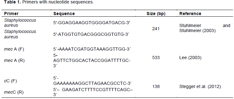
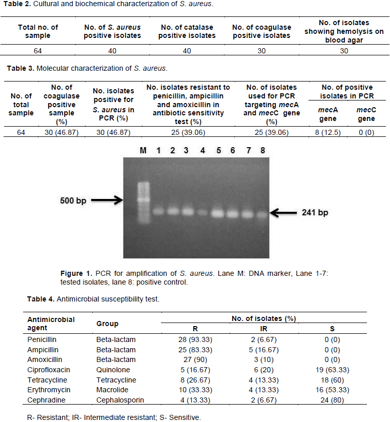
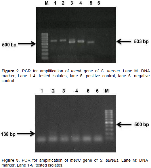
Staphylococcus aureus is an opportunistic pathogen causing dental infection and systemic infections in human body. This organism decreases susceptibility to several types of antibiotics every day and becomes more resistant which is a growing sense of concern in this era. Considering this fact, the study was attempted to characterize the S. aureus from human dental infection and to determine the antibiogram profile of isolates. Sixty four (64) samples were collected from the patients with dental infection who visited different dental clinics and hospitals in Mymensingh, Bangladesh for treatment. Isolation and identification of S. aureus were conducted by using cultural, morphological, and biochemical characteristics. Polymerase chain reaction was performed for final confirmation of S. aureus followed by detection of methicillin resistant S. aureus (MRSA) targeting mecA and mecC genes. Antibiotic susceptibility test of isolated bacteria was tested against seven antibiotics by disk diffusion methods. Forty isolates among 64 samples were found positive for S. aureus based on cultural characteristics. Among them 30 isolates were found positive in coagulase test. Depending on the result of coagulase test, all the 30 isolates were subjected to antibiotic sensitivity test and among them 25 were 100% resistant to penicillin, ampicillin and amoxicillin. All the 25 isolates were subjected to polymerase chain reaction (PCR) to identify methicillin resistant gene mecA and mecC. Eight isolates were positive for mecA gene while no isolates were positive for mecC. The present findings conclude that S. aureus is prevalent in dental infections and contain methicillin resistant genes.


Key words: Dental infection, Staphylococcus aureus, antibiotic resistance, methicillin resistant S. aureus (MRSA).


 INTRODUCTION

Human oral cavity acts as a growth medium for pathogenic microorganisms due to its moistures, temperature and nutrient content such as lipid, carbohydrate and protein (Mohapatra et al., 2012).  There are several types of dental infections which occur in a patient’s oral cavity such as tooth decay, periodontal disease, dental ache, dental plaque, dentin hypersensitivity,   dental    abscess,      dental     calculus,
 
hyperdontia, malocclusion, acid erosion, acute necrotizing ulcerative gingivitis, dental fluorosis, tooth impaction, etc. Staphylococcus aureus is a putative pathogen of many oral diseases, such as oral mucositis, periodontitis, peri-implantitis, endodontic infections and even dental caries (Gibson et al., 2000; Heitz-Mayfield and Lang, 2010; Poeschl et al., 2011; Passariello et al., 2012).
 
S. aureus is a Gram-positive, non-motile, non-spore forming grape like clusters and the most important coagulase positive pathogen from staphylococci due to combination of toxic mediated virulence, invasiveness and antibiotic resistance (Loir et al., 2003). Some strains of S. aureus have developed drug resistance (Faden, 2018). Methicillin-resistant S. aureus (MRSA) (Rajaduraipandi et al., 2006) is the strains of S. aureus that obtained resistance to beta-lactam antibiotics, which incorporates such as penicillin, amoxicillin, ampicillin, methicillin, oxacillin, cephalosporins, etc. (David and Daum, 2010). The propensity of S. aureus to acquire antibiotic resistance has prompted worldwide dissemination of clone expressing various antimicrobial resistances. Several hospital and nonhospital bacterial diseases are caused due to MRSA strains and sometimes lead to death (Bannerman and Peacock, 2007; Moussa et al., 2011; Peters et al., 2013; Faden, 2018).
 
Contaminations due to S. aureus, including the MRSA strains has long been common in Bangladesh. Because, indiscriminate use of antibiotics being a typical practice, hospital environments are not sufficiently clean as well as congestion of patients and attendants support  spread of the infectious agents including S. aureus (Khan et al., 2007). The possible presence of S. aureus is particularly important in dental caries because of its increased resistance (Vellappally et al., 2017). So it is very logical to check the status of microbial resistance against commonly used antibiotics for the treatment of dental infections occurred by S. aureus. Considering the fact that the present study aimed to characterize S. aureus in human dental infections collected from the patients of different clinics and hospital of Mymensingh district, Bangladesh.


 MATERIALS AND METHODS

Sample collection
 
A total of 64 samples of human dental infection were collected from different private clinics and Mymensingh Medical College Hospital (MMCH), Mymensingh during the period from January to March, 2018. The samples were collected directly by rubbing the infected teeth with sterile cotton buds and immediately placed them into nutrient broth and transferred to the laboratory within 1 to 2 h.
 
Isolation and identification of bacteria
 
Each nutrient broth containing samples were incubated at  37°C  for 4 h for enrichment. Samples were then streaked on Staphylococcus species specific growth media mannitol salt agar (HiMedia, India) and incubated at 37°C for 48 h to get single colonies. The suspected colonies were further identified as S. aureus after culturing onto mannitol salt agar (MSA) and 5% sheep blood agar. Gram’s staining and biochemical test including coagulase and catalase tests were performed for further confirmation of S. aureus (Cheesbrough, 1985).
 
Antimicrobial susceptibility test
 
Antibiotic susceptibility test was conducted using disk diffusion method or Kirby-Bauer (Bauer et al., 1966). All the coagulase positive isolates of S. aureus were subjected to antibiotic sensitive test against seven antibiotics namely penicillin (10 µg), ampicillin (20 µg), amoxicillin (30 µg), ciprofloxacin (5 µg), tetracycline (30 µg), erythromycin (15 µg) and cephradine (30 µg) (Himedia, India) which were generally prescribed by physicians in the study areas. The zone of inhibition produced by S. aureus against each antibiotic was measured and interpreted as resistant, intermediate resistant and susceptible according to standards of Clinical Laboratory and Standards Institute (CLSI, 2016).
 
Molecular characterization of S. aureus
 
For final confirmation, all the coagulase positive isolates of S. aureus were subjected to molecular test as methods described by Stuhlmeier and Stuhlmeier (2003). Boiling method was followed for genomic DNA extraction (Dashti et al., 2009). Furthermore, penicillin and penicillin like (ampicillin and amoxicillin) antibiotic resistant isolates of S. aureus were subjected polymerase chain reaction (PCR) for detection of mecA and mecC genes using previously published primers (Lee, 2003; Stegger et al., 2012). The list of primers used in the study is shown in Table 1.


 RESULTS AND DISCUSSION

Out of 64 samples of dental infection, 40 (62.5%) samples were found positive for S. aureus by observing cultural and morphological characteristics (Table 2). S. aureus growth in MSA appeared as golden yellow pigmented colonies and Gram’s staining exhibited Gram positive, cocci shaped and arranged in grape like cluster under microscope. These findings are similar to the findings of Ray and Ryan (2003). The percentage of S. aureus isolated from human dental infection is more frequent as the findings of Ohara-Nemoto et al. (2008) reported 46.4%. McCormack et al. (2015) and Kim and Lee (2015) reported 18 and 36.6% S. aureus in human oral and perioral infections that is comparatively lower than the current results.  
 
All the 40 isolates which were assumed positive for S. aureus were subjected to catalase and coagulase test where all of the isolates found positive for catalase test and 30 of them for coagulase test. Bubble formation in catalase and curd like clot formation in coagulase test indicated that the samples were positive for S. aureus indicating their ability to breakdown the hydrogen-peroxide to release free oxygen and plasma coagulation by activation of prothrombin (Karmakar  et  al.,  2016).  Of
 
the 40 isolates of S. aureus, 30 of them created β-hemolytic character in blood agar. The findings of the present study were higher than the findings of Karmakar et al. (2016) who reported 40% of β-hemolysis in blood agar.
 
Dental patients usually take antibiotics primarily to treat post­operative and secondary infections.  All 30 coagulase positive isolates of S. aureus were subjected to antibiotic susceptibility test and the results revealed that 93.33% isolates were found resistant to penicillin followed by 90% to amoxicillin, 83.33% to ampicillin and 33.33% to erythromycin where 80% sensitive to cephradine, 63.33% to ciprofloxacin, and 60% to erythromycin (53.33%) (Table 3). Kim and Lee (2015) reported that S. aureus isolated from the periodontal patients showed 88% (36/41) isolates resistant to penicillin and ampicillin which is comparable to the present study. They also observed the susceptibility rate and found 98% isolates susceptible to cefotetan, 95% to ciprofloxacin and 90% to tetracycline and erythromycin that is comparatively higher than the current study. Similar antimicrobial susceptibility results were reported by previous authors (Rajaduraipandi et al., 2006; Khan et al., 2007; Kim, 2012; Naeem et al., 2012). The higher resistant rate to commonly used antibiotics indicates indiscriminate or haphazard use that may have effect on treatment cost, poor prognosis as well as enhance  the   bacterial   infection   and   growth   virulent pathogens.
 
Coagulase positive isolates of S. aureus were finally confirmed by molecular test using polymerase chain reaction (PCR) and all them were found to be positive (Figure 1) which showed the relevance of the statement of Ohara-Nemoto et al. (2008). A total of 25 isolates found resistant to penicillin, ampicillin and amoxicillin which were further conducted to molecular characterization to detect mecA and mecC genes (Table 4). Out of 25 isolates, 8 were found positive for mecA and no isolates found mecC positive (Figures 2 and 3). It is a new gene which is emerging in the world especially in European countries and spreading among human and animals throughout the world which follows the statement of Paterson et al. (2014) but not for Bangladesh. The prevalence of mecA gene was 12.5% which was higher than the findings of Ayepola et al. (2015) who reported 2.4%. Smith et al. (2003) reported 6% MRSA positive isolates in oral infection. Another study conducted by Renvert et al. (2008) in Sweden observed similar finding connected to patients of periodontal infections. According to Kurita et al. (2006), dental patients are not only responsible for spreading MRSA, health professional may transmit this pathogen through their instruments. There are on established guideline for controlling MRSA but Centers for Disease Control and Prevention (CDC) recommends some standard precautionary measure  that might help to reduce the spread of MRSA in dental patients (Harte, 2010). The present results revealed that mecA positive but not mecC positive isolates of S. aureus are associated with dental infection in Mymensingh city of Bangladesh.


 CONCLUSION

Prevalence of S. aureus in dental patients is quite high and showed resistance to commonly used antibiotics as well as carried MRSA. Despite these results,  the  sample

size of this study is not sufficient and study period was too short to uncover actual picture of MRSA involved in dental infection in Mymensingh, Bangladesh. Large scale studies could be done both in hospitalized patients and in community to identify prevalence of MRSA, genome analysis, identification of toxin gene and other antibiotic resistant gene. Regular brushing of teeth, keeping up oral cleanliness and consulting with dental doctors to check up the teeth once in a month should be taken to maintain a distance from dental infections.


 CONFLICT OF INTERESTS

The authors have not declared any conflict of interests.


 ACKNOWLEDGEMENT

The author would like to acknowledge the Ministry of Education, Bangladesh for funding this research work. 


 REFERENCES

Ayepola OO, Olasupo NA, Egwari LO, Becker K, Schaumburg F (2015). Molecular characterization and antimicrobial susceptibility of Staphylococcus aureus isolates from clinical infection and asymptomatic carriers in Southwest Nigeria. Plos One 10(9):e0137531.
Crossref


Bannerman T, Peacock S (2007). Staphylococcus, Micrococcus, and other catalase positive cocci. In: Murray, P., Baron, E., Jorgensen, J., Landry, M., Pfaller, M. (Eds.), Manual of Clinical Microbiology 9th ed. ASM Press, Washington, DC, pp. 390-411.


Bauer A, Kirby W, Sherris JC, Turck M (1966). Antibiotic susceptibility test¬ing by a standardized single disk method. American Journal of Clinical Pathology 45(4):493-496.
Crossref


Cheesbrough M (1985). Medical laboratory manual for tropical coun¬tries. In: Microbiology. 1st edition, English Language Book Society, London, pp 400-480.


Clinical and Laboratory Standards Institute (CLSI) (2016). Performance standards for antimicrobial susceptibility testing. 26th edition, CLSI supplement M100s. Clinical and Laboratory Standards Institute, Wayne, Pennsylvania.


Dashti AA, Jadaon MM, Abdulsamad AM, Dashti HM (2009). Heat treatment of bacteria: A simple method of DNA extraction for molecular techniques. Kuwait Medical Journal 41(2):117-122.


David MZ, Daum RS (2010). Community-associated methicillin-resistant Staphylococcus aureus: epidemiology and clinical consequences of an emerging epidemic. Clinical Microbiology Reviews 23(3):616-687.
Crossref


Faden A (2018). Methicillin-resistant Staphylococcus aureus (MRSA) screening of hospital dental clinic surfaces. Saudi Journal of Biological Sciences.
Crossref


Gibson J, Wray D, Bagg J (2000). Oral staphylococcal mucositis: A new clinical entity in orofacial granulomatosis and Crohn's disease. Oral Surgery, Oral Medicine, Oral Pathology, Oral Radiology, and Endodontology 89(2):171-176.
Crossref


Harte JA (2010). Standard and transmission-based precautions: an update for dentistry. The Journal of the American Dental Association 141(5):572-581.
Crossref


Heitz‐Mayfield LJ, Lang NP (2010). Comparative biology of chronic and aggressive periodontitis vs. peri‐implantitis. Periodontology 53(1):167-181.
Crossref


Karmakar A, Dua P, Ghosh C (2016). Biochemical and molecular analysis of Staphylococcus aureus clinical isolates from hospitalized patients. Canadian Journal of Infectious Diseases and Medical Microbiology 2016:9041636.
Crossref


Khan AH, Shamsuzzaman AKM, Paul SK, Alam MM, Mahmud MC (2007). Antimicrobial susceptibility and coagulase typing of MRSA strains at Mymensingh Medical College. Bangladesh Journal of Medical Microbiology 1(2):56-60.
Crossref


Kim GY, Lee CH (2015). Antimicrobial susceptibility and pathogenic genes of Staphylococcus aureus isolated from the oral cavity of patients with periodontitis. Journal of Periodontal and Implant Science 45(6):223-228.
Crossref


Kim Y (2012). Multiple antimicrobial resistance patterns of Staphylococcus aureus isolated from periodontitis patients in Seoul, Korea. Korean Journal of Oral and Maxillofacial Pathology 36:317-339.


Kurita H, Kurashina K, Honda T (2006). Nosocomial transmission of methicillin-resistant Staphylococcus aureus via the surfaces of the dental operatory. British Dental Journal 201(5):297-300.
Crossref


Lee JH (2003). Methicillin (oxacillin)-resistant Staphylococcus aureus strains isolated from major food animals and their potential transmission to humans. Applied and Environmental Microbiology 69(11):6489-6494.
Crossref


Loir LY, Baron F, Gautier M (2003). Staphylococcus aureus and food poisoning. Genetics and Molecular Research Journal 2(1):63-76.


McCormack MG, Smith AJ, Akram AN, Jackson M, Robertson D, Edwards G (2015). Staphylococcus aureus and the oral cavity: An overlooked source of carriage and infection? American Journal of Infection Control 43(1):35-37.
Crossref


Mohapatra SB, Pattnaik M, Ray P (2012). Microbial association of dental caries. Asian Journal of Experimental Biological Sciences 3(2):360-367.


Moussa IM, Al-Qahtani AA, Gassem MA, Ashgan MH, Ismail DK, Ghazy AI, Shibl AM (2011). Pulsed-field gel electrophoresis (PFGE) as an epidemiological marker for typing of methicillin-resistant Staphylococcus aureus recovered from King Saudi Arabia (KSA). African Journal of Microbiology Research 5(12):1492-1499.
Crossref


Naeem M, Adil M, Naz SM, Abbas SH, Khan I, Khan A, Khan MU (2012). Resistance and sensitivity pattern of Staphylococcus aureus; a study in lady reading hospital peshawar. Journal of Postgraduate Medical Institute 27(1):42-47.


Ohara-Nemoto Y, Haraga H, Kimura S, Nemoto TK (2008). Occurrence of staphylococci in the oral cavities of healthy adults and nasal oral trafficking of the bacteria. Journal of Medical Microbiology 57(1):95-99.
Crossref


Passariello C, Puttini M, Iebba V, Pera P, Gigola P (2012). Influence of oral conditions on colonization by highly toxigenic Staphylococcus aureus strains. Oral Diseases 18(4):402-409.
Crossref


Paterson GK, Harrison EM, Holmes MA (2014). The emergence of mecC methicillin-resistant Staphylococcus aureus. Trends in Microbiology 22(1):42-47.
Crossref


Peters PJ, Brooks JT, McAllister SK, Limbago B, Lowery HK, Fosheim G, Guest JL, Gorwitz RJ, Bethea M, Hageman J, Mindley R, McDougal LK, Rimland D (2013). Methicillin-resistant Staphylococcus aureus colonization of the groin and risk for clinical infection among HIV-infected adults. Emerging Infectious Diseases 19(4):623-629.
Crossref


Poeschl PW, Crepaz V, Russmueller G, Seemann R, Hirschl AM, Ewers R (2011). Endodontic pathogens causing deep neck space infections: clinical impact of different sampling techniques and antibiotic susceptibility. Journal of Endodontics 37(9):1201-1205.
Crossref


Rajaduraipandi K, Mani KR, Panneerselvam K, Mani M, Bhaskar M, Manikandan P (2006). Prevalence and antimicrobial susceptibility pattern of methicillin resistant Staphylococcus aureus: A multicentre study. Indian Journal of Medical Microbiology 24(1):34-38.
Crossref


Ray C, Ryan KJ (2003). Sherris medical microbiology: an introduction to infectious diseases. Appleton and Lange Paramount Publishing Business and Professional Group, 237.


Renvert S, Lindahl C, Renvert H, Persson GR (2008). Clinical and microbiological analysis of subjects treated with Brånemark or AstraTech implants: a 7‐year follow‐up study. Clinical Oral Implants Research 19(4):342-347.
Crossref


Smith AJ, Robertson D, Tang MK, Jackson MS, MacKenzie D, Bagg J (2003). Staphylococcus aureus in the oral cavity: a three-year retrospective analysis of clinical laboratory data. British Dental Journal 195(12):701-703.
Crossref


Stegger Á, Andersen PS, Kearns A, Pichon B, Holmes MA, Edwards G, Laurent F, Teale C, Skov R, Larsen AR (2012). Rapid detection, differentiation and typing of methicillin‐resistant Staphylococcus aureus harbouring either mecA or the new mecA homologue mecALGA251. Clinical Microbiology and Infection 18(4):395-400.
Crossref


Stuhlmeier R, Stuhlmeier KM (2003). Fast, simultaneous, and sensitive detection of staphylococci. Journal of Clinical Pathology 56(10):782-785.
Crossref


Vellappally S, Divakar DD, Al Kheraif AA, Ramakrishnaiah R, Alqahtani A, Dalati MHN, Anil S, Khan AA, Varma HPR (2017). Occurrence of vancomycin-resistant Staphylococcus aureus in the oral cavity of patients with dental caries. Acta Microbiologica et Immunologica Hungarica 64(3):343-351.
Crossref


          */?>