Full Length Research Paper

ABSTRACT
The increasing trends of insect associated bacterial infection in humans are severely hampered by disparaging number of bacteria obtained with the culture-based technique. This study therefore determined how the analysis of the 16S rDNA sequences would compare in terms of precision and reliability to the most adoptable culture-based technique. Results obtained depict enhanced accuracy of molecular technique over the cultural method as only 249 (69%) of the total isolates were correctly identified by the cultural method to represent a total of 114 (31%) discrepant species while 100% correct identification was observed with the molecular technique. The most predominant of these bacterial isolates from both the external surfaces and the gut environment was Escherichia coli 43 (20.8%) and 24 (15.5%) respectively. The Gram positive organisms isolated were Staphylococcus aureus, Bacillus subtilis, Staphylococcus epidermidis, and Enterococcus faecalis with a prevalence rate of 8 (3.8%), 14 (6.7%), 8 (3.8%) and 9 (4.3%) from external surfaces and 2 (1.3%), 6 (3.9%), 2 (1.3%) and 7 (4.5%) from gut environment respectively. The least isolated organisms in the external surfaces were Serratia marscencens and Citrobacter werkmanii with a distribution rate of 3(1.4%) while Citrobacter freundii 2(1.3%) was the least isolated organism from cockroach gut environment. This study therefore showed that the molecular analysis of the 16S rDNA sequences is more efficient than culture-based technique for the identification of bacterial contaminants of cockroaches because occurrences of misidentification are very much abated by this method.
Key words: Bacterial contaminants, Cockroaches (Periplaneta americana), cultural technique, molecular technique.
INTRODUCTION
Cockroaches are dorso-ventally flattened insects known for being abundantly notorious and obnoxious non biting insect pest (Mba and Kelly, 2003) and have been reported to be on this planet earth for more than three million years (Zurek and Schal, 2004). These insect pests are among the few insects, serving as vectors for some of the most devastating diseases affecting humans (Czajka et al., 2003), as a result of their nocturnal and filthy habits that make them the ideal carriers of pathogenic microorganisms (Pai et al., 2005; Blazar et al., 2011).
Generally, cockroaches are found in moist shady areas in yards, hollow trees, wood piles and mulch and so, these insects are commonly contaminated with various microorganisms (Popoola, 2019). Their persistent and ubiquitous association with humans, animals, foods, refuse and excreta make them potential mechanical and biological vectors for the dissemination of pathogenic and multi drug resistant bacteria (Graczyk et al., 2001; Rahuma et al., 2005).
These insect pests have been implicated in the transmission of serious diseases such as dysentery, typhoid fever and food poisoning (Mba and Kelly, 2003) and have also been reported to harbor other pathogenic bacteria including Shigella species, Klebsiella species among others (Pai et al., 2005; Fakoorziba et al.,2010; Brown and Alhassan, 2015). The role of these insects in the epidemiology of different infections has been reported (Pai et al., 2005; Fakoorziba et al., 2010) and according to Czajka et al. (2003), an outbreak of nosocomial disease due to extended spectrum beta lactamase producing Klebsiella pneumoniae in neonatal wards was attributed to infestation of the neonatal wards with cockroaches, and the continuous presence of these insects in every home are mainly due to complexity of building structures, furniture as well as the emergence of insecticide resistant strains (Zurek and Gorham, 2008).
Certainly, there is a diverse collection of microorganisms from different sources globally (Stefani et al., 2014; Michaela et al., 2016), and cockroaches without doubts is a potential reservoir that could affect humans either by direct transmission of antibiotic resistant strains through their droppings or body parts. Conversely, despite the ubiquitous presence of microorganisms on cockroaches, identification of microorganism in this insect pest has been based largely on the use of the traditional cultural technique especially in developing world like Nigeria. This study was therefore aimed at comparing molecular and cultural techniques for the identification of bacterial contaminants of cockroaches.
MATERIALS AND METHODS
Collection of cockroaches
Prior to the commencement of the collection of cockroaches, visits were made to different places (houses, offices and shops) in Ago-Iwoye for awareness on the aim of this study and the importance of the study to the community; and, a total of two hundred (200) cockroaches were randomly collected from different parts of Ago iwoye using sticky trap method and sterile hand gloves. A transparent cylindrical plastic container with an opening of 11 cm in diameter and a lid was used to trap the cockroaches. The inside of the container was lined with blotting paper smeared with petroleum jelly and the trap kept in the kitchen or other strategic part of the house overnight, uncovered. The following day, live cockroach specimens were immediately transported to Microbiology Laboratory Unit of Olabisi Onabanjo University, Ago-Iwoye, Nigeria for analysis.
Identification of the sampled cockroaches
DNA of the sampled cockroaches were extracted from the leg of the insects using M-N Nucleospin tissue kit according to manufacturer’s instruction while approximately 2 ng of the extracted DNA was amplified and sequenced using the mitochondrial cytochrome oxidase subunit II (COII) with the aid of the following primer pairs for forward and reverse sequence (5’-AGAGTTTGATCCTGGCTCAG3’ and 3’-AAGGAGGTGATCCAGCCGCA5’). The sequenced isolates were aligned in MEGA 7.0 bioinformatic software and then subjected to blasting in order to identify the sequences. The sequences with the highest bit score were chosen as the appropriate name of the cockroach.
Microbiological and molecular processing of cockroaches
The external surface of the cockroaches were prepared by suspending each cockroach into a sterile universal bottle containing 2 ml buffered peptone water and then shaken to dislodge organisms attached to it in order to produce a homogenate specimen, after which the samples were inoculated onto MacConkey Agar, Xylose Lysine Deoxycholate Agar, and Eosin Methylene Blue Agar and then incubated at 37°C aerobically for 24 h. Also, prior to the isolation of bacterial isolates from the cockroaches gut, the cockroaches were aseptically removed from the container, decontaminated with 70% ethanol before being sacrificed using absolute chloroform-soaked cotton wool. The cockroaches were thereafter placed into 5 ml of sterile normal saline tube to remove ethanol residues before being placed in a wax tray, fixed with pins and dissected under aseptic conditions. Cockroach gut was removed and kept in 2 ml buffered peptone water for 30 min to produce homogenate specimens (Bala and Sule, 2012). Precisely, 0.1 ml of gut homogenate samples of each cockroach was separately inoculated onto MacConkey agar, Xylose Lysine Deoxycholate agar, and Eosin methylene blue agar using spread plate method and then incubated at 37°C aerobically for 24 h. All the isolates obtained in this study were identified first by biochemical test and then by sequencing the hypervariable 16S rDNA sequences using the following primer pairs for forward and reverse sequence (5’- TCCTACGGGAGGCAGCA3’ and 3’ TCACGGCACGAGCTGAC5’). The sequenced isolates were aligned in MEGA 7.0 bioinformatic software and then subjected to blasting in order to identify the organisms. The organisms with the highest bit score were chosen as the isolated organism.
Statistical analysis
The prevalence of bacterial isolates of cockroaches were calculated using the formula n/N, where n = number of specific organisms and N = total number of organisms.
RESULTS
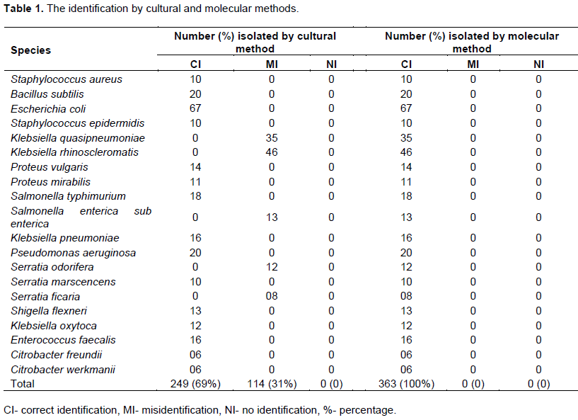
Table 1 shows the result of the identification of bacterial species by both cultural and molecular methods. Except for thirty-five (35) Klebsiella quasipneumoniae, forty-six (46) Klebsiella rhinoscleromatis, thirteen (13) Salmonella enterica, twelve (12) Serratia odorifera and eight (8) Serratia ficaria that were initially misidentified as Klebsiella pneumoniae, Salmonella typhimurium, Serratia marscencens respectively; all the remaining 249 (69%) were correctly identified by the cultural method. Conversely, the molecular technique identified all the isolated organisms resulting in 100% correct identification. The number of discrepant species identified by the cultural method represents a total of 114 (31%). These discrepant species include K. quasipneumoniae, K. rhinoscleromatis, Salmonella enterica, S. odorifera and S. ficaria. Of these discrepant species, K. rhinoscleromatis carries the highest percentage (40.4%) followed by K. quasipneumoniae (30.7%) while the least of the discrepant species was S. ficaria with prevalence rate of 7.02%.
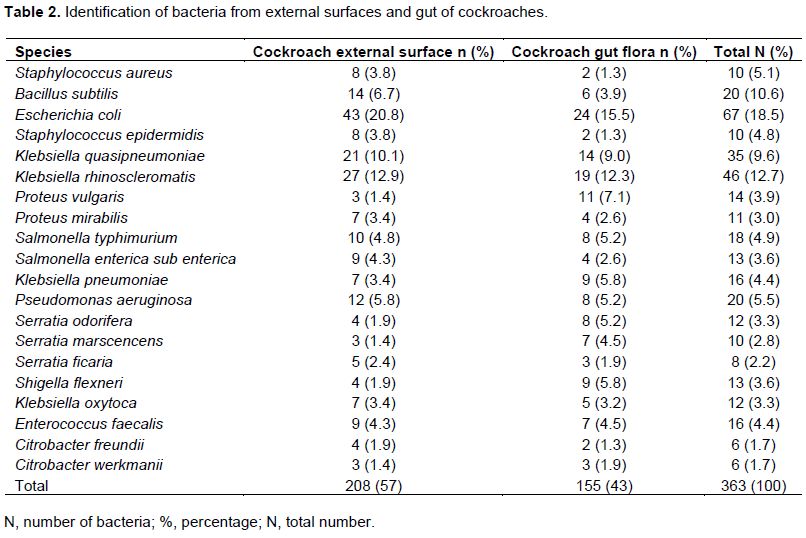
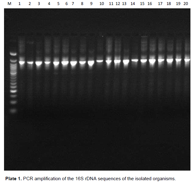
The identification of bacterial isolates from the external surfaces and gut of cockroaches is depicted in Table 2. As shown in Table 2, a total of 363 bacteria 208 (57%) from cockroach external surfaces and 155 (43%) from cockroach gut belonging to 20 bacterial species were isolated from both the cockroach external surfaces and gut. The most predominant of these bacterial isolates from both the external surfaces and gut was Escherichia coli having a prevalence rate of 43 (20.8%) and 24 (15.5%) respectively to depict a total percentage representation of 67 (18.5%) of the total isolates obtained. Staphylococcus aureus, Bacillus subtilis, Staphylococcus epidermidis, and Enterococcus faecalis were the only Gram positive bacterial isolates recovered with a prevalence rate of 8 (3.8%), 14 (6.7%), 8 (3.8%) and 9 (4.3%) from external surfaces and 2 (1.3%), 6 (3.9%), 2 (1.3%) and 7 (4.5%) from gut environment respectively. Generally, Gram negative bacteria were more represented in both external surfaces and gut environment. The least isolated organisms were Serratia marscencens 3 (1.4%) and Citrobacter werkmanii 3(1.4%) from the external surfaces while Citrobacter freundii 2 (1.3%) was the least isolated organism from cockroach gut environment. All the isolates recovered from the cockroach external surfaces were also recovered from the cockroach gut. Despite the almost equilibrium of the isolates of the external surfaces and the gut, the percentage prevalence of the isolates from the external surfaces were found to be significantly higher than those from the gut environment (tvalue = P<0.05). The PCR amplification of the 16S rDNA sequences of the isolated Organisms is depicted in Plate 1.
DISCUSSION
The role of cockroaches as vectors in the transmission of infectious agent has been well documented (Pai et al., 2005; Al-marjani, 2008). These insect vectors which are popular in human dwellings and hospital facilities were appropriately identified molecularly using COII gene and blast analysis of the sequenced cockroaches’ revealed significant similar index of 98% Periplaneta americana. This observation is not unexpected because COII gene has been previously used for the identification of such insects (Liu and Beckenbach, 1992). In a study by Jinfu and Chaohui (2002), COII genes are important in insect nomenclature.
In this study, the bacterial contaminants of cockroaches identified by cultural and molecular methods were represented by bacterial genera belonging to Staphylococcus spp, Bacillus spp, E. coli, Klebsiella spp, Proteus spp, Salmonella spp, Pseudomonas spp, Serratia spp, Shigella spp, Enterococcus spp and Citrobacter spp. This observation may not be unconnected to the fact that this important insect pest has close association with different wastes including garbage, sewage, sanitary waste among others, thus making them important carriers and transmitter of infectious agents including the multi-drug resistant bacterial strains (Tachbele et al., 2006). The identification of these bacterial isolates was more enhanced in terms of sensitivity and specificity with molecular method as compared to the cultural method to emphasize significant reliability of the former technique than the traditional method (Bhattacharya and Mondal, 2010).
Of the bacterial contaminants of cockroaches, the most frequently identified were Gram negative bacilli, explicitly in the family Enterobacteriaceae (Chaichanawongsaroj et al., 2004; Tachbele et al., 2006). Klebsiella spp 109 (30%) which were the most encountered bacterial isolates in cockroaches has been quoted in a related study as a major contaminant of insects (Kassiri et al., 2014). The second most predominant in this study is E. coli 67 (18.5) which corroborate the findings of other studies (Al-marjani, 2008; Tachbele et al., 2006). The findings of E. coli from cockroaches is an indication that these insects have been in contact with human faeces contaminated materials, and hence could be a health challenge to human (Kassiri et al., 2014) due to the possibility of these organisms causing infections including diarrhea, uremic syndrome, thrombocytopaenic purpura among others (Ejimadu et al., 2015).
The isolation of higher rate (57%) of bacterial isolates from the external surfaces compared to gut (43%) of cockroach goes in agreement with other studies (Adeleke et al., 2012; Tachbele et al., 2006). In contrary to this observation, Tachbele et al. (2006) and Ejimadu et al. (2015) reported higher isolation of bacteria from gut of cockroaches rather than the external surfaces. The relatively high external surface carriage rate may be related to filthy habits of cockroach which involves crawling and movement on different wastes thereby carrying microorganisms on their surface. Our findings however show that cockroaches are ubiquitous contaminants of several bacterial contaminants. Hence, it is important to view these organisms as an important vector that could transmit infectious agents to humans. This study therefore showed that the molecular analysis of the 16S rDNA sequences is more efficient than culture based technique for the identification of bacterial contaminants of cockroaches because occurrences of misidentification are very much abated by this method.
CONFLICT OF INTERESTS
The authors have not declared any conflict of interests.
REFERENCES
|
Adeleke MA, Oyebamiji AA, Hassan AO, Adeyi AO, Wahab AA, Olaitan JO (2012). Biolarvicidal efficacies of entomopathogenic microorganisms isolated from the breeding sites of mosquitoes in Osogbo, Southwestern Nigeria. African Entomology 20(2):290-294. Crossref |
||||
| Al Marjani MF (2008). Study Of Extended-Spectrum ßlactamases Producing Enterobacteria isolated from Cockroaches (Periplaneta americana) of Central Medicine City Hospital. Al-Mustansiriyah Journal of Science 19:9-16. | ||||
| Bala AY, Sule H (2012). Vectorial potential of cockroaches in transmitting parasites of medical importance in Arkilla, Sokoto, Nigeria. Nigerian Journal of Basic and Applied Science 20(2):111-115. | ||||
|
Brown C, Alhassan A (2015). Multiple-antibiotic-resistant bacteria from cockroaches trapped from a public hospital and a nearby students' hostel in Accra, Ghana. International Journal of Biological and Chemical Sciences 8(4):1859-1864. Crossref |
||||
|
Blazar JM, Lienau EK, Allard MW (2011). Insects as vectors of foodborne pathogenic bacteria. Terrestrial Arthropod Reviews 4(1):5-16. Crossref |
||||
|
Bhattacharya S, Mondal AS (2010). Clinical microbiology in the intensive care unit: strategic and operational characteristics. Indian Journal of Medical Microbiology 28(1):5-10. Crossref |
||||
| Czajka E, Pancer K, Kochman M (2003). Characteristics of bacteria isolated from body surface of German cockroaches caught in hospitals. PrzeglÄ…d Epidemiology 57(4):655-662. | ||||
| Chaichanawongsaroj N, Vanichayatanarak K, Pipatkullachat T, Mongkol PM, Somkiatcharoen S (2004). Isolation of Gram-negative bacteria from cockroaches trapped from urban environment. The Southeast Asian Journal of Tropical Medicine and Public Health 35(3):3-8 | ||||
| Ejimadu LC, Goselle ON, Ahmadu YM, James R (2015). Specialization of Periplaneta Americana (American Cockroach) and Blattella germanica (German cockroach) towards intestinal parasites: A Public Health Concern. Journal of Pharmaceutical and Biological Sciences 10:23-32. | ||||
|
Fakoorziba MR, Eghbal F, Hassanzadeh J, Moemenbellah-Fard MD (2010). Cockroaches (Periplaneta americana and Blattella germanica) as potential vectors of the pathogenic bacteria found in nosocomial infections. Annals of Tropical Medicine and Parasitology 104(6):521-528. Crossref |
||||
|
Graczyk TK, Knight R, Tamang L (2005). Mechanical transmission of human protozoan parasites by insects. Clinical Microbiology Reviews 35:128-132. Crossref |
||||
| Jinfu W, Chaohui H (2002). Molecular divergence of the mitochondrial cytochrome oxidase ii gene in three mosquitoes. Journal of the American Mosquito Control Association 18(4):301-306. | ||||
|
Kassiri H, Kassiri A, Kazemi S (2014). Investigation on American cockroaches medically important bacteria in Khorramshahr hospital, Iran. Asian Pacific Journal of Tropical Disease 4:201-203. Crossref |
||||
|
Liu H, Beckenbach AT (1992). Evolution of the mitochondrial cytochrome oxidase II gene among 10 orders of insects. Molecular Phylogenetics and Evolution 1(1):41-52. Crossref |
||||
| Mba CE, Kelly D (2003). Preliminary trials of environment friendly cockroach traps in Zaria Northern Nigeria. Journal of Tropical Biosciences 3:7-10. | ||||
| Michaela JD, Irene RG, Alieda VE, Cindy D, Kristina K, Anne-Kathrin S, Guanghui W, Marie AC, Vivienne D, John W, Reiner H, Beatriz G, Stefan S, John T, Martin JW, Nick C, Dik M, Neil W (2016). Diversity of STs, plasmids and ESBL genes among Escherichia coli from humans, animals and food in Germany, the Netherlands and the UK. | ||||
|
Pai HH, Chen WC, Peng CF (2005). Isolation of bacteria with antibiotic resistance from household cockroaches (Periplaneta americana and Blattella germanica). Acta Tropica 93(3):259-265. Crossref |
||||
| Popoola OD (2019). Molecular characterization of extended spectrum beta lactamases and their genes in enterobacteria isolated from periplaneta americana (cockroaches). An unpublished Ph.D thesis in the Department of Microbiology, Olabisi Onabanjo University, Ago Iwoye. | ||||
|
Rahuma N, Ghenghesh KS, Ben Aissa R, Elamaari A (2005). Carriage by the housefly (Musca domestica) of multiple-antibiotic-resistant bacteria that are potentially pathogenic humans, in hospital and other urban environments in Misurata, Libya. Annals of Tropical Medicine and Parasitology 99:795-802. Crossref |
||||
| Stefani S, Ilaria G, Immacolat A, Carla C, Patrizia M, Simona N, Moreno (2014). Susceptibility Testing: Twenty-Fifth Informational Supplement; M100-S25. Clinical and Laboratory Standards Institute, Wayne, PA, USA. | ||||
| Tachbele E, Erku W, Gebre-Michael T, Ashenafi M (2006). Cockroach-associated food-borne bacterial pathogens from some hospitals and restaurants in Addis Ababa, Ethiopia: Distribution and antibiograms. Journal of Rural and Tropical Public Heath 5:34-41. | ||||
|
Zurek L, Schal C (2004). Evaluation of the German cockroach (Blattella germanica) as a vector for verotoxigenic Escherichia coli F18 in confined swine production. Veterinary Microbiology 101:263-267. Crossref |
||||
|
Zurek L, Gorham JR (2008). Insects as vectors of food borne pathogens. In: Hoboken VJG, John NJ, editors. Wiley Handbook of Science and Technology for Homeland Security. Wiley and Sons, Inc; New York. Crossref |
||||
Copyright © 2024 Author(s) retain the copyright of this article.
This article is published under the terms of the Creative Commons Attribution License 4.0


