Review
ABSTRACT
Carotid artery injuries with pseudo-aneurysm are uncommon but associated with central neurologic dysfunction. We present a 26-year-old man with a giant bleeding pseudo-aneurysm of right common carotid artery managed by emergency sternotomy, neck exploration and repair of the aneurysm. On the right side of the neck was a 10 × 8 cm mass occupying almost the entire posterior triangle. It was pulsatile, tender with sinus discharging serosanguinous fluid. Positive thrill and bruit were demonstrable over the mass. Conventional Computerized Angiography (CTA) and Distal Subtraction Angiography (DSA) showed a 1 cm defect in the lateral wall of the distal right common carotid artery (RCCA), complete circle of Willis and massive blood clot at the site of the defect. In middle-and low-income settings where the technical know-how and resources for stenting including cerebral oximeter are not available, expertise in open surgical approach becomes the only way to save life.
Key words: Diagnosis, equipment, surgery, treatment, pseudoaneurysm, carotid artery, rupture.
INTRODUCTION
Aneurysm is defined as localized artery enlargement more than 150% of the segmental artery diameter (Nwafor et al., 2012, 2015). Extra-cranial carotid artery pseudo-aneurysms and true aneurysms are extremely rare, altogether accounting for only 0.4-4% of all peripheral artery aneurysms (Cogbill et al., 1994; Fabian et al., 1996). Pseudo-aneurysm in particular has the propensity to enlarge and rupture (contained or uncontained, put compression on the surrounding structures like trachea, oesophagus, stellate ganglion, recurrent laryngeal nerve (Diego et al., 2015; Akhiwu et al., 2018). Other complication that makes surgical treatment mandatory is the atheroembolism with associated cerebrovascular accident/stroke. According to Attigah et al. (2009) and Malikov et al. (2010) classification the types 1 and 2 are very difficult to treat surgically, hence the need for multidisciplinary approach with neurosurgeons, neurophysician, otorlarngyogologist, maxillofacial surgeon, vascular surgeon and physiotherapist as well as radiologist and histopathologist. Open and endovascular or hybrid treatment approaches are recognized in the literature. Ultimately, the type of treatment will depend on the mode of presentation, surgeon/interventionist experience, aneurysm location or accessibility and aetiology (Welleweerd et al., 2015; Zhang et al., 1999).
CASE SUMMARY
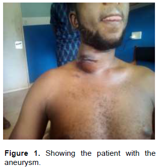
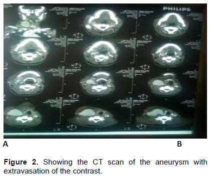
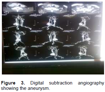
The patient is O. K., a 26-year-old tailor apprentice. He is a Christian of Roman Catholic denomination. He presented with progressive right sided neck swelling; 6 weeks prior (Figure 1). He was cut in the right side of the neck with machete by a co-worker following a misunderstanding. There was profuse bleeding around the site. On raising alarm, he got assistance by some village security men who helped him put pressure at the site and took him to hospital. Wound was sutured to arrest haemostasis and he received 2 units of blood during resuscitation. Patient’s clinical condition remained uneventful. Swelling insidiously developed around the wound and progressively increased in size. With increase in size, a sinus developed within the wound and continued to discharge serosanguinous fluid few days prior to referral to our service. Our review noted the above findings. Patient was moderately pale. Haemoglobin was 8.9 g. In addition, a mass 10 × 8 cm, tender, pulsatile with bruit was found in the posterior triangle with scarification mark and a sinus discharging serosanguinous fluid. Conventional computerized tomographic scan and digital subtraction angiography showed a 1 cm defect in the lateral wall of the distal right common carotid artery, complete circle of Willis and massive blood clot at the site of the defect. Patient was worked up for emergency surgery (Figure 2 to 5).
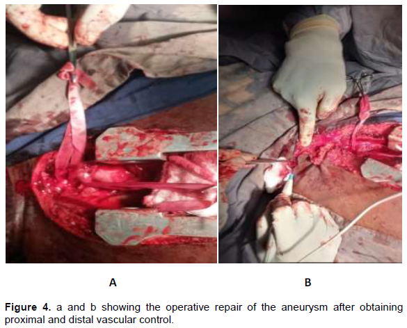
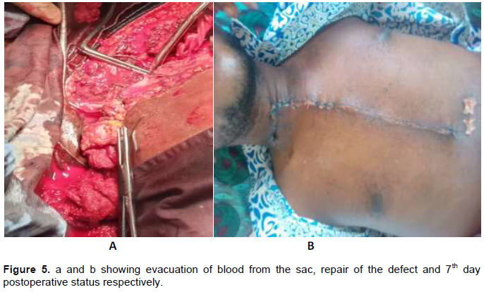
Proximal and distal control of right common carotid artery was done, bleeding defect isolated and controlled with digital pressure before closing with 5-0 prolene. Duration of right innominate artery clamping was 15 min and common carotid artery clamping was 10 min respectively. This is regarded as warm ischemic time. Patient was transfused 3 units of blood intra-operatively and estimated blood loss was about one liter. Wound was closed in layers with redivac drain in the neck and mediastinal drains in the chest. Analgesics, antibiotics, anticoagulants, heamatinic and physiotherapy were instituted postoperatively. His recovery was uneventful.
Operative detail
Under general anaesthesia, positioned supine, a median sternotomy was performed. The aortic arch was identified after careful dissection. The exposure was extended superiorly to isolate the left innominate vein and innominate artery. A vascular clamp was applied to the latter. 5000 unit of heparin was administered intravenously. The aneurysmal sac was entered by detaching the sternal and clavicular attachment of sternocleidomastoid muscle. The haematoma was evacuated and a defect on the right common carotid artery (RCCA) identified. Digital pressure was applied to the point and vascular clamp applied proximally and distally to the defect. The rent was then closed with 5-0 prolene. Haemostasis was achieved using cautery and suture ligation. After removing all the vascular clamps, no surgical bleeding was noticed. Mediastinal and pericardial drains including neck drains were instituted and wound closed in layers. Estimated blood loss intraoperatively was 1 L. Immediate postoperative status was satisfactory.
DISCUSSION
Trauma and prior carotid surgery such as endarterectomy are the most common causes of carotid artery pseudo-aneurysms. Other causes include atherosclerosis, vasculitis and collagen vascular diseases (Chen et al., 2019; Martin et al., 1991). The index case was caused by penetratingtrauma. The location of the aneurysms was equally distributed between the internal and common carotid arteries. The external carotid aneurysm is usually inconsequential as it can be ligated with impunity. In the index case surgical repair was safe and effective with no significant morbidity or mortality and good midterm stroke prevention strategies. In the developed world, endovascular techniques with stent-graft exclusion have emerged as an efficient and safe alternative to open surgical resection (DuBose et al., 2008). However, in low-income setting like ours, with absence of technical know- how and resources for endovascular intervention, expertise in open surgical approach becomes the only way to save life.
Historically, extra-cranial carotid aneurysm repair has undergone evolution from the time of Astley Cooper who performed the first ligation in 1805. Treatment strategiesin open approach involve resection and reconstruction with end-to-end anastomosis, with interposition graft using autologous external artery conduit, saphenous vein or exogenous prostheses like expanded polytetrafluoroethylene (ePTFE) or Dacron graft. Other methods are resection with intracranial and extracranial bypass or outright ligation as a damage control measures in uncontained ruptured (Zhang et al., 1999). Our patient was treated by lateral arteriography.
Usually, a complete work up will help in determining the cause, location, morphologic features, evaluation of surrounding structures, vascular anatomic information and treatment planning. Ultrasonography, computerized tomographic scan and digital subtraction angiography are the main diagnostic tools in evaluating patients with pseudoaneurysms of common carotid artery of either side or both. These were deployed in the index case.
In the modern era, surgical repair of carotid artery injuries is associated with mortality rates of 0 to 22% and postoperative progression of neurologic deficit of 0 to 21% (Martin et al., 1991). Endovascular approaches are increasingly used more frequently in carotid injuries. Stenting has most commonly been used for high extracranial internal carotid lesions as pseudoaneurysms (Martin et al., 1991; DuBose et al., 2008). Compared with surgical treatment of carotid injuries, with an associated mortality rate of up to 22%, carotid stenting appears to be much lower at 0.9% (DuBose et al., 2008; Coldwell et al., 2000; Berne et al., 2008; Biffl et al., 2001). In addition, stroke rates associated with carotid stenting of trauma, at 3.5%, appear comparable to those after operative repair (0-21%) (Chen et al., 2019). In the low-income setting, the technical know-how and resources for stenting are absent. Furthermore, the use of cerebral oximeter in the course of surgery is a cutting-edge technology that is currently not available in the developing country like ours.
CONCLUSION
Though extra-cranial carotid aneurysm is rare, it usually presents many challenges in its management, especially when it presents as emergency in the form of rupture, either contained or uncontained. Depending on the type, according to Attigah and Malikove classification a multidisciplinary team approach (surgeon, radiologist, haematologist, anaesthesist including physiotherapist and nurses) is very important in the management. Stenting and or hybrid procedures are not possible in low-income settings such as ours. This justifies an emphasis to gain expertise with an open approach.
CONFLICT OF INTERESTS
The authors have not declared any conflict of interests.
REFERENCES
|
Akhiwu BI, Peter SD, Njem JM, Ojo EO (2018). Giant Extracranial Carotid Aneurysm Causing Acute Airway Obstruction. Journal of Advances in Medicine and Medical Research 28(1):1-7. |
|
|
Attigah N, Kulkens S, Zausig N (2009). Surgical therapy of extracranial carotid artery aneurysms: Long-term results over a 24-year period. European Journal of Vascular and Endovascular Surgery 37(2):127-133. |
|
|
Berne JD, Reuland KR, Villarreal DH, McGovern TM, Rowe SA, Norwood SH (2008). Internal carotid artery stenting for blunt carotid artery injuries with an associated pseudoaneurysm. The Journal of Trauma 64(2):398-405. |
|
|
Biffl WL, Moore EE, Offner PI, Burch JM (2001). Blunt carotid and vertebral arterial injuries. World Journal of Surgery 25(8):1036-1043. |
|
|
Chen Z, Chen L, Zhang J, Yongjun L (2019). Management of Extracranial Artery Aneurysms: 6-year Case series. International Medical Journal of Experimental and Clinical Research 25:4933-4940. |
|
|
Cogbill TH, Moore EE, Meissner Ml (1994). The spectrum of blunt injury to the carotid artery: a multicenter perspective. The Journal of Trauma-Injury Infection and Critical Care 37(3):473-479. |
|
|
Coldwell DM, Novak Z, Ryu RK (2000). Treatment of posttraumatic internal carotid arterial pseudoaneurysms with endovascular stents. Journal of Trauma, Injury, Infection and Critical Care 48(3):470-472. |
|
|
Diego R, Stefanov S, Riera del Moral L., Álvarez J, Riera de Cubas L (2015). Endovascular Treatment of Posttraumatic Pseudoaneurysm of the Common Carotid Artery, Hindawi. Case Reports in Vascular Medicine Volume. Article ID 427040, 4 pages |
|
|
DuBose J, Recinos G, Teixeira PGR, Inaba K, Demetriades D (2008). Endovascular stenting for the treatment of traumatic internal carotid injuries: expanding experience. The Journal of Trauma 65(6):1561-1566. |
|
|
Fabian TC, Patton IH, Croce MA, Minard,Kudsk GKA, Pritchard FE (1996). Blunt carotid injury: importance of early diagnosis and anticoagulant therapy. Annals of Surgery 223(5):513-525. |
|
|
Malikov S, Thomassin JM, Magnan PE (2010). Open surgical reconstruction of the internal carotid artery aneurysm at the base of the skull. Journal of Vascular Surgery 51:323-329 |
|
|
Martin RF, Eldrup-Jorgensen J, Clark DE, Bredenberg CE (1991). Blunt trauma to the carotid arteries. Journal of Vascular Surgery 14(6):789-795. |
|
|
Nwafor IA, Eze JC, Ezemba N, Anyanwu CH (2012). The Challenges Facing the Management of Arterial Aneurysms in University Nigeria Teaching Hospital, Enugu, Nigerian Journal of Medicine 21(4):438-440. |
|
|
Nwafor IA, Eze JC, Ezemba N, Ngene CI, Akpan AF (2015). Giant Pseudoaneurysm of a splanchnic artery: A case report. Journal of Vascular Medicine and Surgery 3:201. |
|
|
Welleweerd JC, Den Ruijter HM, Nelissen BGL, Bots ML, Kappelle LJ, Rinkel GJE, De Borst GJ (2015). Management of extracranial carotid artery aneurysm. European Journal of Vascular and Endovascular Surgery 50(2):141-147. |
|
|
Zhang Q, Duan ZQ, Xin SJ, Wang XW, Dong YT (1999). Management of Extracranial Carotid Artery Aneurysms: 17 Years' Experience. European Journal of Vascular and Endovascular Surgery 18(2):162-165. |
|
Copyright © 2024 Author(s) retain the copyright of this article.
This article is published under the terms of the Creative Commons Attribution License 4.0


