International Journal of
Biodiversity and Conservation

  • Abbreviation: Int. J. Biodivers. Conserv.
  • Language: English
  • ISSN: 2141-243X
  • DOI: 10.5897/IJBC
  • Start Year: 2009
  • Published Articles: 679

Full Length Research Paper

In vitro micropropagation of Rhinacanthus nasutus (L) Kurz

Nissar Ahmad Reshi
  • Nissar Ahmad Reshi
  • Plant Tissue Culture Laboratory, Department of Studies in Botany, University of Mysore, Manasagangotri, Mysore, Karnataka, 570 006, India.
  • Google Scholar
Sudarshana M. S.
  • Sudarshana M. S.
  • Plant Tissue Culture Laboratory, Department of Studies in Botany, University of Mysore, Manasagangotri, Mysore, Karnataka, 570 006, India.
  • Google Scholar
Girish H. V.
  • Girish H. V.
  • Plant Tissue Culture Laboratory, Department of Studies in Botany, University of Mysore, Manasagangotri, Mysore, Karnataka, 570 006, India.
  • Google Scholar


  •  Received: 11 November 2016
  •  Accepted: 17 January 2017
  •  Published: 30 September 2018

 ABSTRACT

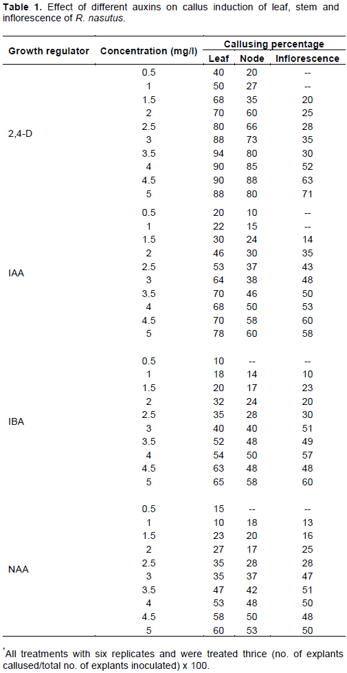
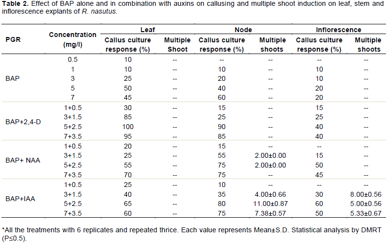
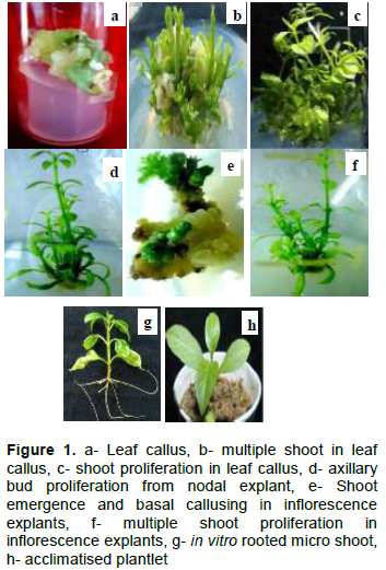
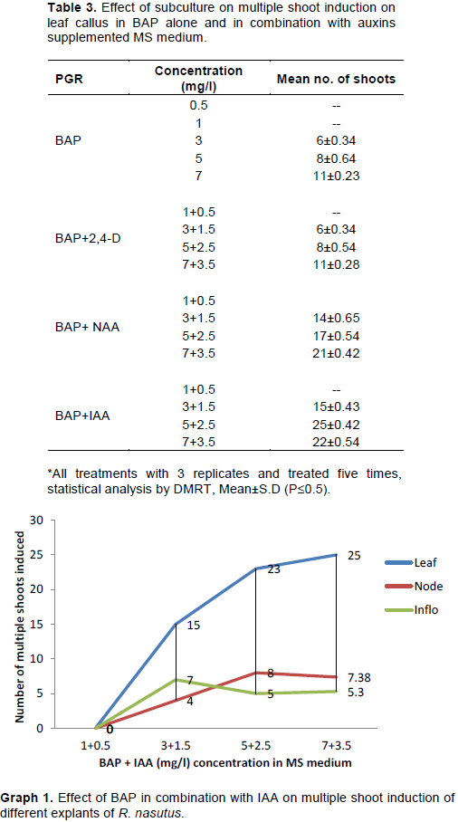
The study was aimed to standardise a protocol for the in vitro mass propagation of Rhinacanthus nasutus (L) Kurz, an anticancer shrub. Leaf, node and inflorescence explants were inoculated onto the Murashige and Skoog’s (MS) medium enriched with different combinations and concentrations of growth regulators. Maximum callusing percentage was achieved in leaf explants in MS medium supplemented with 5 mg/l 2,4-D. Multiple shoots were achieved from leaf, node and inflorescence explants with maximum of 25±0.42 (5 mg/l BAP + 2.5 mg/l IAA), 11±0.87 (5 BAP + 2.5 mg/l IAA) and 8±0.56 (3 mg/l BAP + 1.5 mg/l IAA) shoots, respectively. For in vitro rhizogenesis, elongated micro shoots were aseptically transferred to the half strength MS liquid medium with maximum number of 8±0.89 roots per shoot achieved in 1 mg/ml IAA fortified MS medium. The in vitro rooted micro shoots were acclimatised under laboratory conditions for two weeks by transferring to polycups containing sterile soil, sand and vermiculite (1:1:1). After two weeks, hardened plantlets were transferred to the green house for two weeks and then finally to the garden with 95% survivability.


Key words: Rhinacanthus nasutus, multiple shoots, leaf callus, node, inflorescence, rhizogenesis.  

Abbreviation: MS, Murashige and Skoog; BAP, 6-benzylaminopurine; 2,4-D, 2,4-dichlorophenoxyaceticacid; IAA, indole-3 acetic acid; IBA, idole butyric acid; NAA:- naphthalene acetic acid; Kn, kinetin.  

 INTRODUCTION

Ever since the the existence of human being, plants have been relied upon for medicinal purposes. In the last few decades, there has been surge in the public interest for the herbal medicine due to the ill effects associated with synthetic medicine. The pressure on pharmaceutical industries to meet the demands has led to the over exploitation of medicinal wealth. Unrestricted harvesting of plants from the wild which is the main source of raw material  is  the  cause  of  loss  of  genetic  diversity  and habitat destruction. Approximately, there are 50,000 medicinal plant species used and two third of this number is collected from the wild (Edwards, 2004). Growing public interest in herbal medicine, industrial resurgence, growing world population, fast urbanisation and increasing anthropogenic activities has put the natural habitat of medicinal plants in dwindling mode and the wild population of medicinal plants has drastically reduced (Saha et al., 2007).  Therefore, the  management  for  the conservation of the traditional medicinal plants is the matter of urgency to negate the threat of extinction in future. Hence, ex situ conservative measures need to be strategized and standardised for the mass propagation of medicinal plants to cater the rising pharmaceutical demands while simultaneously negating the threat.
 
Rhinacanthus nasutus is a multipurpose ethnomedicinal shrub, belonging to the family, Acanthaceae. The plant is 1 to 2 m high; leaves are oblong, 3 to 8 cm in length, narrowed and pointed at both ends. The plant is available in India, Taiwan, Thailand, South china, Ceylon and Madagascar. The plant is collected throughout the year for its roots and leaves which are used by tribals for the treatment of various ailments. Traditionally, it is used to treat cancer, rheumatism (Chopra et al., 1956), eczema (Atsusi and Yoshioki, 1993), pulmonary tuberculosis, influenza virus, liver diseases, peptic ulcers, helminthiasis, scurvy, inflammation, hypertension and obesity (Kernan et al., 1997), leprosy, dhobie’s itch, ring worm, poison toxicity (Muthukumaraswamy et al., 2003), herpes simplex virus, measles virus, polio virus (Subramaniam, 2006). Pharmacologically, the plant has been reported to possess cytotoxic (Wu et al., 1988); antiviral (Sendl et al., 1996); antitumor (Kongkathip et al., 2003), antiproliferative (Gotoh et al., 2004), anticancer (kupradinun et al. 2009), antimicrobial (Puttarak et al., 2010), anti-inflammatory (Nisarat et al., 2010), antidiabetic (Rao and Naidu, 2010) and antioxidant activities (Upendra et al., 2010). Natural resurgence of R. nasutus is through stem cuttings which is plagued with low rooting percentage and seeds are non-viable (Rao and Naidu, 2010). From last few years, R. nasutus seldom appears in its natural habitat of low hills of Nilgiri of Western Ghats of Tamil Nadu. At this point, micropropagation through plant tissue culture holds a significant promise for true to type and mass propagation of medicinally important plants. It is a potent biotechnological tool which has opened a vast area of research for biodiversity conservation. Owing to the alarming rate at which the R. nasutus is harvested and high pharmaceutical demands, a study was taken up to standardize a protocol for the mass propagations of R. nasutus using leaf, node and inflorescence explants. 


 MATERIALS AND METHODS

Rhinacanthus nasutus plants were collected from Nilgiri Hills of Western Ghats of Tamil Nadu and maintained in the botanical garden of Department of Botany, University of Mysore, Mysuru, India. Explants like leaf, node and inflorescence were collected from the garden and washed under running tap water for 10 min to remove the soil and dust particles. The explants were then treated with bavistin (5% w/v) for 5 min followed by dipping in 70% alcohol for 30 s and were then washed with sterile double distilled water. Inside the laminar air flow explants were treated with 0.01% HgCl2 for 5 min followed by washing thrice with sterile double distilled water. The surface sterilised explants were cut into small pieces (1 to 2 cm) and inoculated on the MS medium fortified with different combinations and concentrations of growth regulators (Tables 1 and  2).   The   pH  of   the   medium   was   adjusted  by  using  1 N NaoH/HCL to 5.8 prior to autoclaving at 121°C for 15 min. The cultures were incubated at 22±2°C with 16 h photoperiod under light florescent tubes with light intensity of 25 μmol/s2/m2 for 4 weeks.    In vitro regenerated shoots were excised aseptically after attaining a height of 3 to 5 cm and transferred to half strength MS liquid medium fortified with different concentrations of auxins (Table 4) for in vitro rhizogenesis. The data was collected after 4 weeks, measuring root lengths and number of roots per shoot. After 4 weeks, well rooted micro shoots were hardened in laboratory for 2 weeks by transferring to the polycups containing sterile soil, sand and vermicompost in the ratio of 1:1:1. The hardened plantlets were then transferred to the green house for two weeks and then to the garden. Statistical analysis was carried out by using SPSS 16 version (DMRT).


 RESULTS

Callus was initiated from all the explants tested (leaf, node and inflorescence) on MS medium enriched with different concentrations and combinations of auxins alone and in combination with BAP (Table 1). Callus was initiated from the cut edges of leaf and from the basal portions of nodal and inflorescence explants within three weeks of inoculation. Out of the auxins used, 2,4-D (2 to 5 mg/l) was found to be the best for callus induction. Leaf explants almost transformed into complete callus within 3 to 4 weeks of inoculation (Figure 1a). On combination of auxins with BAP (Table 2), BAP + 2,4-D (5 mg/l+2.5 mg/l) was found to be effective in callus induction. Differences in callus appearances and morphology were observed among the three explants with callus from leaf discs was soft creamy whitish, node with nodular light brown callus and inflorescence with light creamy and nodular callus. The calli in some leaf cultures turned dark brown and developed roots. The formation of roots was least found in nodal and inflorescence calli. After 5 weeks, calli derived from explants were subcultured for proliferation on suitable callus inducing growth regulator supplemented MS medium. Post subculture, most of the calli cultures of node and inflorescence turned brown, hard and eventually stopped proliferation; however, leaf callus proliferated well and appeared creamy and soft. Callus with creamy and soft appearance from leaf explants was further subcultured (Figure1b) on suitable regeneration MS medium supplemented with BAP alone and in combination with IAA, IBA and NAA (Table 3). BAP+IAA (7 +3.5 mg/l) was found to be the best combination to induce indirect multiple shoots with maximum of (25±0.54) shoots per culture (Figure 1c). The regenerated shoots from callus were allowed to grow on regeneration medium for better growth. Axillary bud elongation was achieved in BAP alone and in combination with different auxins (Table 2); however, bud proliferation was achieved with maximum number of 11±0.87 shoots per node in BAP+IAA (5+2.5 mg/l) combination (Figure 1d). This concentration was found to be very specific for the proliferation of axillary buds, as no other combinations like BAP+IAA, BAP+NAA, BAP+IBA induced axillary bud proliferation  although  bud elongation was achieved in all in clusters, multiple shoots (8±0.56) were achieved in BAP+IAA (3 +1.5 mg/l) combination with profuse basal nodular  callusing   (Figure   1e).  In   the  fourth  week  of incubation, vegetative buds started emerging from flower buds which was later confirmed when well-developed shoots  emerged out in most of the cases (Figure 1f). The effect of BAP in combination with IAA on the multiple shoot induction in leaf, nodal and inflorescence explants is represented in Graph 1. For in vitro rhizogenesis, the well-developed elongated shoots (3 to 5 cm) derived from leaf callus, node and inflorescence explants were aseptically excised and transferred to half strength liquid MS medium fortified with different auxins (Table 4). Data were collected for days to root initiation, percent of shoots forming roots, number of roots per shoot and root length. Roots were induced in 3 weeks of time with maximum root induction (85%, Figure 1g) in half strength MS liquid medium supplemented with IAA at 1 mg/l concentration followed by IBA at 2 mg/l (62%). At higher auxin concentration basal callusing was reported with stunted shoot growth. Fully developed rooted plantlets were hardened in the laboratory for 2 weeks by transferring to the polycups containing autoclaved mixture of garden soil, sand and vermiculite (1:1:1) (Figure 1h). After two weeks of hardening, plantlets were transferred to greenhouse conditions and finally to the garden with maximum survivability (95%). 


 DISCUSSION

The overall aim of the present investigation was to standardise an in vitro system for the establishment of high frequency callusing and regeneration  from  different explants of R. nasutus. Explants showed different callus responses at various concentrations and combinations of auxins and cytokinin BAP, when used alone and in combinations. Best callus induction was reported in leaf explants with 2,4-D at 4 mg/l as compared to other explants tested. Similar responses of higher callus induction from leaf explants have been reported in other plant species such as Cichorium intybus (Nandagopal and Ranjitha, 2006) and Clematis gouriana (Raja and Krishna, 2007). In contrary, other investigations carried out by Perera and Ozias (1991) have shown that stem explants callused more than leaf explants. During the callus formation, some replicates of leaf explant formed roots and the same is in confirmation with the results of Chuhan and Singh (1995). The combination of BAP with 2,4-D produced high percentage of callus than other combinations tested. The results are in concurrence with the studies carried out by Davendra et al. (2009) and Tola et al.  (2015), whereas contradicts the one carried out by Chaitali et al. (2014), who reported BAP+IBA as best combination for callus induction. Callus induction at the proximal ends of the nodal and inflorescence explants in the present study is in confirmation with the results of Beena et al. (2003). According to Marks and Simpson (1994), basal callus formation might result from auxin accumulation in the tissues that stimulate cell proliferation.  Different  explants  responded  differently to multiple shoot induction against different concentrations and combinations of growth regulators tested. There was a significant difference in multiple shoot induction when different auxins were used in combination with BAP. In this study, IAA in combination with BAP induced maximum multiple shoots in leaf callus followed by BAP+NAA and BAP+IBA. However, in nodal and inflorescence explants, only BAP+IAA induced multiple shoots. These studies contradict the studies carried on Rotula aquatica (Martin, 2003) where addition of different auxins to the medium containing BAP produced same number of multiple shoots in nodal explants. The synergistic effect between BAP and auxins have been reported in many medicinal plants like Santolina canescens (Casodo et al., 2002), Bupleurum fruticosum (Fraternale et al., 2002) and Curcuma species (Salvi at al., 2002). The main observation of their investigation was that low auxin concentration with high cytokinin modifies the frequency of shoot induction and growth and auxins at higher concentrations facilitates the more callus formation, which is in line with the current investigations. However, contrasting results have also been reported by Nair and Seeni (2003) where addition of auxins in combination with BAP decreased the percentage of shoot induction and proliferation.
 
Several researchers (Bairu et al., 2008; Venkatachalam et al., 2007; Vuylsteke and Lanhe, 1985) observed that BAP at 3 to 5 mg/l is the most efficient concentration for in vitro shoot proliferation and the same is in line with the current results. In our study, BAP at higher concentration; beyond 5 mg/l did not enhance fresh weight or number of shoots in nodal explants. At concentration of 6-8 mg/l, the number of shoots produced was less when compared with concentration of 5 mg/l. Higher concentrations of cytokinin tend to have an adverse effect on the multiplication rate and morphology of the culture in nodal explants (Jafari et al., 2011; Strosse et al., 2004).
 
The success of in vitro micropropagation depends on rooting percentage and survival of hardened plantlets in natural conditions. In the current study, IAA at 1 mg/l was found effective for in vitro rooting followed by IBA and NAA. IBA and NAA showed poor shoot quality with intervening callus. Auxins at lower concentrations (2 mg/l and below) have been reported to be effective in in vitro rooting in earlier studies by Turker et al. (2001) and Mandal and Gupta (2001). de-Klerk et al. (1997) reported in their studies that rate of auxin uptake varies from species to species. The current study results are in concurrence with the studies carried out by Van der Krieken et al. (1993) who reported in their study that IAA is taken four times faster than IBA. Subsequently, the efficacy of rooting in the presence of IAA may be due to its faster intake. The superiority of IAA over other auxins for root induction has also been reported in Cajanus cajon (Dayal et al., 2003), Murraya konini (Rout, 2005) and Quill resiliencies (Fleck et al., 2009). Of the in vitro rooted shoots transferred to small cups, 95% survived in the natural habitat.


 CONCLUSION

The present study describes the standardised protocol for callus induction and mass propagation of R. nasutus from leaf derived callus, nodal and inflorescence explants. Wild population depletion due to reckless anthropogenic approach can be neutralised by adopting in vitro techniques of mass propagation. Callus cell lines can be established for the mass production of biologically viable secondary metabolites and hence the near future threat to such medicinal plants could be kept at bay and thus could conserve the valuable taxa.


 CONFLICT OF INTERESTS

The authors have not declared any conflict of interests.


 ACKNOWLEDGEMENT

The authors are thankful to the University of Mysore for providing all the necessary facilities to carry out this research.


 ABBREVIATIONS

MS, Murashige and Skoog; BAP, 6-benzylaminopurine; 2,4-D, 2,4-dichlorophenoxyaceticacid; IAA, indole-3 acetic acid; IBA, idole butyric acid; NAA:- naphthalene acetic acid; Kn, kinetin.


 REFERENCES

Atsusi K, Yoshioki H (1993). Isolation and identification of an antifungal naphthopyran derivative from Rhinacanthus nasutus. Journal of Natural Products 56:292-294.
Crossref


Bairu MW, Strik AW, Dolezal K, Staden VJ (2008). The role of topolins in micro propagation and somaclonal variation of banana cultivars 'williums' and 'grand naine' (musa spp. Aaa). Plant Cell, Tissue and Organ Culture 95(3):373-379.
Crossref


Beena MR, Martin PK, Kirti PB, Hariharan M (2003). Rapid in vitro propagation of medicinally important Ceropegia candelabrum L. Plant Cell, Tissue and Organ Culture 72:285-288.
Crossref


Casodo JP, Navarro MC, Utrilla PM, Martinez A, Jimenez A (2002). Micropropagation of Santolina canescens and in vitro volatiles production by shoot explants. Plant Cell, Tissue and Organ Culture 69:147-153.
Crossref


Chaitali N, Malti S, Preeti K (2014). Callus induction and plant regeneration from leaf explants of Spilanthes acmella Murr. - an endangered medicinal plant. Annals of Biological Research 5(9):66-71.


Chopra RN, Nayar LS, Chopra CI (1956). Glossary of Indian Medicinal Plants. Council of Scientific and Industrial Research, New Delhi, pp. 212-219.


Chuhan RS, Singh MB (1995). Induction of somaclonal variants from different explants of bread wheat for resistance to Karnal bunt. Proceedings of the Indian National Science Academy 61:479-486.


Davendra NK, Subhash B, Seetharam NY (2009). Callus growth and plant regeneration in Momordica dioica (Roxb.) Willd. Cucurbitaceae. American-Eurasian Journal of Sustainable Agriculture 3:743-748.


Dayal S, Lavanya M, Devi P, Sharma KK (2003). An efficient protocol for shoot regeneration and genetic transformation of pigeon pea Cajanus cajan (L.) Mill sp.) using leaf explants. Plant Cell Reports (21:1072-1079.
Crossref


de-klerk GJ, Brugge JT, Marinova S (1997). Effectiveness of indoleacetic acid, indolebutyric acid and naphthaleneacetic acid during adventitious root formation in in vitro in Malus jork. Plant Cell, Tissue and Organ Culture 49:39-44.


Edwards R (2004). No remedy in sight for herbal ransack. New Scientist 181(6):10-11.


Fleck JD, Schwambach J, Almeida EM, Yendo ACA, de-Costa F, Gosmann G, Fett-Neto GA (2009). Immuno adjuvant saponin production in seedlings and micropropagated plants of Quillaja brasiliensis. In Vitro Cellular & Developmental Biology - Plant 45:715-720.
Crossref


Fraternale D, Giamperi L, Ricci D, Rocchi LBM (2002). Micropropagation of Bupleurum fruticosum; the effect of triacontanol. Plant Cell, Tissue and Organ Culture 69:135-140.
Crossref


Gotoh A, Sakaeda T, Kimura T, Kimachi T, Takemoto Y, Iida A, Iwakawa S, Hirai M, Tomita H (2004). Antiproliferative activity of Rhinacanthus nasutus (L.) KURZ extracts and the active moiety rhinacanthin-C. Biological and Pharmaceutical Bulletin 27:1070-1074.
Crossref


Jafari N, Othman YR, Khalid N (2011). Effect of Ben- zylaminopurine (BAP) Pulsing on in vitro Shoot Multi- plication of Musa acuminata (Banana) cv. Berangan. African Journal of Biotechnology 10(13):2446-2450.


Kernan MR, Sendl A, Chen LJ, Jolad DS, Blanc P, Murphy TJ, Stoddent AC, Nanakor W, Rozhon JL (1997). Two new lignin's with activity against influenza virus from the medicinal plant, Rhinacanthus nasutus. Journal of Natural Products 60:635-637.
Crossref


Kongkathip N, Kongkathip B, Siripong P, Sangma C, Luangkamin S, Niyomdecha M, Kongsaeree P (2003). Potent antitumor activity of synthetic 1, 2 naphthoquinones and 1, 4-naphthoquinones. Bioorganic & Medicinal Chemistry 11(14):3179-3191.
Crossref


Kupradinun P, Siripong P, Chanpai R, Piyaviriyagul S, Rungsipipat A, Wangnaitham S (2009). Effects of Rhinacanthus nasutus on colon carcinogenesis in mice. Asian Pacific Journal of Cancer Prevention 10(1):103-106.


Mandal AKA, Gupta SD (2001). Direct shoot organogenesis and plant regeneration in safflower. In Vitro Cellular and Developmental Biology Plant 37:50-54.
Crossref


Marks TS, Simpson ES (1994). Factors effecting shoot development in apically dominant Acer cultivars in vitro. The Journal of Horticultural Science and Biotechnology 69:543-551.


Martin KP (2003). Rapid in vitro multiplication and ex vitro rooting of Rotula aquatica - A rare rhoeophytic woody medicinal plant. Plant Cell Report 21:415-420.
Crossref


Muthukumaraswamy S, Mohan RV, Kumaresan S, Chelladurai V (2003). Herbal remedies of Palliyar tribe of Grizzled Giant Squirrel wildlife sanctuary, Western Ghats, Srivilliputhur, Tamil Nadu for poisonous bites. Journal of Economic and Taxonomic Botany 27:761-764.


Nair GL, Seeni S (2003). In vitro micropropagation of Calophyllum apetalum (Clusiaceae), an endemic medicinal tree of Western Ghats. Plant Cell, Tissue and Organ Culture 75:169-174.
Crossref


Nandagopal S, Ranjitha Kumari DB. (2006). Adenine sulphate induced high frequency high shoot organogenesis in callus and in vitro flowering of Cichorium intybus – A potent medicinal plant. Acta Agriculturae Slovenica 87(2):415-425.


Nisarat SN, Fiebich T, Efferth MJ, Prieto T, Heinrich M (2010). Traditionally used Thai medicinal plants: in vitro anti-inflammatory, anticancer and antioxidant activities. Journal of Ethnopharmacology 130(2):196-207.
Crossref


Perera SC, Ozias Atkins P (1991). Regeneration from sweet potato protoplasts and assessment of growth conditions for flow sorting of fusion mixtures. Journal of the American Society for Horticultural Science116:917-922.


Puttarak P, Charoonratana T, Panichayupakarananta P (2010). Antimicrobial activity and stability of Rhinacanthins rich Rhinacanthus nasutus extract. Phytomedicine 17:323-332.
Crossref


Raja Naika H, Krishna V (2007). Plant regeneration from callus cultures of Clematis gouriana Roxb. A rare medicinal plant. Turkish Journal of Botany 32:99-103.


Rajini T, Sharad T (2006). In vitro morphogenesis of Rauvolfia serpentina through cotyledons. Plant Cell Biotechnology and Molecular Biology 21(4):231-236.


Rao PV, Naidu DM (2010). Rhinacanthus nasutus: A plant with potential activity in radical scavenging capacity. Current Trends in Biotechnology and Pharmacy 17:323-327.


Rout GR (2005). Direct plant regeneration of curry leaf tree (Murraya koenigii Konig.) an aromatic plant. In Vitro Cellular and Developmental Biology Plant 42:133-136.
Crossref


Saha S, Mori H, Hattori K (2007). Synergistic effect of kinetin and benzyl adenine plays a vital role in high frequency regeneration from cotyledon explants of bottle gourd (Lagenaria siceraria) in relation to ethylene production. Breed Science 15(7):197-202.
Crossref


Salvi ND, George L, Eapen S (2002). Micropropagation and field evaluation of micropropagated plants of turmeric. Plant Cell, Tissue and Organ Culture 68:143-151.
Crossref


Sendl A, Chen LJ, Jolad DS, Stoddart C, Rozhon E, Kernan M (1996). Two new naphthoquinones with antiviral activity from Rhinacanthus nasutus. Journal of Natural Products 59:808-811.
Crossref


Strosse H, Van-den-Houwe I, Panis B (2004). Banana Cell and Tissue Culture Review. Science Publishers, Inc., Gainesville.


Subramaniam A (2006). Promising plants in the development of new life saving drugs for viral and fungal diseases. Current Science 90:480-486.


Sudha CG, Seeni S (1996). In vitro propogation of Rauwolfia micrantha, a rare medicinal plant. Plant Cell, Tissue and Organ Culture 44:243-248.
Crossref


Tola BG, Kahia J, Onguso J, Peter KN (2015). Standardization of in vitro sterilization and callus induction protocol for leaf explants of anchote: Coccinia abyssinica. International Journal of Research and Development in Pharmacy and Life Sciences 4(2):1427-1433.


Turker AU, Camper ND, Gurel E. (2001). In vitro culture of common mullein (Verbascum thapsus L). In Vitro Cellular & Developmental Biology – Plant 37:40-43.
Crossref


Upendra RM, Sreenivasulu M, Chengaiah B, Ravikrishna D, Jaganmohan KR, Sangeetha K, Chetty MC (2010). Rhinacanthus nasutus-A comprehensive review. International Journal of Pharmaceutical Research and Development 2(7):115-123.


Van-der Krieken WM, Breteler H, Visser MHM, Mavridou D (1993). The role of conversion of IBA into IAA on root regeneration in apple: introduction of test system. Plant Cell Report 12:203-206.
Crossref


Venkatachalam L, Sreedhar VR, Bhagyalakshmi N (2007). Micropropagation in Banana using high levels does not involve any genetic change as revealed by RAPD and ISSR markers. Plant Growth Regulation 51(3):192-205.
Crossref


Vinothkumar D, Murugavelh S, Senthikumar S (2011). Clonal propagation of Wattakaka volubilis through nodal explant culture. Ceylon Journal of Science 40(1):53-58.
Crossref


Vuylsteke D, Lanhe E (1985). Feasibility of in vitro propagation of Bananas and Plantains. Tropical Agriculture 62(20):323-328.


Wu TS, Hsu HS, Wu LP, Teng HC, Wu CY (1998). Rhinacanthin-Q, a naphthoquinone from Rhinacanthus nasutus and its biological activity. Phytochemistry 49:2001-2003.
Crossref


          */?>