ABSTRACT
Pleomorphic adenoma of the salivary gland is described as a slow growing painless benign tumor characterized histologically by its mixed appearance with epithelial and myoepithelial components. It occurs in both the major and minor salivary glands with the parotid gland as the commonest site for the major gland while the palate is the commonest site for the minor glands. This study was conducted to evaluate the clinical presentation of pleomorphic adenoma seen in the University of Nigeria Teaching Hospital Ituku/Ozalla, Enugu, Nigeria. Records of all the patients with pleomorphic adenoma seen in the clinics within a six year period (January 2009 to December 2014) were retrospectively reviewed. Relevant information retrieved from the patients’ file. Data was analyzed using SPSS version 23; qualitative variables were compared using chi-square test. A total of 37 cases (prevalence = 41.1%) of pleomorphic adenoma of the major and minor salivary gland met the required criteria for inclusion in the study. Majority of the cases occurred in females. The mean age of the patients was 38.2 and standard deviation was 16.4. The mean duration of the tumor at presentation was 39.4 months. Parotid gland was the commonest site. Postoperative complication seen was facial nerve palsy. Most of the tumors occurred on the left side. Pleomorphic adenoma present as a painless swelling is seen more in our environment among women. The peak age of occurrence is in the 4th decade of life. Surgical excision is the mainstay of treatment. Many of the patients presented years after the onset of tumor growth.
Key words: Pleomorphic adenoma, salivary gland, benign tumors.
Salivary gland tumors are rare when compared to with other tumors in the head and neck regions and studies have shown that they make up about 3- to 6% of the head and neck tumors (Everson and Cawson, 1985; Araya et al., 2015). Majority of them are benign tumors and occurs mostly in the major salivary glands with the parotid gland being the most affected gland while the submandibular and sublingual glands are less affected (Rai et al., 2011; Patil et al., 2014). Eneroth, (1971) reported that 80% of pleomorphic adenoma of the parotid gland, are found within the superficial lobe. Within the mouth, the palate constitutes the commonest site (60%) of the minor salivary glands tumors while other sites less affected are the lips, tongue and buccal mucosa, also about 80- to 90% of benign salivary gland tumors are pleomorphic adenoma (Rai et al., 2011). Most studies favors female predilection (Everson and Cawson, 1985; Ito et al., 2009; Araya et al., 2015).
Pleomorphic adenoma, also known as mixed tumor, presents as a slow growing, painless, non-tender swelling. World Health Organization (WHO) described it as a well-defined tumor characterized by its mixed appearance and basically an epithelial tumor with complex structural organization having epithelial and myoepithelial components in either a mucoid, myxoid, chondroid or osteiod connective tissue arranged in different patterns (Eveson et al., 2005; Rousseau and Badoual, 2011). Treatment of this tumor includes surgical excision with safe margin but other options have been reported (Nzegwu et al., 2011; AL-Khtoum et al., 2013; Elumelu et al., 2014). Studies on salivary gland tumors have been reported in Enugu, south eastern part of Nigeria but none was focused on pleomorphic adenoma histopathological variant (Ezeanolue, 1999; Nzegwu et al., 2011).
Objective
The objective of this study was to evaluate the clinical presentation of pleomorphic adenoma seen in the Otorhinolaryngology and Oral and Maxillofacial Surgery services of University of Nigeria Teaching Hospital, Ituku-Ozalla, Enugu.
This is a six-year retrospective study of all patients with pleomorphic adenoma of the salivary glands seen at the Oral and Maxillofacial Surgery and the Otorhinolaryngology Departments of the University of Nigeria Teaching Hospital, Ituku-Ozalla, Enugu from January 2009 to December 2014. Records of all the patients with salivary gland lesions were sorted and their case files retrieved from the health record department. Relevant demographics, clinical, histopathological and treatment information were obtained from the patients’ record. Those included in this study were all patients with complete data who presented to these clinics within the study period and have histological confirmation of the excised surgical specimen as pleomorphic adenoma. Other histopathological variants and cystic swellings were excluded. Those cases without their histopathology report seen and those with incomplete records were excluded from the study.
Data computation and analysis
Data were computed and analyzed using Statistical Package for Social Sciences (SPSS) version 23, Chi-square test was used to compare qualitative variables, p-value of 0.05 or less was considered as significant.
Ethical clearance
Ethical approval was obtained from the Health Research Ethics Committee of the University of Nigeria Teaching Hospital. The study was limited by poor record keeping, improper documentation and patients’ loss to follow-up as many cases were excluded due to these reasons.
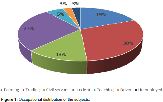
In the period under study, 90 patients were treated for salivary gland lesions. Only 37 (41.1%) of them met the inclusion criteria. Fifty three were excluded because of the following reasons: incomplete data, missing histopathological report and other pathological types such as adenocystic carcinoma, mucoepidermoid carcinoma, canaliculi cell adenoma, cystic lesions and those that declined surgery. The 37 eligible cases were further analyzed and reported herein. Sex distribution showed that 11 (29.7%) male and 26 (70.3%) female made up the study group. The various occupations of the subjects are as shown in Figure 1 with traders constituting the greatest majority. The patients’ mean age at presentation was 38.2 years and standard deviation (SD) of 16.35 (range 10 to 73 years), while the mean duration of the tumor at presentation was 39.43 months (range 4 to 156 months). Male subjects (48 (SD 18.3) years) were significantly older than the females (34.1 (SD 13.8) years); p = 0.016.

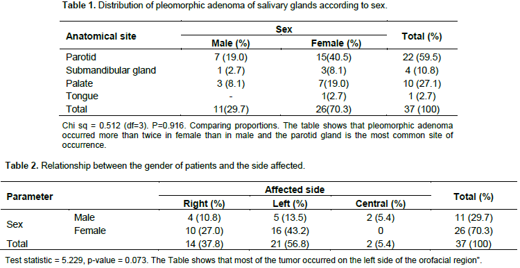
Similarly, disease duration was longer in males but insignificant (56.4 (SD 43.8) Months versus 32.3 (SD 31.6) Months; p = 0.068). Most of the tumors involved the parotid gland, while the anterior two third of the tongue was the site in one subject (Table 1). Tumors in the parotid gland was most common in women (15; 57.7%) than in men 7 (63.6%)). Three (30%) of the tumor in the palate were in males, while 7 (70%) were in females. Three of the four tumors in the submandibular gland occurred in females. Most of the tumor seen occurred on the left side of the face (Table 2). The treatment of choice in this study was surgical excision. Nineteen (86.4%) cases affecting the parotid gland had superficial parotidectomy, while 3 (13.6%) total parotidectomy. Four (100%) of the submandibular gland tumor were all treated by gland excision. Three patients (8.1%) had postoperative facial nerve palsy as the postoperative complications after their parotid gland surgery.
Pleomorphic adenoma is a common benign salivary gland tumor that shows different cell morphology and arrangements. Etiology of pleomorphic adenoma is not clear but studies have suggested that exposure to radiation and Simian virus (S40) may be implicated in causing its development (Jain et al., 2015). Report on prevalence of pleomorphic adenoma varies. Ninety cases of salivary gland lesions were seen within the 6 years study period with pleomorphic adenoma accounting for 37 (41.1%) of them. This finding is similar to a study studies reported in Maiduguri, Nigeria where prevalence of 44.3% was report (Otoh et al., 2005; Nzegwu et al., 2011). Published report on pleomorphic adenoma among Jordanians shows a prevalence of 48.8% which is also similar to the finding of the present study (AL-Khtoum et al., 2013). However, some other studies reported higher prevalence which could be attributed to the longer study period of those authors (Jude and Olu-Eddo, 2014).
The sex distribution of pleomorphic adenoma in this study shows lower male prevalence than female in a ratio of 1:2.4 which is different from previously reported studies in Enugu, Maiduguri and Lagos Nigeria where distribution ratios of 1.4:1, 2:1 and 8:7, respectively were reported (Odukoya, 1990; Otoh et al., 2005; Nzegwu et al., 2011), but is similar to some other African and Western studies which demonstrated higher frequencies in females than in males (Chizonga et al., 1995; Califano and Eisele, 1999; Eisele and Johns, 2001; Araya et al., 2015). The reasons for this finding may be attributed to laissez faire attitude of men towards their health care in Africa since pleomorphic adenoma is almost asymptomatic except for the observed swelling. On the other hand women seem to be more concern about their facial appearance than men hence tends to seek for treatment earlier. A mean age of 38.2 year was found in this study. This is similar to other studies reported in Enugu (35 years), Benin (37.1), Maiduguri (30.4 years) and Lagos Nigeria (31 years) as they are all within the same decade (Odukoya, 1990; Otoh et al., 2005; Nzegwu et al., 2011; Jude and Olu-Eddo, 2014).
It is also comparable to some other studies in African and Middle East but appears significantly lower in some European studies (Everson and Cawson, 1985; Chizonga et al., 1995; AL-Khtoum et al., 2013). This demonstrates that pleomorphic adenoma occurs mostly within the fourth decade of life in our environment. Men were found to be significantly older than women in this study. This may be due to sudden realization of potentials of malignant transformation of the tumor in addition to the earlier stated reasons. From this study, the mean duration of the tumor at presentation was 39.4 months (range 4 to 156 months) which is much higher than that of a Jordanian study with a mean duration of 18±7 months. This finding may be due to the slow growth rate of the tumor and its apparent symptomless presentation, hence, not perceived as life-threatening. Also, failure of the health care delivery system and out of pocket rather than insurance driven payment system may have contributed in making many of the patients to delay presenting.
Parotid gland and the palate were the most common sites of the tumor. This finding concurs with similar previous studies which reported high frequencies in the parotid gland as compared to other glands (Everson and Cawson, 1985; Otoh et al., 2005; Nzegwu et al., 2011; Patil et al., 2014). Palate is the most common site for tumors involving the minor salivary glands (Nzegwu et al., 2011; Sahoo et al., 2013; Jain et al., 2015). In contrast, Odukoya (1990) reported higher frequency of pleomorphic adenoma (68.9%) in the minor salivary glands as against 21.1% in the major glands. This study also shows that the tumor occurs rarely in the tongue, sublingual and submandibular glands (Rai et al., 2011; Nzegwu et al., 2011; AL-Khtoum et al., 2013; Patil et al 2014). The left side is the most common side affected by this tumor in the present study. The reason for this is unknown. In this study, gland excision was the commonest treatment option carried out for those tumors affecting the parotid and mandibular glands. For those that occur in the parotid gland, majority (86.4%) had superficial parotidectomy with preservation of facial nerve when possible due to the location of the tumor at the superficial lobe while others had total parotidectomy.
Excision of the tumor with safe margin was performed for the minor glands. The rational for this was to completely remove the tumor and satellites thereby preventing micro extensions which may lead to recurrence. Other treatment options like enucleation and radiotherapy were not utilized even though reported in other studies (AL-Khtoum et al., 2013; Elumelu et al., 2014). Treatment outcome in this study was good as no case of recurrence has been recorded within the study period. Facial nerve injury was the most common complication seen after surgical excision of the parotid gland and this occurred only in 3 patients. No case of malignant transformation was seen in our study, however, previous researches have shown that long standing pleomorphic adenoma may transform to malignant tumor (carcinoma ex pleomorphic adenoma) in any of the affected gland and this risk is also believed to increase in recurrent cases (Freeman et al., 2003; To et al., 2003; Rowley et al., 2006; Jain et al., 2015).
Pleomorphic adenoma is a relatively common salivary gland tumor in our environment. It occurred more in females than males in this study which appear similar to many other studies. The age distribution, treatment option and the site of occurrence are also similar to other previously reported studies but most men present at an older age than the women in this study. However, majority of the patients present long after the onset of this tumor which may be due to its slow growing, painless nature and patients’ attitude towards their health condition.
The authors have not declared any conflict of interests.
REFERENCES
|
AL-Khtoum N, Qubilat AR, Al-Zaidaneen, Al-Sarhan M, AL-Qudah A (2013). Clinical characteristics of pleomorphic adenoma of salivary glands among Jordanian patients. J. Pak. Med. Assoc. 63(3):358-360.
|
|
|
|
Araya J, Martinez R, Niklander S, Marshall M, Esguep A (2015). Incidence and prevalence of salivary gland tumors in Valparaiso, Chile. Med. Oral Patol. Oral Cir. Buccal. 20(5):e532.
Crossref
|
|
|
|
|
Califano J, Eisele DW (1999). Benign salivary gland neoplasms. Otolaryngol. Clin. North America 32(5):861-873.
Crossref
|
|
|
|
|
Chizonga MM, Lopez Perez VM, Portilla Alvarez AL (1995). Pleomorphic adenoma of the salivary glands. Clinicopathological studies of 206 cases in Zimbabwe. Oral Surgery, Oral Medicine, Oral Pathol. Oral Radiol. Endodontol. 79(6):747-749.
|
|
|
|
|
Eisele DW, Johns ME (2001). Salivary gland neoplasm in Head and Neck surgery. Otolayingology, Ed, BJ Bailey. Philadelphia, Lippincott Williams and Wilkins. pp. 1279-1297.
|
|
|
|
|
Elumelu TN, Folasire AM, Ntekim AI Oboh O (2014). Salivary gland tumors in Nigeria patients - a 10 year review. J. Cancer Biol. Res. 2(3):1054-1059.
|
|
|
|
|
Eneroth CM (1971). Salivary gland tumors in the parotid gland, submandibular gland and the palate region. Cancer 27:1415-1418.
Crossref
|
|
|
|
|
Everson JW, Cawson RA (1985). Salivary gland tumor. A review of 2410 cases with particular reference to histologic type, age and sex distribution. J. Pathol. pp. 14651-14658.
|
|
|
|
|
Everson JW, Auclair PL, Gneep DR, El-Naggar AK (2005). Tumours of the salivary glands. In: Barnes L, Eveson JW, Reichart P, Sidransky D, editors. World Health Organization Classification of Tumours: Pathology and genetics of head and neck tumours. Lyon: IARC. pp. 209-281.
|
|
|
|
|
Ezeanolue BC (1999). Salivary gland neoplasms: a descriptive analysis of the pattern seen in Enugu. West Afr. J. Med. pp. 179-182.
|
|
|
|
|
Freeman SR, Sloan P, de Carpentier J (2003). Carcinoma ex Pleomorphic adenoma of the nasal septum with adenoid cystic and squamous carcinomatous differentiation. Rhinology 41:118-121.
|
|
|
|
|
Ito FA, Jorge J, Vargas PA, Lopes MA (2009). Histopathological findings of pleomorphic adenomas of the salivary glands. Med. Oral Patol. Oral Cir. Bucal. 14:57-61.
|
|
|
|
|
Jain S, Hasan S, Vyas N, Shah N, Dalal S (2015). Pleomorphic adenoma of the parotid gland: report of a case with review of literature. Ethiop. J. Health Sci. 25(2):189-194.
Crossref
|
|
|
|
|
Jude UO, Olu-Eddo AN (2014). Salivary gland tumors, a twenty year retrospective study. Afr. J. Med. Health Sci. 13(10):24-29.
Crossref
|
|
|
|
|
Nzegwu MA, Njeze NR, Ugochukwu AI, Amu C, Okolugbe N, Okoye L.O., Mba AU, Uguru CC (2011). A review of salivary gland neoplasms in Eastern Nigeria for A five-year period from January 1, 2000 to December 31st 2004. Adv. Biores. 2(1):28-32.
|
|
|
|
|
Odukoya O (1990). Pleomorphic Adenoma of the salivary glands in Lagos, Nigeria. Clinicopathological analysis of 45 cases. Central Afr. J. Med. 36(10):251-256.
|
|
|
|
|
Otoh EC, Johnson NW, Olasoji H, Danfilo IS, Adeleke OA (2005). Salivary gland neoplasms in Maiduguri, north-eastern Nigeria. Oral Dis. 11(6):386-391.
Crossref
|
|
|
|
|
Patil P, Burde K, Naikmasur VG, Thorawat A (2014). Pleomorphic adenoma of submandibular gland: A case report with review of literature. Dental Res. J. 11(3):411-414.
|
|
|
|
|
Rai S, Sodhi SPS, Sandhus SV (2011). Pleomorphic adenoma of submandibular gland: An uncommon occurrence Natl J Maxillofac Surg. 2(1):66-68.
Crossref
|
|
|
|
|
Rousseau A, Badoual, C (2011). Head, neck: Salivary gland tumors: an overview Atlas Genet. Cytogenet. Oncol. Haematol. 15(6):533-541.
Crossref
|
|
|
|
|
Rowley H, Murphy M, Smyth D, O'Dwyer TP (2006). Recurrent pleomorphic adenoma: Uninodular versus multinodular disease. Irish J. Med. Sci. 169(3):201-203.
Crossref
|
|
|
|
|
Sahoo NK, Rangan MN, Gadad RD (2013). Pleomorphic adenoma palate: Major tumor in a minor gland. Ann Maxillofac. Surg. 3(2):195-197.
Crossref
|
|
|
|
|
To EW, Tsang WM, TSe GM (2003). Mucoepidermoid carcinoma ex pleomorphic adenoma of the submandibular gland. Am. J. Oto- laryngol. 64:253-257.
Crossref
|
|