ABSTRACT
The objective of this study was to detect the presence of Mycobacterium bovis (M. bovis) in whey. A total of 233 cow milk samples were analyzed together with 26 tank milk samples that came from dairy herds of several states of the Mexican Republic (Querétaro, San Luis Potosí, Guanajuato, Hidalgo, Coahuila). DNA was obtained from whey and used for polymerase chain reaction-multiplex (PCR-M). Tuberculosis complex was first identified through the detection of gene RD1. Positive samples were subjected to a second PCR-M with the primers for gene RD9 to identify M. bovis. Samples were bacteriologically cultured using conventional techniques for the isolation of mycobacteria. Cohen’s Kappa test (k) and Pearson’s Chi2 were carried out for statistical analysis. A 150 bp amplification product of the RD1 region was obtained, which corresponds to the tuberculosis complex, in 34/233 (14.59%) of the individual milk samples and in 4/26 (15.38%) of the tank milk samples. PCR-M with primer RD9, of the 34 individual samples and the 4 tank milk samples, gave an amplification product of 200 pb, which is the expected product for M. bovis. By bacteriological culture, six isolates were obtained; four in individual whey samples and two from tank milk samples, which were then classified by biochemical tests as M. bovis. The concordance between RD1, RD9 PCR-M and bacteriological culture was low, but there was a significant difference between diagnostic techniques with a P = 0.000. The results showed the potential of the PCR-M as a confirmatory test for the diagnosis of tuberculosis in cattle, as well as the advantage of using whey samples, that may be a possible source of infection for the herd and/or humans.
Key words: Bovine tuberculosis, whey, polymerase chain reaction-multiplex (PCR-M), RD1, RD9, milk, isolation.
Mycobacterium bovis, is the etiological agent of bovine tuberculosis (TB), which infects a wide range of mammal species, including humans (Daza et al., 2017). In developing countries, TB is an important zoonosis, especially for high risk populations such as workers in dairy farms and slaughterhouses, veterinarians and persons that consume fresh milk or cheese produced with non-pasteurized milk that comes from infected herds (Franco et al., 2013; Bapat et al., 2017). Tuberculosis in humans by M. bovis is less frequent in countries where the milk is pasteurized and bovine tuberculosis control and eradication programs are implemented. High prevalence in cattle facilitates airborne or digestive exposure to the bacilli, increasing the public health risk (Milián et al., 2012; Bapat et al., 2017). TB causes elevated economic loss to the cattle industry due to increased costs of control and eradication programs, as well as direct loss caused to the herd due to retention of carcasses in the slaughterhouse, reduction of 17 to 20% of milk production, 15% of calves’ production and causing 20% premature discards (Boland et al., 2010; Iturra, 2016). Natural infection in bovines is direct or indirect, with respiratory and oral the main infection routes. M. bovis is mostly shed through expectoration and is considered that elimination through milk is less than 2% depending on the degree of infection the cow has and that the mammary lymph nodes and udder develop granulomatous lesions. M. bovis infected milk may contaminate milking equipment, floors, bedding, and containers used for retaining or storage of milk. Likewise, insufficient processing of milk may help the dissemination of the disease to other herds when the calves are fed with milk contaminated with M. bovis (Boland et al., 2010; Iturra, 2016). Confirmation diagnosis of TB is carried out by bacteriological culture of tissue samples with granulomatous lesions, nasal swabs and milk; although there is an inconvenience, the test takes four weeks for the development of the bacterial colonies and three weeks more for the typification by biochemical methods (Pérez et al., 2002; Clavijo et al., 2004; Michel et al., 2015).
Diagnostic techniques based in DNA amplification of M. bovis by polymerase chain reaction (PCR) have been amply described (Ramírez et al., 2004; Bapat et al., 2017). Diverse primers have been evaluated to amplify gene fragments that are specific of the Mycobacterium tuberculosis complex (MPB70, IS6110, IS1081) (Talbot et al., 1997; Diaz, 2013; Sweetline et al., 2017).
Comparative analysis between genomes allows the study of the evolution of a virulent strain to an attenuated variant. Even though M. tuberculosis as well as M. bovis and M. bovis BCG have a high degree of genome conservation, the presence of polymorphism regions which allow their differentiation has been detected (Diaz, 2013). Through of genomic hybridization studies, Mahairas et al. (1996), described that there were genetic differences or regions of differentiation (RD1 to RD16) in the M. tuberculosis genome, which allow to differentiate at the genetic level between species of the tuberculosis complex. Talbot et al. (1997), used primers for the RD1 region in a PCR-M test that allows the differentiation between pathogenic strains of the tuberculosis complex and the BCG strain. Parsons et al. (2002), assessed region RD9 to be used for PCR-M and conclude that this region allows for the differentiation between M. bovis and M. tuberculosis. Bovine tuberculosis is a disease that requires control and to have alternate diagnostic methods is desirable if they allow a more sensitive, specific, and quick diagnosis, when compared with the bacteriological culture. Likewise, if different types of samples can be used, such as bovine whey, it would facilitate the establishment of handling and control measures for the disease. The objective of this study was to detect the presence of M. bovis in bovine whey samples through a multiplex PCR and bacteriological culture.
Sample collection
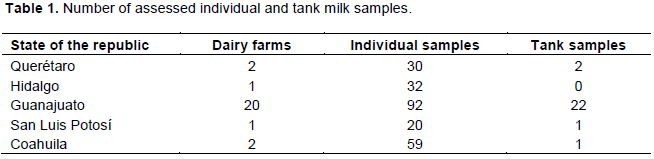
A convenience sampling was carried out with cooperating producers, in dairy herds with and without backgrounds of tuberculosis, as well as with or without mastitis problems, to determine the quality and safety of milk in relation to the shedding of M. bovis. A total of 259 milk samples were taken (233 individual samples from cows in different herds and 26 tank milk samples from different dairy herds in different states of the Mexican Republic) (Table 1). Each sample was approximately 50 ml of milk obtained prior to milking. The samples were collected in 50 ml falcon type sterile tubes and transported to the laboratory in an icebox with refrigerants. Milk samples were processed in the following 24 to 48 h after they were obtained.
Obtaining whey
In a falcon tube, 10 ml of milk were placed and 50 μl of 1% chymosin solution was added. The samples were incubated in a double boiler at 37°C for 30 min, and centrifuged at 2500 rpm for 10 min. Whey was collected in 15 ml falcon type tubes and maintained in the freezer at -20°C until use. (Gurrola, 2017).
DNA extraction from whey
Two milliliters of whey were placed in microtubes and centrifuged at 12000 rpm for 15 min, supernatants were removed and to the pellet, 500 μl TE 1X and 100 μl of lysozyme (10 mg/ml) were added. The product was incubated at 37°C for 24 h; then, 75 μl of Proteinase K (10 mg/ml) and SDS (10%) were added, and incubated at 64°C for 10 min; a further 100 μl of NaCl 5M and CTAB (5 M)/NaCl (5 M) were added and incubated at 64°C for 10 min; finally, 750 μl of Chloroform-Isoamyl Alcohol (24:1) was added and centrifuged at 12000 rpm for 5 min. Aqueous phase was then transferred to a new 2 ml tube, 250 μl of Guanidine Isothiocyanate (5 M) and 250 μl Ammonium Acetate (7.5 M) were added and kept at 8°C, for 20 min; 500 μl of Chloroform-Isoamyl Alcohol (24:1) was added and centrifuged at 12000 rpm for 5 min. The aqueous phase was transferred to a 1.5 ml microtube and centrifuged at 12000 rpm for 5 min. The supernatant was eliminated and 500 μl of isopropyl alcohol was added to the pellet and incubated at -20°C overnight. This was centrifuged at 12000 rpm for 15 min, the supernatant was eliminated, and then washed with 70% ethanol for 5 min. The DNA pellet was allowed to dry at ambient temperature and hydrated with 100 μl of milli-Q water and stored at -20°C, until use in PCR-M (Ramírez et al., 2004).
Multiplex polymerase chain reaction RD1Test (PCR-M RD1)
The reaction was done using the following primers for the RD1 region: ET1 (5´-AAG-CGG-TTG-CCG-CCG-ACC-GAC-C-3´), ET2 (5´-CTG-GCT-ATA-TTC-CTG-GGC-CCG-G-3´) and ET3 (5´-GAG-GCG-ATC-TGG-CGG-TTT-GGG-G-3´), that amplify a 150 bp product of the tuberculosis complex, which allows differentiation between pathogenic strains of the tuberculosis complex from the BCG vaccine strains that amplify a 200 bp product (Talbot et al., 1997). For each sample, the following were used: 12.5 μl PCR Master-Mix (4 mM MgCl2, 0.4 mM of each dATP, dGTP, dCTP, dUTP, 0.05 U DNA polymerase), 1 μl of each of the primers ET1 (5 pmol), ET2 (25 pmol), ET3 (5 pmol), 5 μl nuclease-free water, and 10 μl of DNA (6 ng/μl.) obtained from whey. M. tuberculosis H37Ra (ATCC # 25177) and M. bovis BCG (ATCC # 35734) DNA were included as positive controls. The thermocycler program was as follows: one cycle at 94°C/5 min, followed by 35 cycles at 94°C/40 s, 65°C/40 s, 72°C/40 s, and a final extension at 72°C/4 min, holding the reactions at 4°C until transferred out. The amplification products were visualized in 2% agar gel stained with ethidium bromide. The samples that came out positive to PCR-Multiplex RD1 were subjected to PCR-Multiplex RD9.
RD9 PCR-M
The following primers were used: RD9 FF (5´-GTG-TAG-GTC-AGC-CCC-ATC-C-3´), RD9 Int (5´-CAA-TGT-TTG-TTG-CGC-TGC-3´) and RD9 FR (5´-GCT-ACC-CTC-GAC-CAA-GTG-TT-3´) that amplify a 300 bp product of M. tuberculosis and a 200 bp product of M. bovis (Parsons et al., 2002). For each sample, the following were used: 12.5 μl PCR Master Mix; 1 μl of each of the primers RD9 FF (5 pmol), RD9 Int (25 pmol), RD9 FR (5 pmol); 5 μl of nuclease-free water and 10 μl DNA (6 ng/μl) obtained from whey. As positive controls, M. tuberculosis H37Ra (ATCC #25177) and M. bovis BCG (ATCC #35734) DNA were used. The same thermocycler program described earlier was used. The amplification products were visualized in 2% agar gel stained with ethidium bromide.
Bacteriological culture
The whey samples were cultured in duplicate with the technique described by Perez et al. (2002) using Lowenstein Jensen and StoneBrink culture media, and incubated at 37°C during nine weeks. Obtained bacterial growth was stained with Ziehl Neelsen and typified by biochemical methods (niacin test, catalase, tween 80 hydrolysis; nitrate reduction, pyrazinamidase) as well as pigmentation production and growth rate.
Statistical analysis
Statistical analysis was carried out using the STATA® 7.0 software package (StataCorp LP, College Station, TX, USA). Cohen’s Kappa test (k) or inter-rater agreement index was used to establish the association between the results obtained with the RD1, RD9 PCR-M and culture. Differences between diagnostic techniques were tested using Pearson’s Chi2 (c2).
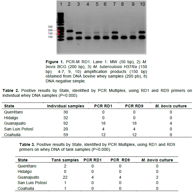
A 150 bp amplification product was obtained from the RD1 PCR-M tuberculosis complex in 34/233 (14.59%) individual milk samples (Figure 1 and Table 2) and 4/26 (15.38%) tank milk samples (Table 3).
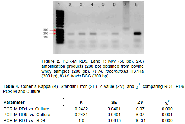
In all cases of PCR-M amplification using RD9 primers of the 34 individual samples and 4 tank samples, a 200 bp band was obtained, which was the expected product for M. bovis strains (Figure 2 and Tables 2 and 3).
Six isolates were obtained by bacteriological culture, four in the individual whey samples and two in the tank samples. The isolates were cultured for four weeks and were positive to the Ziehl Neelsen stain, classifying them as Acid-Fast Bacillus (AFB). Using biochemical methods strains were classified as M. bovis (Tables 2 and 3).
The concordance between RD1, RD9 PCR-M and bacteriological culture was low, but there was a significant difference between diagnostic techniques with a P = 0.000 (Table 4).
M. bovis isolation percentage in this study was 1.7% in individual milk samples and 7.6% in tank milk samples. To have isolates positive to AFB depends on many factors, amongst them that the agent is being shed in the sample that is obtained, and its viability and adaptability; even if the percentage of isolates that were obtained in the study may be considered of low importance, it confirms the shedding of viable AFB that can be recovered in whey which could be the origin of infections in animals and humans (Boland et al., 2010; Michel et al., 2015; Bapat et al., 2017). The probability of obtaining positive cultures from bovine milk samples is generally low; less than 2% of the cows that have tuberculosis shed the bacillus by milk. This occurs when the animal has a generalized infection and open lymph nodes, as well as involvement of the udder and the mammary lymph nodes. Conversely, the elimination of mycobacteria by aerosols and nasal discharge occurs intermittently (Clavijo et al., 2004).
A study carried out in Brazil (Franco et al., 2013) detected, respectively 7 and 9% presence of Mycobacterium species, in individual and tank milk samples, identifying other mycobacteria (Mycobacterium fortuitum, Mycobacterium flavescens, Mycobacterium smegmatis, Mycobacterium vaccae) besides M. bovis, as possible milk contaminants. Bacteriological culture results of this study may be considered low in comparison to those reported by Franco et al. (2013), yet the difference in all cases was the identification of M. bovis and the lack of sample contamination with environmental mycobacteria.
The presence of pathogenic microorganisms in food and food-borne illnesses represent essential and growing public health problems due to the increase in frequency, the surging of new transmission forms, growth of vulnerable population groups and the social and economic impact they have (Yánez et al., 2008; Bapat et
al., 2017). Whey is a byproduct of the cheese industry and represents 80 to 90% of the total processed milk volume. It contains 40 to 80% protein concentrates, which allow an ample use of these byproducts, mainly in the food industry, such as for the substitution of other ingredients and components in drinks, yogurt, spreadable cheeses, curd, sausages, bakery, confectionery, and even in the pharmaceutical industry. It can be used as a food source for pigs and bovines, and therefore it is considered necessary that it receives special treatment to eliminate a possible load of pathogenic agents to avoid them becoming a potential source of infection for humans, as well as animals.
Doran et al. (2009), described a case of bovine tuberculosis in Ireland in which a family of six were affected due to the consumption of non-pasteurized milk and derivatives. In the farm, all adult bovines, as well as 80% of the calves that were fed from a milk storage tank, tested positive to the tuberculin test, later presented tuberculous lesions, and M. bovis was isolated from udder and milk. There are regions in Mexico where more than 28% of milk or cheeses produced with non-pasteurized milk are consumed raw, and whey is used as feed supplement for ruminants and pigs and therefore there is a high risk of contamination with M. bovis (Gurrola, 2017). It is important to underline that in Mexico there are not many well documented studies of bovine tuberculosis in humans that detect the origin in consumption of non-pasteurized milk. Yet the danger is latent due to the consumption of non-pasteurized milk habits. Therefore, control measures must be taken for milk products, in relation to possible infectious agents that could be contaminating the product, as well as the containers that are used for it.
Biotechnology advances have allowed the development of alternative diagnostic methods that provide advantages in relation to efficiency, sensitivity, and reduction of detection time (Ramírez et al., 2004; Sweetline et al., 2017). These methods are quick because they are based in the determination of nucleic acids and have the property of being specific.
PCR needs the correct selection of the target sequence to be identified in the microorganism genome of interest. In this study, PCR-M was carried out to diagnose bovine tuberculosis with a sensitivity of 95% and a specificity of 100% (Talbot et al., 1997; Ramírez et al., 2004; Das et al., 2007), based on the identification of two Difference Regions, 1 and 9 (RD) of the tuberculosis complex. In this study, 257 bovine cattle milk samples were analyzed using PCR-M of RD1 and RD9, of which the presence of M. bovis DNA was detected in 14.8% of them. The use of the genetic markers of regions RD1 and RD9, in PCR-M for the diagnosis of bovine tuberculosis in DNA samples obtained from whey has allowed the establishment of a sensitive and specific technique to detect the presence of M. bovis DNA that takes less time than the bacteriological culture, which is considered the confirmation proof for the diagnosis of bovine tuberculosis, but it has the inconvenience that the time for the growth of mycobacterial colonies is slow and its sensitivity is below 50% (Sweetline et al., 2017).
The use of conventional techniques for bacteriological culture and PCR in milk samples are key tools to determine milk quality in relation to the presence of M. bovis, since it allows the detection of animals shedding the bacterium in milk (Sweetline et al., 2017). A control program should be implemented to eliminate cows with tuberculous mastitis, including the appropriate training of dairymen and population in general about the risks when consuming raw milk and its derivatives, when they come from tuberculosis infected herds. With this, the infection risk for humans and replacement calves should be reduced.
The results in this study show the potential that the use of PCR-M with RD1 and RD9 primers has as a diagnostic method for bovine tuberculosis, as well as the advantage of using whey samples that may be a possible source of infection for herds and/or humans.
The authors have not declared any conflict of interests.
REFERENCES
|
Bapat PR, Dodkey RS, Shekhawat SD, Husain AA, Nayak AR, Kawle AP, Daginawala HF, Singh LK, Kashyap RS (2017). Prevalence of zoonotic tuberculosis and associated risk factors in Central Indian populations. Journal of Epidemiology and Global Health 7(4):277-283.
Crossref
|
|
|
|
Boland F, Kelly G, Good M, More S (2010). Bovine tuberculosis and Milk Production in Infected Dairy Herds in Ireland. Preventive Veterinary Medicine 93(2-3):153-161.
Crossref
|
|
|
|
|
Clavijo AM, de Rolo M, Alfro C, Corso M (2004). Todo lo que usted debe saber sobre la tuberculosis bovina. Revista Digital CENIAP.
|
|
|
|
|
Das S, Chandra-Das S, Verma R (2007). Occurrence of RD9 Region and 500 bp Fragment among Clinical Isolates of Mycobacterium tuberculosis and Mycobacterium bovis. Medical Microbiology and Immunology 51(2):231-234.
Crossref
|
|
|
|
|
Daza BCA, Lechinski DPC, Trevizan GS, Junquiero FMM, Garcia RM (2017). Diagnosis of Mycobacterial in Bovine Milk: an overview. Revista do Instituto de Medicina Tropical de São Paulo 59:1-13.
|
|
|
|
|
Diaz ACI (2013). Regiones de diferencia en genomas de cepas de Mycobacterium bovis prevalentes en Chile. Tesis de Licenciatura Valdivia-Chile.
|
|
|
|
|
Doran P, Carson J, Costello D, More SJ (2009). An outbreak of tuberculosis affecting cattle and people on an Irish dairy farm, following the consumption of raw milk. Irish Veterinary Journal 62(6):390-397.
Crossref
|
|
|
|
|
Franco JMM, Paes CA, Garcia RM, Figueiredo PJC, Barreto SCA, Fujimara LQC, Garcia MR, Paganini LFJ (2013). Occurrence of mycobacteria in bovine milk samples from both individual and collective bulk tanks at farms and informal markets in the southeast region of Sao Paulo, Brazil. BMC Veterinary Research 9:85-90.
Crossref
|
|
|
|
|
Gurrola MME (2017). Detección de Mycobacterium bovis por medio de PCR-Multiplex a partir de muestras de suero de leche. Tesis de Licenciatura. FMVZ-UNAM. Mexico. P 47.
|
|
|
|
|
Iturra HLC (2016). Evaluación del impacto económico de la erradicación de tuberculosis bovina en predios lecheros infectados. Tesis de Licenciatura. FCV; Universidad de Chile. Santiago de Chile. P 45.
|
|
|
|
|
Mahairas GG, Sabo PJ, Hickey MJ, Singh DC, Stover CK (1996). Molecular analysis of genetic differences between Mycobacterium bovis BCG and virulent M. bovis. Journal of Bacteriology 178:1274-1282.
Crossref
|
|
|
|
|
Michel LA, Geoghegan C, Hlokwe T, Raseleka K Getz MW, Marcotty T (2015). Longevity of Mycobacterium bovis in raw and traditional souring milk as a function of storage temperature and dose. PLoS ONE 10(6):1-12.
Crossref
|
|
|
|
|
Milián SF, García CL, Romero TC (2012). Diversidad Genética y distribución regional de las cepas de Mycobacterium bovis del ganado en México. Revista Mexicana de Ciencias Pecuarias 3(4):459-471.
Crossref
|
|
|
|
|
Parsons LM, Brosch R, Cole, ST, Somoskövi A, Loder A, Bretzel G, van Soolingen D, Hale YM, Salfinger M (2002). Simple Approach for Identification of Mycobacterium tuberculosis Complex Isolates by PCR-Based Genomic Deletion Analysis. Journal of Clinical Microbiology 40(7):2339-2345.
Crossref
|
|
|
|
|
Pérez A, Reniero A, Forteis A, Meregalli S, López B, Ritacco V (2002). Study of Mycobacterium bovis in milk using bacteriological methods and the polymerase chain reaction. Revista Argentina de Microbiología 34(1):45-51.
|
|
|
|
|
Ramírez CIC, Santillán FMA, Arriaga DC, Arellano RB, Morales AJF (2004). Empleo de una PCR-multiplex para diferenciar caprinos vacunados con M. bovis BCG, de infectados con M. bovis de campo. Revista Mexicana De Ciencias Pecuarias 42(3):419-428.
|
|
|
|
|
Sweetline AN, Ronald BS, Kumar TM, Kannan P, Thangavelu A (2017). Molecular identification of Mycobacterium tuberculosis in cattle. Veterinary Microbiology 198:81-87.
Crossref
|
|
|
|
|
Talbot E A, Williams DL, Frothingham R (1997). PCR identification of Mycobacterium bovis BCG. Journal of Clinical Microbiology 35:566–569.
|
|
|
|
|
Yánez E, Mattar S, Durango A (2008). Determinación de Salmonella spp. por PCR en Tiempo Real y método convencional en canales de bovinos y en alimentos de la vía pública de Montería, Córdoba. Asoc Colomb Infectolo 12(4):247-254.
|
|