Full Length Research Paper

ABSTRACT
The purpose of this study was to investigate the phenotype, genotype and relationships of vancomycin-resistant Enterococcib (VRE) strains isolated from Minjiang River of Fujian province, residence district pond, university fish farm, cesspool and sewer. PCR was used to confirm Enterococci and amplify antibiotic resistance genes. Relationships between different strains were determined by repetitive extragenic palindromic elements-PCR (REP-PCR). A total of 17 VRE strains were isolated from water environment, among which 9 strains were isolated from cesspool, 6 strains were isolated from sewer and 2 strains were isolated from university fish farm. VRE was not isolated from Minjiang River or residence district pond. Analysis on the antibiotic resistance gene showed that 3 strains belong to genotype VanB, 9 strains belong to VanC1 and 5 strains belong to genotype VanC2/3. Genotype VanA was not detected. REP-PCR analysis showed that 17 strains were divided into type A1 (n=6), type A2 (n=1), type A3 (n=2), type B (n=3), type C (n=1), type D (n=3) and type E (n=1). These results demonstrated that the antibiotic resistant phenotype was consistent with the genotype of 17 VRE strains. VRE strains isolated from water sources were resistant to multiple antibiotics. The majority of the strains isolated in this study shared high similarity.
Key words: Enterococci, antibiotic resistance, vancomycin, repetitive extragenic palindromic elements-PCR
INTRODUCTION
Enterococci are normal human and animal intestinal flora and can be isolated from a variety of natural environments, including soil, water, plants, animals, birds, insects and human excrement. Enterococci are opportunistic pathogens, which can not only cause human urinary tract, skin and soft tissue infections, but also lead to serious infections, e.g., bacteremia, endocarditis and meningitis (Coque et al., 1996). In recent years, drug resistance of enterococci and spread of resistance genes has become a major concern. Particularly, the occurrence of vancomycin-resistant enterococci (VRE) breaks the last line of human defense against intractable drug-resistant strains. Drug resistant bacteria may spread from hospitals or farms to the surrounding environment through a variety of means. As a medium of resistant gene dissemination, water contains a massive resistance gene pool, which provides opportunities for the pathogens or opportunistic pathogens to obtain large number of resistant genes. Infection with these bacteria could cause outbreak of epidemics and their treatment would be extremely difficult. Therefore, in this study we investigated the prevalence of VRE in different water sources. We also analyzed the phenotype, genotype and relationships of the VRE strains in order to provide basic information of VRE in the water environment in China.
MATERIALS AND METHODS
Bacterial strains
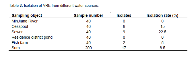
A total of 200 water samples were collected from different sites of Minjiang River of Fujian province (n=40), residence district pond (n=40), university fish farm (n=40), cesspool (n=40) and sewer (n=40). All VRE was isolated from these samples. Only one isolate was selected from each positive sample. The university fish farm was connected to the sewer. The waste water in the cesspool was emitted from central laboratory of university and flew into the university fish farm and sewer.
Antibiotics
Six kinds of reference antibiotics: vancomycin (VAN), erythromycin (ERY), chloramphenicol (CHL), ciprofloxacin (CIP), oxytetracycline (OTC) and ampicillin (AMP) were purchased from China’s National Institute for the Control of Pharmaceutical and Biological Products.
Quality control strain
Enterococcus faecalis ATCC29212 was purchased from China’s National Institute for the Control of Pharmaceutical and Biological Products.
Main reagents and instruments
Enterococcus growth medium and tryptone soy agar were purchased from OXOID (UK). MH broth was purchased from Beijing Shuanxuan microbial culture products factory. Bacterial genomic DNA extraction kit, PCR reagents and agarose for electrophoresis were purchased from Shanghai Sangon (China).
Primers for PCR amplification
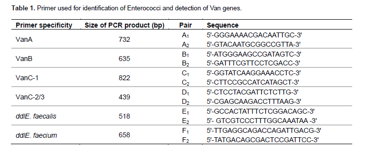
Primers for identification of Enterococcus and detection of drug resistant genes were shown in Table 1 (Dongyou et al., 2005; Reiko et al., 2000). Primers for REP-PCR were 5′-IIIICGICGICATCIGGC-3′and 5′-ICGICTTATCIGGCCTAC-3′ (James et al., 1991).
Isolation of VRE
Water samples were inoculated onto selective agar plates containing 6 μg/mL of vancomycin. After incubation at 35°C for 24 h, colonies growing on the plates were confirmed by broth microdilution method.
Strain identification
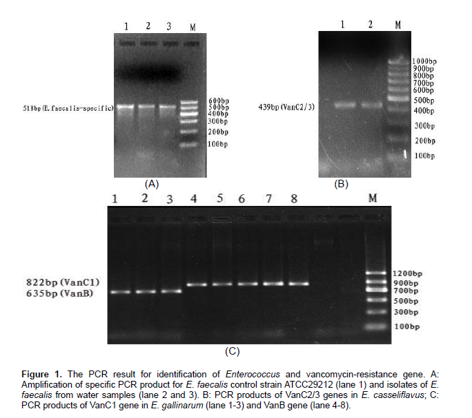
All the strains were identified by PCR method using E. faecalis, Enterococcus faecium, Enterococcus gallinarum and Enterococcus casseliflavus-specific primers ddlE Faecalis, dd E.faecium, VanC-1, VanC-2/3.
Making stocks of the strains
Strains of Enterococcus were inoculated in MH broth. Fresh bacterial culture was added with sterile glycerol (final concentration: 20%) and stored in freezer (-20°C) for future uses.
Antimicrobial susceptibility testing
Antimicrobial susceptibility testing was performed according to the protocol recommended by Clinical and Laboratory Standards Institute (CLSI). Minimum inhibitory concentration (MIC) was determined for the isolated Enterococcus strains using minimal broth microdilution method.
Plasmid extraction and genomic DNA preparation
Plasmid was extracted as previously described (Li and Zhang, 2006). Meanwhile, Genomic DNA was isolated with commercial kit from Shanghai Sangon (China).
PCR amplification
The volume of PCR was 20 μL containing 10 μL of 2×PCR Master, 0.5 μL of forward and reverse primers, 2 μL of template and 7 μL of water. The parameters for amplification of products (<500 bp) were 93°C pre-denaturing for 2 min, followed by 35 cycles of 93°C for 30 s, 53°C for 30 s, 72°C for 1 min and a final extension at 72°C for 1 min. The parameters for amplification of products (>500 bp) were 93°C pre-denaturing for 2 min, followed by 35 cycles of 93°C for 1 min, 53°C for 1 min, 72°C for 1 min and a final extension at 72°C for 2 min. PCR products were detected by electrophoresis in 1.5% agarose gel containing 0.5% ethidium bromide (100 V for 45 min). Automatic gel image analysis system was used to detect the band and take images.
REP-PCR detection of the similarity between VRE strains
The volume of PCR was 25 μL containing 2.5 μL of 10×buffer, 1 μL of 25 mmol/L MgCl2, 0.2 mM dNTP, 2U Taq, 50 pmol/L of forward and reverse primers and 4 μL of template DNA. The parameters PCR were 95°C pre-denaturing for 7 min, followed by 30 cycles of 90°C (denaturing) for 30 s, 40°C (annealing) for 1 min and 65°C (extension) for 8 min. PCR products were detected by electrophoresis in 1.5% agarose gel containing 0.5% ethidium bromide (90 V for 60 min). Automatic gel image analysis system was used to detect the band and take images.
Typing methods
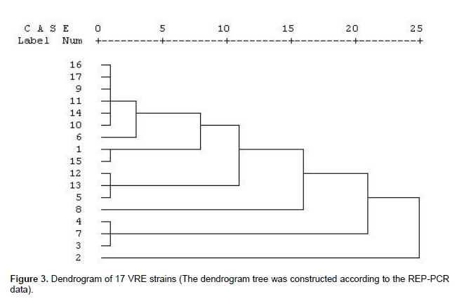
Different types of strains contain differences in more than 3 bands. Different subtypes of strains contain difference in 1-2 bands. Identical subtype indicated that these strains were from the same clone. According to the REP-PCR fingerprints of electrophoresis, each band was converted to binary datamatri x. “1” indicated “presence” and “0” indicated “absence” of a band. Binary data for all the PCR bands was entered into SPSS statistical software. Dendrogram was constructed by selecting Hierarchial Cluster Analysis process and between group average linkage methods to analyze the relationship between strains.
RESULTS
Isolation of VRE from different water sources
VRE was isolated from 9 cesspool samples with a prevalence of 22.5% (9/40), 6 sewer samples with a prevalence of 15% (6/40) and 2 fish farm samples with a prevalence of 5% (2/40) (Table 2). VRE was not isolated from samples of Minjiang River and residence district pond. PCR detection with species-specific primers showed that 3 strains were E. faecalis, 9 strains were E. gallinarum and 5 strains were E. casseliflavus.
Antimicrobial susceptibility test for 17 VRE strains
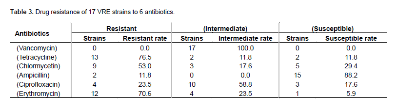
The MIC of vancomycin for 17 VRE strains was 8-32 μg/mL. A total of 13 strains were resistant to tetracycline, with the highest resistance rate of 76.5%, followed by the resistance rate of 70.6% (12/17) to erythromycin (Table 3).
Determination of drug resistance genes in VRE
PCR detection of the drug resistance gene showed that 3 strains belonged to genotype VanB, 9 strains belong to VanC1 and 5 strains belong to genotype VanC2. Genotype VanA was not detected (Table 4).

Detection of genetic similarity among 17 VRE strains
REP-PCR results showed that 3-9 bands (with a size range of 200-2000bp) could be amplified from the genomic DNA of each individual VRE strain. Based on the pattern of the PCR bands, these 17 VRE strains were classified into 5 types designated as type A, B, C, D and E. Type A was further divided into A1, A2 and A3 subtypes. Type B, C, D and E did not have subtypes. The majority of the strains were type A, with 6 strains belonging to type A1, 1 strain belonging to A2 and 2 strains belonging to A3. Three strains belonged to type B and 3 strains belonged to type D. Type C and type E had 1 strain. Figure 1 showed the PCR band pattern for each strain. Dendrogram of 17 strains was constructed based on the REP-PCR band patterns (Figure 3). As shown in Figure 2, strains 9, 10, 11, 14, 16 and 17 had identical PCR band pattern (type A1), indicating that these strains had 100% homology. Strains 5, 12 and 13, strains 3, 4 and 7, and strains 1 and 15 had identical PCR band pattern, indicating that these strains had 100% homology. Strains 9, 10, 11, 14, 16 and 17 were isolated from cesspool and all these strains had drug resistance genotype VanC1. Strain 3 (isolated from cesspool), strains 4 and 7 (isolated from sewer) had the same drug resistance genotype VanC2. Strains 5, 12 and 13 were isolated from sewer and all these strain had identical drug resistance genotype VanC1. Strain 15 (isolated from university fish farm) and strain 1 (isolated from cesspool) had genotype VanB. These results suggested that clonal dissemination occurred between these strains.

DISCUSSION
Enterococcus is a clinically common opportunistic pathogen. Widespread application of antibiotics caused the resistance of Enterococcus to multiple antibiotics. Enterococcus has become an important pathogen causing nosocomial and secondary infections. In the United States, approximately 12% of nosocomial infections were caused by Enterococci (Edmond et al., 1999). Studies have shown that antibiotic-resistant bacteria or genes were disseminated via stool, water or food. In addition, antibiotic-resistant bacteria or genes in animals or humans can be further directly or indirectly, continuously or intermittently disseminated through various channels and ecological environments (Witte, 2000). Many researchers have considered Enterococcus as an indicator bacterium to investigate animal, human and environmental bacterial resistance to antimicrobial drugs. All the 17 strains isolated in this study have low level resistance to vancomycin. Strains with high level resistance to vancomycin were not isolated. VRE strain can be resistant to multiple antibiotics. Our results showed that VRE was isolated from 22.5% (9/40) of the cesspool samples, 15% (6/40) of sewer samples and 5% (2/40) of university fish farm samples. VRE was not isolated from Minjiang River and residence district pond. These results indicate that cesspool and sewer are severely contaminated, while Minjiang River and residence district pond are not contaminated with VRE.
The resistance rate of VRE to tetracycline and erythromycin was 76.5 and 70.6%, respectively, which was consistent with resistance rate reported in previous studies (Yi et al., 2008; Yu and Xu, 2008). Genotypic analysis for the drug resistant genes showed that 3 strains belonged to type VanB, 9 strains belonged to type VanC1 and 5 strains belonged to type VanC2. Genotype VanA was not detected. The genotype of drug resistant genes was consistent with the phenotype of VRE.
The highest isolation rate of VRE was observed in cesspool, indicating that VRE is more frequently present in severely contaminated environments. The waste water in the cesspool was emitted from the central laboratory building of the university, which includes plant pathology laboratory, veterinary pharmacology laboratory, aquatic veterinary laboratory and animal biochemistry laboratory. The wastes from these laboratories may contain a variety of toxic chemicals, bacteria and drug residues. VanB-type Enterococcus strains in university fish farm may come from either cesspool or the surrounding human and animal contacts. REP-PCR analysis showed that the VRE strains isolated from sewer had high level of genetic similarity to those isolated from cesspool. In addition, one VanB-type VRE strain from fish farm was genetically identical to one VanB-type VRE strain isolated from cesspool, indicating that clonal dissemination occurred between these two environments.
CONFLICT OF INTERESTS
The authors have not declared any conflict of interests.
ACKNOWLEDGEMENTS
This research was supported by National Natural Science Foundation of China (Grant No. 31272606) and Researching Center of Water source and water safety, Fujian Agriculture and Forestry University, Fujian province, China.
REFERENCES
|
Coque TM, Tomayko JF, Ricke SC, Okhyusen PC, Murray BE (1996). Vancomycin-resistant enterococci from nosocomial, community, and animal sources in the United States. Antimicrob. Agents Chemother. 40:2605-2609. |
|
|
Dongyou Liu, Chinling Wang, Edwin JS (2005). PCR amplification of a species-specific putative transcriptional regulator gene reveals the identity of Enterococcus faecalis. Res. Microbiol. 156:944-948. |
|
|
Edmond MB, Wallace SE, McClish DK,Pfaller MA,Jones RN,Wenzel RP (1999). Nosocomial bloodstream infections in United States hospitals: A three-year analysis. Clin. Infect. Dis. 29:239-244. |
|
|
James V, Thearith K, Lupski R (1991). Distribution of repetitive DNA sequences in eubacteria and application to fingerprinting of bacterial genomes.Nucl. Acids Res. 19(24):6823-6831. |
|
|
Li S, Zhang Z (2006). Detection and analysis of molecular epidemiology in vancomycin-resistant enterococci. Chin. J. Lab. Diagn. 10(4):372-375. |
|
|
Reiko K, Ritsuko M, Joseph WC (2000). Simple and Reliable Multiplex PCR Assay for Surveillance Isolates of Vancomycin-Resistant Enterococci. J. Clin. Microbiol. 3092-3095. |
|
|
Witte W (2000). Ecological impact of antibiotic use in animals of different complex microflora: Environment. Int. J. Antimicrob. Agents 14:321-325. |
|
Copyright © 2024 Author(s) retain the copyright of this article.
This article is published under the terms of the Creative Commons Attribution License 4.0


