ABSTRACT
The present study was carried out to isolate, purify and elucidate the structure of low molecular weight bioactive compounds from sea anemone, Gyrostoma helianthus of the Red Sea environment. The obtained results indicated that the ethanolic crude extract of the sea anemone, G. helianthus showed inhibition activity against acetylcholinesterase (AchE). The 0.5 kD fraction inhibited the activity of the AchE that indicated the presence of low M.W active compound(s) of less than 0.5 kD. Two active fractions were obtained after BioGel P2 fractionation of the 0.5 kD of the G. helianthus. The first active fraction inhibited the activity of the acetylcholinesterase, while the other fraction did not. High-performance liquid chromatography (HPLC) technique aided by semipreparative columns was used to isolate the target compounds in a pure form for the structure elucidation, one dimensional nuclear magnetic resonance (NMR) analysis (1H and 13C-NMR) and DEPT were carried out to elucidate the structure of the isolated compound which was tentatively identified as N,N'-bis-(1-methyl-pyridin-2-yl)-hydrazine.
Key words: Sea anemone, Gyrostoma helianthus, Red Sea, structure elucidation, acetylcholinesterase (AchE) inhibitor.
The study of marine organisms as a source of biologically active compounds is considered a very productive field, having already led to the discovery of various new pharmacological tools and medicines (Bhakuni, 1994; Munro et al., 1999; Faulkner, 2001).
The work of Bergman and Feeney at the beginning of the1950s initiated the study of marine natural products, and in the last few decades, an appreciable number of new compounds have been isolated from marine organisms (Bhakuni, 1994; Faulkner, 2000a, b; Faulkner, 2001). Many authors explained this ecological success on the basis of the ability of marine invertebrates to synthesize secondary metabolites, which have an important defensive role against predation (Jimenez et al., 2002).
It is increasingly realized that many marine toxins are very site specific in their actions and hence are of value as biological tools. As a result, there is an increasing exploration of toxins from marine sources, especially those from invertebrates (Walker and Masuda, 1990). Moreover, the very high potencies of some marine toxins are attained through strong, highly selective interactions, frequently with specific sites on excitable membranes. Investigating the mechanism of toxin action has revealed a great deal about the physiology of affected tissues and systems (Hall and Strichartz, 1990). The improvement in isolation and chemical identification techniques, the collaboration between chemists and pharmacologists, and most recently, the interest of pharmaceutical industries have been important determinants in the development of marine natural products research (Faulkner, 2000b).
The search for novel low molecular weight natural products from marine organisms is of crucial importance for different applications as drugs and pesticides. Of special interest is the Red Sea environment, which is unique in its life forms and considered as one of the richest marine areas in bio-diversity and richer than similar areas in the eastern Pacific and Atlantic (Gomaa and Aboul-Enein, 2000).
Therefore, the objective of the present study was to isolate, purify and elucidate the structure of low molecular weight bioactive compounds from sea anemone, Gyrostoma helianthus of the Red Sea environment using the bioassay guided fractionation technique.
Organism under study
Sea anemone, G.helianthus was collected from the vicinity of Hurghada, Red Sea, Egypt.
Sampling locations
Samples of sea anemone, G. helianthus were collected from Hurghada vicinity at latitude from 27°00’N to 27°45’N and longitude from 33°30’E to 34°00’E. The locations of the sampling sites were as follows:
Shaab AbuShaar, Shaab AbuGalawa, Shaab AbuSadaf, ElFanadir Island, Gafton Kabier Island, Gafton Saghier Island, Dishet AbuMinqar, Magawish Kabier Island and Magawish Saghier Island.
Sample preparation
Sea anemone, G. helianthus samples were collected by trained divers working in the areas known to harbor the specific species under study. The collected samples were kept in seawater during the sampling trip around the vicinity of Hurghada. Upon arrival at the laboratory of Hurghada Marine Station, National Institute of Oceanography and Fisheries, the samples were washed with distilled water followed by gentle centrifugation to remove excess water. Samples were weighed and counted and the wet weight was recorded. Samples were also extracted in the laboratories of the Hurghada Marine Station of the National Institute of Oceanography and Fisheries.
Extraction of bioactive material form marine organism
Samples were extracted using a modified technique of Gomaa et al. (2000). Washed intact marine organism samples were weighed and equal amounts of absolute ethanol were added (1:1 w/v) and blended for 3 mins. The extracted samples were centrifuged at 2570 g for 10 min and the supernatant was removed and kept for further extraction. The residues were subjected to a second extraction with absolute ethanol and to a third extraction with 50% aqueous ethanol.
For defatting and partial depigmentation of sea anemone, G. helianthus ethanolic crude extract, as a step of purification, the supernatants were mixed with an equal volume with n-hexane. Defatting with n-hexane was repeated for 3 times. The residues were also blended with equal amounts (w/v) of n-hexane. Both aqueous ethanolic and hexane extracts were evaporated under reduced pressure at 40°C.
Inhibitory effect on acetylcholinesterase (AchE)
The in vitro AchE inhibition activity of the ethanolic crude extract and the other fractions was determined according to Ellman et al. (1961) using reagent kits purchased from Qunica Clinica Aplicada S.A., Spain.
Isolation, purification and identification of G. helianthus AchE inhibitor
Fractionation and purification of the G. helianthus ethanolic crude extract was carried out using different chromatographic techniques, as described in the following sections, followed the AchE inhibition activity guided fractionation protocol.
Molecular weight exclusion ultrafiltration
Crude extract of G. helianthus was filtered through membrane filters with cut off 10, 5, 3, 1 and 0.5 k Dalton (76 mm in diameter, Millipore Corporation, Bedford, MA, USA). Ultrafiltration was performed under pressure of nitrogen gas (40 kg.cm-2).
Biogel P2 gel filtration
Gel filtration column chromatography was prepared using a 3.5 x 80 cm Omni LC column, packed with BioGel P2 (BioRad Laboratories, Richmond, CA, USA), to reach a bed height of 75 cm and a bed volume of 728 ml (Shimizu et al., 1975; Buckley et al., 1975; Hall 1982; Gomaa, 1990). 15 ml of Milli Q water was used to redissolve 5 g of the less than 0.5 k Dalton freeze-dried filtrate and then applied on the top of the BioGel P2 column. The column was eluted with 2 bed volumes of milli Q water using a peristaltic pump to provide a flow rate of 48 ml/h, and 5 ml fractions were collected.
Buckley spot plate technique (Buckley et al., 1975) was used to detect fluorescence and quenching activity of the collected fractions along with their reaction with ninhydrin. Aliquots of all the collected fractions, 5 µl each, were spotted on 10x10 cm silica gel TLC plates (precoated, type 60 F254, with fluorescent indicator, aluminum backed, E. Merck, Germany). Fluorescent and quenching activity was observed under long wave (366 nm) and short wave (254 nm) UV. Spot plates were sprayed with 1% ninhydrin in ethanol to detect the presence of the free a-amino groups (purple color) or the substituted a-amino group (yellow color) in the active fractions.
Active fractions, detected by in vitro AchE, were compared with any physical property that may appear by Buckley spot test. Percent bed volume was calculated for each fraction to correlate between the position(s) of the active fractions with the calculated percent bed volume.
High-performance liquid chromatography (HPLC)
Different HPLC techniques were tested to compare between the active and nonactive fractions to locate the peak that may account for the detected activity (AchE inhibition). The HPLC system used was Perkin-Elmer series 200 pump system equipped with a Perkin-Elmer series 200 UV absorbance detector set at wavelength of 203, 254 and 365 nm. Different mobile and stationary phases were used to find out the best method for the separation of the toxic compounds. Data were collected and integrated with a Totalchrom Navigator Chromatography Manager.
The analytical column chromatography, Spheri 5 silica (100 x 4.6 mm; 5 mm) (Applied Biosystems Inc. Hoster City, A 94404 USA, Brownlee columns) was used to separate the AchE inhibitor. The mobile phase system (H2O : MeOH : Acetonitrile) gradient program as shown in the Table 1, UV detector wavelength at 254 nm and the flow rate was 1 ml/min. Also, the semi preparative column hyperprep HS/Silica (250 x 10 mm; 12 mm) Thermo Hypersil was used to obtain enough materials for the structure elucidation.
Structure elucidation
1D nuclear magnetic resonance (NMR) (1H and 13C) analysis and EI-MS was carried out to elucidate the structure of the isolated bioactive compound.
Organism under study
To identify the collected sea anemone samples from the Red Sea, specimens of the collected sea anemone species were sent to Prof. Dan Hartog of the National Museum of the Netherlands who was assigned by the British Museum of the Natural History as the best expert in this field. According to Prof. Dan Hartog, the scientific name of the species under study was confirmed as G. helianthus or Entacmaea quadricolor
Crude extract bioactivity
According to Gomaa et al. (2000), the crude aqueous ethanolic extract of G. helianthus when intra peritoneally (i.p.) injected in the 20 g male mice showed symptoms of neurotoxicity. Gomaa et al. (2000) also showed that the part of the neurotoxicity observed in the mouth assay was due to reversible inhibition of the AchE in the brain and blood of the mice, which was confirmed by Gomaa and Aboul-Enien (2000) after using the in vitro AchE assay. The same assay was used in this study to track the bioactive compound during the bioassay-guided fractionation technique. AchE inhibition activity was confirmed in this study in the ethanolic crude extract of sea anemone G. helianthus. Different reports showed that sea anemones produce two types of protein toxins: neurotoxins, which act mainly on ion channels (Honma and Shiomi, 2005) and cytolysins (or actinoporins), which exhibit lytic activity on a variety of cells (Anderluh and Macek, 2002; Alegre-Cebollada et al., 2007a; A´ lvarez et al., 2009; Kristan et al., 2009). However, the presence of AchE inhibitors in marine invertebrate has not been reported in the different extracts even in those that exhibit high neuroactivity.
Molecular weight exclusion AchE inhibition activity
In this study, G. helianthus crude extract showed 91.9% inhibition in the AchE activity while the MW exclusion fractions showed lower percent inhibitions (Table 2).
This table shows the effect of purification steps of G. helianthus on the activity of AchE. The more the purification, from the crude extract until less than 5 kD filtrate, the less the inhibition percent of AchE which means the presence of more than one AchE inhibitors in the crude extract. The only previous report detecting the AchE inhibitor in the extract of G. helianthus was that of Gomaa and Aboul-Enien (2000) and Gomaa et al. (2000). However, Mebs et al. (1983) did not detect any AchE inhibitors in G. helianthus collected from the Red Sea, the AchE inhibition activity detected in the less than 0.5kD filtrate indicated the presence of low MW active compound (less than 0.5 kD).

Biogel P2 fractions bioactivity
Using Biogel P2 fractionation and Buckley spot plate, the two distinct chemically active groups reported by Gomaa and Aboul-Enien (2000) were confirmed. The first group (65 to 72% bed volume) showed quenching under short wave (254 nm) while the second (72 to 77% bed volume) showed fluorescence activity under long wave (366 nm).
Only one of these two active groups showed in vitro AchE inhibition activity which confirmed the reported results obtained by Gomaa and Aboul-Enien (2000), while the second group showed no inhibition activity toward AchE. The AchE inhibition activity was detected along the purification steps from the crude to the 0.5 kD filtrate until the first active group of the Biogel P2 fractions, While AchE inhibition activity was not detected in the second active group.
HPLC fractions activity
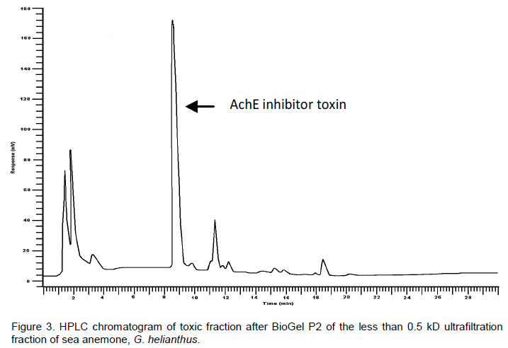
Different HPLC methods were tested to compare between the toxic (AchE inhibitor) and nontoxic fractions to locate the peak that may account for the detected AchE inhibition activity. The best result was obtained when the 254 nm wavelength was used. The first active group, after Biogel P2 fractionation, that showed the inhibition of AchE was tested by the HPLC technique to define the peak responsible for such activity. AchE inhibition and non-inhibition fractions were injected in the HPLC using a spheri 5 silica column (100 x 4.6 mm; 5 mm). Different mobile phase systems were used and the best resolution was achieved using a mixture of H2O : methanol : acetonitrile in a gradient program. The peak responsible for AchE inhibition was observed at 8.5 min retention time. The active fractions of the Biogel P2 have a higher peak area at this retention time while the non-active fractions before and after these active ones showed no peaks or a very small one. Also, the peak areas were proportionally related to the activity level.
HPLC purification of AchE inhibitor
The HPLC technique described in the current study aided by semipreparative columns was used to isolate the target compounds in a pure form for the structure elucidation. Thus, the present study may be a step towards the discovery of novel compounds from the sea anemone G. helianthus.
Figures 1 to 4 illustrate the HPLC chromatograms of the different purification steps of G. helianthus ethanolic crude extract to isolate the AchE inhibitor compound through ultrafiltration steps reaching less than 0.5 kDa filtrate to the Biogel filtration and finally to obtain the pure bioactive compound from the HPLC aided by semi-preparative column. The percent of the peak area of the desired AchE inhibitor bioactive compound in the HPLC chromatograms were 29.9, 51.6, 75.8 and 98% in the crude extract, 0.5 kDa filtrate, AchE inhibition fraction after Biogel P2 and the isolated pure toxin after HPLC, respectively.
Structure elucidation of AchE inhibitor
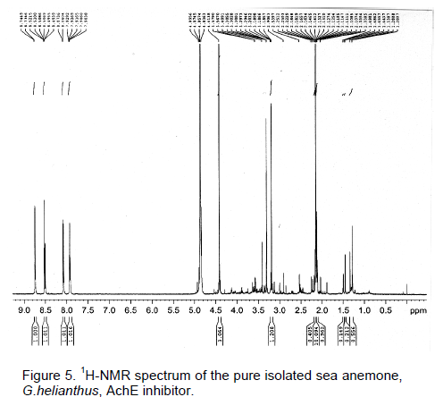
Further purification on a semi-preparative HPLC led to the isolation of one major constituent in a pure form from the first active fraction after Biogel P2. The purity of the isolated bioactive compound was examined by TLC and HPLC where it gave only one spot on TLC and one peak in the HPLC chromatogram (Figure 4). One dimensional NMR analysis 1H, 13C-NMR and DEPT were carried out to elucidate the structure of the AchE inhibitor isolated from the sea anemone, G. helianthus (Figure 5 to 7).
1H NMR (600MHz, MeOH-d4/Acetone-d6) (Figure 5): d ppm 8.74 (2H, br., J = 6.2 Hz, H-6/6'), 8.51 (2H, ddd=td, J = 7.8, 1.0Hz, H-4/4'), 8.07 (2H, br., J = 7.8 Hz, H-3/3'), 7.92 (2H, ddd=td, J = 6.2, 1.3 Hz, H-5/5'), 4.41 (6H, s, 2xN-Me)
13C NMR (150 MHz, MeOH-d4/ Acetone-d6) (Figure 6): d ppm 165.16 (C-2/2'), 146.98 (C-4/4'), 146.43 (C-6/6'), 127.57 (C-5/5'), 127.46 (C-3/3'), 47.41 (2xN-CH3).
EI. MS, m/z (%) (Figure 8): 186.0 [M-C2H6]+, (40%); 171.0 [M-C2H7N]+, (90%); 94.0 [M-C7H10N2]+, (100%); 79.0 [M-C7H11N3]+, (23%); 52.0 [M-C9H14N3] +, (23%) As it was interpreted above, the isolated compound was expected to be 2-subsitituted pyridine-like structure on
the basis of its splitting pattern, d- and J-values in the 1H NMR data (Figure 5). The presence of N-CH3 functionality was deduced from the singlet at 4.41 ppm. Depending on its M.S fragments (Figure 8), the isolated bioactive compound was tentatively identified as a symmetric dimer of 2-amino-N-methylpyridine. The base peak (100%) at m/z 94.0 mu was confirmative evidence for the homolytic cleavage of N-N bond. The fragment was diagnostic for the aminopyridine structure, which was followed by loss of 15 mu of NH to give a relatively weak abundant ion at m/z 79.0 (23%). In its 13C-NMR spectrum (Figure 6), five characteristic carbon resonances were assigned to two pyridine moieties together with an aliphatic C-resonance at 47.41 of two N-CH3 groups. Large down field shift (+16 ppm) of the C-2 to 165.16 (~149 in pyridine) was indicative of the attachment of amino group to C-2 and accumulation of positive charge on the ring nitrogen.
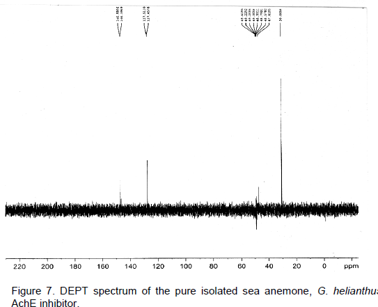
All 13C-resonances were assigned by aid of increment of subsistent additive rule and comparison with structure related compounds (Eberhard and Wolfgang, 1987). The appearance of only four 1H-resonances in the aromatic together with the aliphatic (CH3) regions, respectively and six 13C-resonances (each of two equivalent carbons), was indicative of an asymmetric bis-(2-amino-N-methylpyridine) structure. Finally, DEPT spectrum (Figure 7) confirmed the suggested structure of the AchE inhibitor toxin as N,N'-bis-(1-methyl-pyridin-2-yl)-hydrazine (Figure 9) through the differentiation among C, CH and CH3 resonances.
The study of marine organisms as a source of biologically active compounds is considered a very productive field, having already led to the discovery of various new pharmacological tools and medicines. The inhibition activity towards AchE that was detected in crude extract of the sea anemone, G. helianthus and in the 0.5 kD fraction indicates a presence of low molecular weight active compound of less than 0.5 kD, this hypothesis coincided with the calculated molecular weight (216 D) after the structure elucidation of the isolated compound using mass spectrometer, NMR analysis (1H and 13C-NMR) and DEPT. The isolation of a low molecular weight AchE inhibitor compound in a pure form with known chemical structure will be a step forward to discover a new drug.
The authors have not declared any conflict of interests.
REFERENCES
|
A´ lvarez C, Mancheno JM, Martınez D, Tejuca M, Pazos F, Lanio ME (2009). Sticholysins, two pore-forming toxins produced by the Caribbean Sea anemone Stichodactyla helianthus: their interaction with membranes. Toxicon. 15; 54(8):1135-47
|
|
|
|
Alegre-Cebollada J, Onaderra M, Gavilanes JG, Martınez del Pozo A (2007a). Sea anemone actinoporins: the transition from a folded soluble state to a functionally active membrane-bound oligomeric pore. Curr. Protein Pept. Sci. 8:558-572.
Crossref
|
|
|
|
|
Anderluh G, Macek P (2002). Cytolytic peptide and protein toxins from sea anemones (Anthozoa: Actiniaria). Toxicon. 40:111-124.
Crossref
|
|
|
|
|
Association of Official Analytical Chemist (AOAC). (2007). Official methods of analysis, p. 319, 18th Ed., revised, AOAC, Washington, D.C.
|
|
|
|
|
Bhakuni DS (1994). Bioactive marine alkaloids. J. Indian Chem. Soc. 71: 329-340.
|
|
|
|
|
Buckley LJ, Ikawa M, Sasner Jr. JJ (1975). Purification of two Gonyaulax tamarensis toxins from clams (Mya arenaria) and the identification of STX. Toxic Dinoflagellate Blooms. Proc. Int. Conf. Mass. Sci. Technol. Found. pp. 423-431.
|
|
|
|
|
Eberhard B, Wolfgang V (1987). Carbon-13 NMR spectroscopy, High resolution methods and application in organic chemistry and biochemistry, 3rd Ed. Weinhein, New York, NY, VCH.
|
|
|
|
|
Ellman GL, Courtney KD, Andres V, Featherstone RM (1961). A new and rapid colorimetric determination of Acetylcholinesterase activity. Biochem. Pharmacol. 7: 88-95.
Crossref
|
|
|
|
|
Faulkner DJ (2000a). Marine pharmacology. Antonie van Leeuwenhoek. 77: 135-145.
Crossref
|
|
|
|
|
Faulkner DJ (2000b). Marine natural products. Nat. Prod. Rep. 17: 7-55.
Crossref
|
|
|
|
|
Faulkner DJ (2001). Marine natural products. Nat. Prod. Rep. 18: 1-49.
Crossref
|
|
|
|
|
Gomaa MN (1990). Toxins and toxicity of a blue green alga Aphanizomenon flos-aquae, Ph.D. thesis. Marine-Estuarine-Environmental Science, University of Maryland, College of Park, MP.
|
|
|
|
|
Gomaa MN, Aboul-Enein MN, Naguib KM, Rawi S (2000). Neurotoxicity of sea anemone Gyrostoma helianthus from the Red Sea. J. Egypt. Soc. Toxicol. 22: 75-81.
|
|
|
|
|
Gomaa MN, Aboul-Enein MN (2000). Low molecular weight sea anemone Gyrostoma helianthus neurotoxins from the Red Sea. J. Egypt. Soc. Toxicol. 23: 01-07.
|
|
|
|
|
Hall S (1982). Toxins and toxicity of Protogonyaulax from the Northeast pacific . Ph.D. thesis, University of Alaska, P.196.
|
|
|
|
|
Hall S, Strichartz G (1990). Preface. In: Marine Toxins: Origin, Structure, and Molecular Pharmacology (Hall S, Strichartz G eds.). American Chemical Society, Washington DC. pp. 377.
Crossref
|
|
|
|
|
Honma T, Shiomi K (2005). Peptide toxins in sea anemones: structural and functional aspects. Mar. Biotechnol. 8: 1-10.
Crossref
|
|
|
|
|
Jimenez PC, Fortier SC, Lotufo TMC, Pessoa C, Moraes MEA, de Moraes MO, Costa-Lotufo LV (2002). Biological activity in extracts of ascidians (Tunicata, Ascidiacea) from the northeastern Brazilian coast. J. Exp. Marine Biol. Ecol. 287: 93-101 .
Crossref
|
|
|
|
|
Kristan K, Viero G. Dalla Serra M, Macˇek P, Anderluh G (2009). Molecular mechanism of pore formation by actinoporins. Toxicon. 54: 1125-1134.
Crossref
|
|
|
|
|
Mebs D, Librich M, Reul A, Samejima Y (1983). Hemolysins and proteinase inhibitors from sea anemone of the Gulf of Aqaba. Toxicon. 21: 257-264.
Crossref
|
|
|
|
|
Munro MHG, Blunt JW, Dumdei EJ, Hickford, SJH, Lill RE, Li S, Battershill CN, Duckworth AR (1999). The discovery and development of marine compounds with pharmaceutical potential. J. Biotechnol. 70:15-25.
Crossref
|
|
|
|
|
Shimizu Y, Alam M, Fallon WE (1975). Purification and partial characterization of toxins from poisonous clams, In LoCicero (ed): "Toxic Dinoflagellate Blooms", Proc. Int. Conf. Mass. Sci. Technol. Found, p. 275-285.
|
|
|
|
|
Walker MJA, Masuda VL (1990). Toxins from marine invertebrate. In: Marine Toxins: Origin, Structure, and Molecular Pharmacology (Hall S. and Strichartz G. eds.). American Chemical Society, Washington DC. pp.120-132.
Crossref
|
|