Full Length Research Paper
ABSTRACT
The purpose of this in vitro study was to investigate the linear-elastic fracture toughness of endodontically treated teeth after restoration with prefabricated post systems against root restored with metal cast post-core. Thirty two maxillary canines with similar anatomic characteristic were sectioned to obtain the same length for all specimens. Group I consisted of 8 roots restored with FibreKor system; Group II consisted of 8 roots restored with C-Post system; Group III consisted of 8 roots restored with metal cast post-core (the diameter was similar FibreKor system); and Group IV consisted of 8 roots restored with metal cast post-core (the diameter was similar C-Post system). Specimens were cemented with dual-cure bonding agent (Dual-Cement and Unibond). These specimens were then mounted in acrylic blocks and tested in a universal testing machine (Kratos). Each sample was angled at 135° to the long axis of the root. A constantly increasing force was applied until the root fracture. Means (kilogram force, kgf) and standard deviations for four groups were found to be: Group I, 31.01 (2.08); Group II, 41.32 (3.44); Group III, 49.17 (2.09); Group IV, 47.65 (2.94). The present research indicated that root restored with metal post was statistically significant (p<0.05), more resistant to fracture over root restored with non metal post.
Key words: C-Post system, linear-elastic fracture, endodontic, teeth.
INTRODUCTION
The restoration of endodontically treated teeth is a complex procedure. Endodontically treated teeth, often resulted into the loss of a significant part of the tooth structure, weakness due to the endodontic treatment (Sedgley and Messe, 1992).
There are many therapeutic methods for the restoration of these teeth. Often the decision-making confuses the practitioner. The post and core is the method used most frequently. However, this treatment may weaken the remaining tooth structure. A high failure rate has been reported in the literature. Many authors have reported that the vertical fracture is common in teeth restored with post and core system. This type of fracture requires the tooth extraction (Pontius and Hutter, 2002; Sirimai et al., 1999). Non-metallic post systems have elastic modulus near the dentin. Martha et al. reports that in this system, the fracture occurs in the cervical region of the tooth. Thus the remaining tooth can be restored (Martha et al., 2008).
It is commonly stated that endodontically treated teeth are more susceptible to fracture as the result of the amount of tooth structure that remains after endodontic treatment. The post space preparation is the other important consideration in the resistance to fracture of pulpless teeth. Metallic post systems generally are used when more than half of the coronal tooth structure is lost (Kimmel, 2000; Barkhordar et al., 1989). Selecting the optimum restorative modality is considered the key to restorative success. Fiber-reinforced composite (FRC) posts were introduced in dentistry, and consist of carbon, glass, or quartz fibers embedded in epoxy resin (Bateman et al., 2003). They have bonding ability to dentin and the core material (Sirimai et al., 1999; Newman et al., 2003). Saupe et al. (1996) reported that FRC posts treated teeth had more resistance to masticatory forces. The modulus of elasticity is similar in all components of the tooth restored (Stricker and Göhring, 2006; Freilich et al., 2000). Therefore, all components are a homogeneous block. According to Dallari and Rovatti (1996), Duret et al. (1996) and Fredriksson et al. (1998), non-metallic post reduces stress in the tooth.
Sidoli et al. (1997) compared the in vitro performance and the failure characteristics of the carbon-fiber post system. They concluded that, the specimens restored with the carbon-fiber post system exhibited inferior strength properties to the other post and core system. Dean et al. (1998) observed that there were no differences in fracture resistance among carbon fiber post, tapered stainless steel post, and parallel stainless steel post.
Literature lack consensus as to the best restorative system. The purpose of this study was to determine the null hypothesis of no difference in linear-elastic fracture toughness between non-metallic post system and metal cast post-core.
MATERIALS AND METHODS
Thirty two extracted maxillary canines (selected by the Human Teeth Bank of Universidade de São Paulo – Bauru, School of Dentistry, controlled by the Ethics Committee of the school) were used for this study. The teeth were selected to submit similar dimensions. A digital caliper was used. This device recorded the dimensions vestibule-lingual and mesial-distal to the level of the cervical region of the teeth. Teeth to submit discrepancy greater than one millimeter were discarded.
All teeth were stored in a timol 0.9% saline solution before experimental storage times; prepared specimens were stored in deionised water at 37°C. Teeth were randomly divided into three groups of 8. All teeth were endodontically treated. Crowns were then resected at the cement enamel junction to achieve the length of 12 mm. The root was mounted in acrylic resin block with 9 mm of the root in the block and 3 mm outside the block.
Group I: Fibrekor post and composite core
The roots were prepared for post placement using the armamentarium supply kit manufacture to develop 8 mm post length. The preparation was cleaned using water spray for 15 s. The post space was then dried using paper points. The walls were conditioned for 15 s using 37% H3PO4 gel, and cleaned with water spray for 15 s. The cavity walls was dried using paper points and Unibond dental adhesive was applied in a thin layer and light cured for 30 s. The Fibrekor post was cleaned with alcohol at 96% and silane was applied. Unibond dental adhesive was applied and light polymerized for 30 s. Posts were cemented into the post hole with dual cement and excess cement was removed using hand instruments. Fibrekor was light-cured for 40 s. The remaining dentin surface was acid etched for 15 s and cleaned prior to the application of the Unibond dental adhesive; this was light polymerized for 30 s. The coronal build up was finally completed using a self-polymerizing resin composite (Build-it, Jeneric-Pentron, Inc.) inserted in an adjusted metal matrix. To ensure even distribution of force, a foil metal incisal-lingual bevel was placed in core resin composite.
Group II: C-Post and composite core
The root canal was prepared using drill n°1 or pre-shaping drill following drill n°2 or finishing drill to the preparation depth of 8 mm. The post was a parallel-side stepped post having a coronal diameter 1.5 mm and apical diameter of 1.0 mm. The preparation was conditioned, dried, and dental adhesive was applied, and light cured as in Group I. The carbon fiber post was sandblasted for 2 to 3 s with a micro-etcher and cleaned with water spray for 15 s; C-Pos was then cemented with dual cement and light cured for 40 s. The coronal surface was made as in Group I. To ensure even distribution of force, a foil metal incisal-lingual bevel was cemented in core resin composite.
Group III: Cast post and core with anatomy similar to Fibrekor
The post space preparation was made with Gates Glidden burs n°1 to 4 to a depth of 8 mm and diameter of 1.2 mm at the coronal portion. A direct impression of the root preparation was made with Duralay resin. A matrix partner was used to make the core portion. After custom cast metal post, this was sandblasted for 2 to 3 s with a Micro-etcher and cleaned with water spray for 15 s. The walls of the root were conditioned, dried, and dental adhesive was applied and light cured as in Group I. Post were cemented into the post hole with Dual Cement and light cured for 40 s in bucal, lingual, mesial, and distal faces.
Group IV: Cast post and core with anatomy similar to C-Post
The post space preparation was made with the drill of the Kit of C-Post to that if it got a length of 8 mm. Abundant irrigation was used to guarantee the removal of the debris of the interior of the conduit in all the stages. The impression of the post space was made with pins of the type Pin Jet® and resin Duralay®. A small mark was made with a bur diamond drill on the pin Pin Jet® in a length of 8 mm. Through this mark, it was possible to check the waste of the post space if it correspond to the wanted length and if the pin reached the end of the preparation. The interior of the conduit was lubricated with solid vaseline through the drill of wide n°2, involved in a cotton wick. A small portion of acrylic resin Duralay® was taken into the post space and distributed with a lentule drill; the pin was introduced until the risk reached the cervical limit of the root. It was waited for 2 min and the pin was removed to avoid the fact that the resin adhered to the walls. After the complete polymerization of the resin, the coronary portion of the pin was sectioned with approximately 3 mm of saliency in relation to the cervical level, so that it roots is possible to position the metallic head office described previously. This head office was lubricated with solid vaseline through a brush. Small portions of acrylic resin were placed at the head office, until it filled out the interior completely. The head office was positioned on the resin pin, being careful to adapt from the best possible way to the limits of the root, mainly in the vestibular face, and after 2 min it was removed, to avoid the resin from sticking to the internal walls. After the complete polymerization of the resin, the pattern was taken for inclusion in coating and subsequent foundry in metallic league CuBe2.
After the foundry of the cast post and core, it was adapted to the root, which remove possible bubbles with drills carbide n° 170l in high rotation and refrigeration. Kota® liquid-carbon was used when necessary to obtain a better adaptation of the post.
The fixation of the nucleus in the root was accomplished with Dual resinous cement (Cement®). The root was conditioned with acid phosphoric at 37% for 15 s and washed in water for more 15 s. The excesses of water of the interior of the root were removed with cones of absorbent paper. To avoid the complete drying of the walls, a paper cone was applied soaked in water against the walls of the conduit. The sticker of the type Unibond® was applied with a brush, inside the conduit and photopolymerized for 30 s. The melted metallic nucleus air abraded with oxide of aluminum, in the whole surface of the root portion and it was washed in running water by 30 s. A portion of Dual cement (Cement®), was manipulated for 10 s and applied with a brush on the root portion of the nucleus. The nucleus was seated inside the root with movement of it to improve the distribution of the cement in the walls. After the removal of the excesses, the proof body containing the nucleus was positioned under a device developed to maintain a uniform force during the foundation (Illustration 4.5). The polymerization of the dual cement was gotten through the light application by 40 s in the faces vestibular exam, lingual, mesial and distal, and the chemical polymerization happened at least 5 min after agreement with the manufacturer's specifications. To follow the proof bodies, they were stored in a recipient with water deionised and maintained in a greenhouse to 37° for 24 h.
After sample preparation, the specimens were stored in deionised water at 37° for 24 h before being tested. The specimens were kept slightly above the water level. Thus the specimens were preserved in water absorption.
The specimens were then subjected to a load on a universal testing machine (Kratos). Each sample was angled at 135° to the long axis of the root. A constantly increasing force was applied until the root fracture.
The force at failure was analyzed statistically with analysis of variance. The Method of Student-Newman-Keuls was used to evaluate the comparisons in pairs for the four groups of the study.
RESULTS
There were significant differences in the amount of force required to produce failure among the three groups (p<0.05).
The resistance values to the fracture for Group I, roots restored with the system Fibrekor®, was what presented the smallest medium value, 31.01 kg force (kgf), with a standard deviation of 2.08 Kgf. Group II, roots restored with the system C-Post, obtained a medium value of 41.32 kgf and a standard deviation of 3.44 kgf. Group III, roots restored with cast post and core with cervical box, presented a medium value of 49.17 kgf and deviation pattern of 2.09 kgf.
Results revealed that the roots restored with cast post and core presented larger resistance to the fracture, being statistically significant (tests Student-Newman-Keuls), p<0.05 in relation to the Groups I and II.
Table 2 presents the obtained results of the resistance tests to the fracture accomplished in the machine of Universal tests Kratos, for each samples and the average and standard deviation of each group.
DISCUSSION
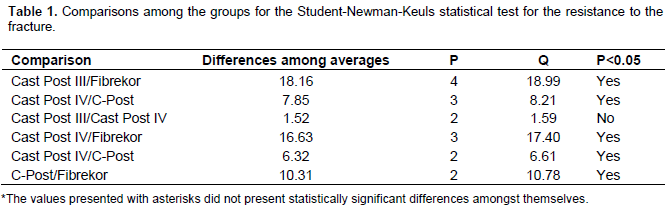
The Fibrekor systems and C-Post are non-metallic pins. It has been speculated in literature that these pins have elastic modulus similar to tooth structure. This similarity can contribute to the restoration that has better resistance to masticatory forces. However, this is not well established in the literature. Many authors report the advantages of non-metallic pins. But when there is little remaining tooth structure, these pins do not show the alleged performance. Fiberglass pins are superior aesthetics to the metal cast post and core. Thus, this study opted to use the canine, a single-rooted tooth. These teeth can better reproduce the clinical conditions. In Group III, eight superior canine teeth received metal cast post and core. The cast post were made with a length of 8 mm inside the root canal and an approximate diameter of 1.2 mm. The root canal in Groups III and I were prepared with the same anatomy (similar to Fibrekor®). Group III achieved the best results. The average achieved by this group was 49.17 kgf. This value was statistically significant (p<0.05) compared to Groups I (Table 1). Group I had an average of 31.01 kgf. Despite the fact that Groups I and III present similar anatomy, the specimens of Group I showed lower results. In this work, comparing Groups IV and Group II, it showed that Group IV required a force significantly larger (average 47.65 kgf) than the necessary force to fracture the roots of the Group II (average of 41.32 kgf) which is statistically significant.
This study failed to demonstrate the superiority of non-metallic pins. In both situations tested, the cast metal post and core showed better results than the Group I (Fibrekor) and Group II (C-Post). The longevity of the restorative treatment is the main goal. The cosmetic may not be the only factor that determines the choice of a therapeutic method. The durability of a restoration must be established by a number of factors, including the ability to withstand the masticatory forces. The resistance to fracture is one of the properties that determines this ability to withstand the masticatory forces. The results obtained in this study seemed to be in agreement with the statement of other authors (Sidoli et al., 1997; Morgano and Brackett, 1999; Sirimai et al., 1999); they obtained results similar to the present study, where the metal post demonstrated larger resistance to the fracture of the remaining root. Already Mc Donald et al. (1990) did not find any difference between metal post and carbon fiber post.
.png)
Group II, presented larger values when compared with Group I, and it was statistically significant, P<0.05.
The fracture type presented by the specimens was not statistically analyzed in the present study. The four groups presented characteristics similar in relationship to the fracture place, in spite of the fact that Groups I and II obtained larger incidence of fracture of the coronary portion and larger displacement of the nucleus than the other two groups. Many researchers have argued that closer fracture of the cervical level can be important to reutilization of the root. However, if a restorative treatment fails to ease, this treatment may not be beneficial to the patient. Clinical studies have shown that the presence of at least 2 mm remaining tooth structure type core used is not adversely influenced. The use of non-metallic pins should be limited to these clinical situations.
No effort was accomplished to simulate the periodontal ligament, during the inclusion of the roots in the samples, because the incorporation of silicon with this objective seems to be a doubtful procedure due to lack of studies that prove that this material presents the same characteristics of the periodontium (King and Setchell, 1990).
The choice of an appropriate system for the restoration of the remaining dental element is a difficult task due to the great amount of systems found at the market. Several clinical situations observed other factor that prevents the use of a unique system. Clinical studies are necessary to evaluate the behavior of these systems.
CONCLUSIONS
Within the parameters of the study design and materials tested, the following conclusions may be made:
1. Group III (cast post and core similar to C-Post) demonstrated significantly higher resistance to fracture than the other three groups;
2. Group II (C-Post), demonstrated more resistance to fracture than Group I (Fibrekor) with statistically significant differences;
3. Group IV (cast post and core similar to Fibrekor) demonstrated significantly higher resistance to fracture than Group I;
4. Group I (Fibrekor) demonstrated significantly less resistance than other groups;
5. No statistically significant difference in resistance to fracture was demonstrated between Group III and Group IV.
CONFLICT OF INTEREST
The authors have not declared any conflict of interest.
REFERENCES
|
Barkhordar RA, Radke R, Abbasi J (1989). Effect of metal collars on resistance of endodontically treated teeth to root fracture. J. Prosth. Dent. 61(6):676-678. Crossref |
||||
|
Bateman G, Ricketts DN, Saunders WP (2003). Fiber-based post systems: A review. Br. Dent. J. 195:43-48. Crossref |
||||
| Dallari A, Rovatti L (1996). Six years of in vitro/in vivo experience with composipost. Compend. 17(20):557-563. | ||||
|
Dean JP, Jeansonne BG, Sarkar N (1998). In vitro evaluation of a carbon fiber post. J. Endod. 24(12):807-810. Crossref |
||||
| Duret B, Duret F, Reynaud M (1996). Long-life physical property preservation and post endodontic rehabilitation with the composipost. Compend. 17(20):550-556. | ||||
|
Fredriksson M, Astbäck J, Pamenius M, Arvidson K (1998). A retrospective study of 236 patients with teeth restored by carbon fiber-reinforced epoxy resin posts. J. Prosthet. Dent. 80(2):151-157. Crossref |
||||
| Freilich MA, Meiers JC, Duncan JP, Goldberg AJ (2000). Fiber-reinforced composites in clinical dentistry. Quintessence Books; P. 63. | ||||
| Kimmel SS (2000). Restoration and reinforcement of endodontically treated teeth with a polyethylene ribbon and prefabricated fiberglass post. General Dent. 6:700-706. | ||||
|
King PA, Setchell J (1990). An in vitro evaluation of a prototype CFRC prefabricated post developed for the restoration of pulpless teeth. J. Oral Rehabil. 17:599-609. Crossref |
||||
|
Martha VA, Marcos VSP, Fathi AID, Arnaldo FC (2008). Virtual Analysis of Stresses in Human Teeth Restored with Esthetic Posts. Mater. Res. 11(4):459-463. Crossref |
||||
| Mc Donald AV, King PA, Setchell DJ (1990). In vitro study to compare impact fracture resistance of intact root-treated teeth. Int. Endod. J. 23(6):307-312. | ||||
|
Morgano SM, Brackett SE (1999). Foundation restorations in fixed prosthodontics: current knowledge and future needs. J. Prosth. Dent. 82(6):643-657. Crossref |
||||
|
Newman MP, Yaman P, Dennison J, Rafter M, Billy E (2003). Fracture resistance of endodontically treated teeth restored with composite posts. J. Prosthet. Dent. 89:360-367. Crossref |
||||
|
Pontius O, Hutter JW (2002). Survival rate and fracture strength of incisors restored with different post and core systems and endodontically treated incisors without coronoradicular reinforcement. J. Endod. 28:710-715. Crossref |
||||
|
Saupe WA, Gluskan AH, Radke RA Jr (1996). A comparative study of fracture resistance between morphologic dowel and cores and a resin-reinforced dowel system in the intraradicular restoration of structurally compromised roots. Quintessence Int. 27:483-491. Pubmed |
||||
|
Sedgley CM, Messer HH (1992). Are endodontically treated teeth more brittle? J. Endod. 18:332-335. Crossref |
||||
|
Sidoli GE, King PA, Setchell DJ (1997). An in vitro evaluation of a carbon fiber based post and core systems. J. Prosthet. Dent. 78:5-9. Crossref |
||||
|
Sirimai S, Douglas NR, Steven MM (1999). An in vitro study of the fracture resistance and the incidence of vertical root fracture of pulpless teeth restored with six post and core systems. J. Prosthet. Dent. 81:262-269. Crossref |
||||
|
Sirimai S, Douglas NR, Steven MM (1999). An in vitro study of the fracture resistance and the incidence of vertical root fracture of pulpless teeth restored with six post and core systems. J. Prosthet. Dent. 81:262-269. Crossref |
||||
|
Stricker EJ, Göhring TN (2006). Influence of different posts and cores on marginal adaptation, fractures resistance, and fracture mode of composite resin crowns on human mandibular premolars: An in vitro study. J. Dent. 34:326-335. Crossref |
||||
Copyright © 2024 Author(s) retain the copyright of this article.
This article is published under the terms of the Creative Commons Attribution License 4.0


