Full Length Research Paper
ABSTRACT
The aim of this study was to investigate and compare the short and long term antibacterial effects of white and gray mineral trioxide aggregate (MTA) and calcium enriched mixture (CEM) on Streptococcus Sanguinis and Enterococcus faecalis which are commonly associated with endodontic infections. The test materials, including white MTA (WMTA), gray (GMTA), and CEM were manipulated strictly in accordance with the manufacturer's instructions. A total of 72 culture plates were prepared and divided into three experimental groups (one group for each of freshly mixed and set WMTA cements; one group for each of freshly mixed and set GMTA cements; one group for each of freshly mixed and set CEM cements. Each group consisted of 24 plates in which 12 plates were cultured by S. sanguinis and the other 12 plates were cultured by E. faecalis. Antibacterial activities of the materials against the S. sanguinis and E. faecalis were evaluated using agar diffusion test (ADT). The materials were tested in form of set (24-h, 1 week and 1 month) and freshly mixed. In the E. faecalis groups, plates containing freshly mixed and set WMTA, GMTA, and CEM cements did not show any antibacterial properties. The antimicrobial activity of freshly mixed GMTA was lower than WMTA and higher that of CEM. The largest mean diameters of inhibition zone of bacterial growth were found in set WMTA cement group at any time period. In conclusion, the origin of MTA as well as the preparation techniques may affect its antimicrobial activities.
Key words: White mineral trioxide aggregate (WMTA), gray mineral trioxide aggregate (GMTA), calcium enriched mixture (CEM), growth inhibition zone, S. sanguinis, antibacterial activity, agar diffusion test, E. faecalis.
INTRODUCTION
Microorganisms and their byproducts are the main etiologic factors for the development and progression of pulpal and periapical disease as well as in endodontic treatment failures (Fouad et al., 2005). Most endodontic treatment failures are attributable to inadequate cleansing of the RC and egress of bacteria and other antigens into the periradicular tissues (Hasan Zarrabi et al., 2009).
Elimination of microbial flora and infected tissues during root canal (RC) treatment by instrumentation, irrigation and intracanal medication has always been an important part of successful endodontic treatments (Sundqvist, 1982). After a RC procedure, because of persistence of inflammation or infection in the bony area around the end of the tooth, then it is necessary to perform an apicoectomy and a root-end filling is placed to prevent re-infection of the root (Bhavana et al., 2015).
Endodontic treatment result will depend on the effective seal to prevent future recontamination as well as successful reduction or elimination of the associated microorganisms (Torabinejad et al., 1995). Antimicrobial testing of biomaterials should consider this effect. The agar-diffusion test (ADT) is the most commonly used technique for evaluating antibacterial property of dental materials (Cobankara et al., 2004). Hence, root-end filling materials not only should have sealing ability and biocompatibility but also should ideally have some antibacterial activity to prevent bacterial and fungal growth (Hasan Zarrabi et al., 2009). However, numerous materials have been recommended as root-end filling materials; none has so far been found to be completely ideal. One of the well-known root-end filling materials is mineral trioxide aggregate (MTA), which has unique properties such as, excellent sealing ability, high alkalinity induction of hard tissue formation, and antibacterial effects (Bhavana et al., 2015; Tziafas et al., 2002). Due to its physical and chemical properties, the use of MTA as a biomaterial for a wide variety of endodontic treatments has been recommended (Torabinejad and Chivian, 1999). In a recent prospective clinical study, MTA showed a high success rate when used as root-end filling material (Saunders, 2008). However, MTA has a delayed setting time and poor handling characteristics, and is expensive to use (Asgary and Kamrani, 2008).
Recently, calcium enriched mixture (CEM) cement, also called new endodontic cement (NEC), has been developed with similar clinical uses to tooth-colored ProRoot MTA but different chemical composition (Asgary et al., 2008). It mainly contains different calcium compounds.
This material has acceptable physical properties and is capable of hydroxyapatite formation over material in normal saline solution (Asgary et al., 2008a). An experimental study carried out on dogs demonstrated that MTA and NEC have the same favorable results as pulp capping materials and were even preferred over calcium hydroxide (Asgary et al., 2008c). This biocompatible cement forms an effective seal when used as root-end filling material and the results is comparable with different root-end filling materials (Asgary et al., 2008a).
In recent years, several studies have been carried out on the antibacterial characteristics of MTA (Bhavana et al., 2015; Asgary et al., 2006); less research has been performed on the antibacterial properties of CEM (Asgary et al., 2007). Moreover, antibacterial properties of root-end filling materials have been rarely compared. Based on data gathered from previous studies, long term antibacterial effects of the materials have been not considered before. Therefore, the aim of this study was to investigate and compare the long term antibacterial effects of white and gray MTA and CEM on Streptococcus Sanguinis and Enterococcus faecalis microorganisms which are commonly associated with endodontic infections using the agar diffusion test (ADT).
MATERIALS AND METHODS
The test materials, including WMTA (Angelus, Londrina, Paraná, Brazil), GMTA (Angelus, Londrina, Paraná, Brazil), and CEM (ShahidBeheshti University of Medical Sciences, Iran) were manipulated strictly in accordance with the manufacturer's instructions. Antibacterial activities of the materials against S. sanguinis and E. faecalis were evaluated using ADT. The materials were tested in form of set (24-h, 1 week and 1 month) and freshly mixed. A total of 72 culture plates were prepared and divided into three experimental groups (one group for each of freshly mixed and set WMTA cements; one group for each of freshly mixed and set GMTA cements; one group for each of freshly mixed and set CEM cements. Each group consisted of 24 plates in which 12 plates were cultured by S. sanguinis and the other 12 plates were cultured by E. faecalis.
Agar diffusion test
The study was conducted on double-layer plates, in which the base layer was made of 10 ml Muller-Hinton agar (MHA) (Difco, USA) poured into 100 ml sterilized Petri dishes. Four uniform cavities (5 mm in diameter and deep, one for each test material) were punched at equidistant points in the agar using Pasteur pipette after 24 h. The cavities were immediately filled by materials after being mixed with amalgam carrier.
Lyophilized samples of S. sanguinis(PTCC 1449) and E. faecalis (PTCC 1394) were provided by Industrial and Scientific Research Organization, Tehran, Iran. The bacteria were dissolved in 0.5 ml Brain-Heart Infusion (Himedia, India) and cultured on the surface of Nutrient agar (Himedia, India) using sterile cotton swabs. Microbial strains were confirmed by gram staining and colony and biochemical properties. Fresh inoculate of each microorganism was prepared by growing an overnight culture until a complete suspension of growth was achieved. Both bacteria strains were diluted to obtain a suspension of approximately 5 X 10⁸ colony forming unit/ml (0.5 in McFarland nephelometer) in sterile Trypticase Soy Browth (TSB). S. sanguinis and E. faecalis suspensions were inoculated with sterile cotton swabs onto MHA (Difco, USA) plates (Asgary et al., 2008a). After pre-diffusion of the three test materials for 2 h at room temperature, all experimental groups were then incubated at 37°C. Antibacterial properties of the materials were evaluated immediately after the insertion of set (24-h, 1 week and 1 month time periods) and freshly mixed material test. To set the test material, all the materials were poured into the cavities of sterile media and were kept closed at 4°C until the experiment. Round of the plates were covered with para-film to prevent evaporation.
Microbial inhibition zones were measured blind using a 0.5 mm precision ruler.
Statistical analysis
To compare the differences among WMTA, GMTA, and CEM; data were analyzed statistically by one-way analysis of variance (ANOVA) and Tukey's honest significant difference (HSD) post-hoc test, using Statistical Package for Social Sciences (SPSS) software version 21. P value < 0.05 was considered as statistically significant. Intra-group differences were analyzed by using repeated measure ANOVA.
RESULTS
In the E. faecalis groups, plates containing freshly mixed and set WMTA, GMTA, and CEM (24-h, 1 week and 1 month) cements did not show any antibacterial properties.
The antibacterial activities of set and freshly mixed test materials determined by the means and standard deviation of growth inhibition zones in millimeters on S. sanguinis are presented in Table 1. The data obtained from the freshly mixed cements revealed that the mean growth inhibition zones of WMTA on S. sanguinis was larger in diameter than that of the CEM and GMTA. Freshly mixed GMTA group was associated with smaller growth inhibition zones compared to the WMTA group (Table 1). However, no significant difference was observed in the mean growth inhibition zones of the two groups (P=0.12).
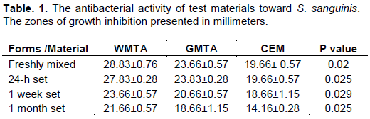
Growth inhibition zones in freshly mixed CEM group was significantly smaller than that of WMTA and GMTA (P= 0.02) (Figure 1).
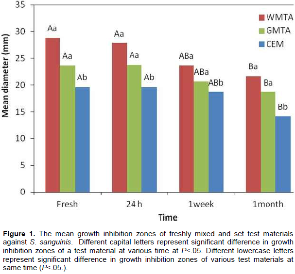
The largest mean diameters of inhibition zones of bacterial growth were found in set WMTA cement group at any time period.
The differences between inhibition zones of bacterial growth of the set CEM cement were significant compared to the GMTA and WMTA (24h, P= 0.025; 1week, P= 0.029; 1 month, P=0.025). Set GMTA group was associated with smaller growth inhibition zones compared to that in the WMTA group at any time period (24 h, 1 week, and 1 month) (Table 1). However, no significant difference was observed in the mean growth inhibition zone of the two groups (P=0.28).
The mean values of growth inhibition zones of WMTA on S. sanguinis were 28.83, 27.83, 23.66, and 21.66 mm when freshly mixed, 24-h, 1 week and 1 month set were used respectively. Increasing the incubation time of the mixed cements resulted in a reduced mean growth inhibition zones in WMTA group. The reduction of the growth inhibition zones was statistically significant in 1 month set WMTA compared to the freshly mixed (P<0.001) and 24-h set WMTA (P= 0.04) (Table and Figure 1).
Similar with what was observed in WMTA group, increasing the incubation time of the mixed cements resulted in a decrease in the mean growth inhibition zones in GMTA and CEM groups. The mean growth inhibition zones of 1 month set GMTA cement was significantly different compared to the freshly mixed GMTA (P= 0.02) and 24-h set GMTA (P= 0.01). The data obtained from changing the incubation time of the CEM cements were similar to those of WMTA and GMTA. The smallest mean growth inhibition zones were seen in 1 month set CEM group. The differences between the mean inhibition zones were statistically significant in 1 month set CEM group compared to the freshly mixed (P= 0.02) and 24-h set CEM cement (P= 0.02).
DISCUSSION
The primarily aim of this study was to evaluate and compare the antibacterial properties of freshly mixed and set WMTA, GMTA, and CEM cements. Based on our knowledge, this is the first study that investigates the more than one month antibacterial effects of white and gray MTA and CEM on S. sanguinis and E. faecalis microorganisms using ADT. Agar diffusion test is the most frequently used method for evaluating in-vitro antibacterial activity of root-end filling materials (Nirupama et al., 2014). This test indicates which materials are more likely to have antibacterial activity within the root canal system via direct comparisons between them (Siqueira et al., 2000). It has been previously demonstrated that the selected bacteria in this study, S. sanguinis and E. faecalis, are frequently associated with endodontic infections or therapy resistant cases (Sundqvist, 1982). In addition, E. faecalis is associated with persistent periradicular lesions after root canal treatment (Portenier et al., 2003).
The data obtained from this study showed that all three tested materials either freshly mixed or set cements were ineffective against E. faecalis. These results are in accordance with the findings of other studies that MTA and CEM cements did not show any antimicrobial activity against E. faecalis (Hasan Zarrabi et al., 2009; Torabinejad et al., 1995). This consideration in agreement with contrast with the findings by Poggio et al. (2017) who found that time had no effect on antibacterial efficacy.
Investigating the antimicrobial activity of the freshly mixed cement showed WMTA cement had the highest antimicrobial activity against S. sanguinis. Furthermore, the antimicrobial activity of freshly mixed GMTA was lower than that for WMTA and higher than that of CEM. These findings suggest that the freshly mixed WMTA (Angelus, Londrina, Paraná, Brazil) contains more potent antibacterial inhibitors such as high pH than GMTA (Angelus, Londrina, Paraná, Brazil) and CEM (ShahidBeheshti, Iran) cements. The antibacterial effect of MTA against the microorganisms has been attributed to its high pH or release of diffusible substrate into growth medium (Asgary et al., 2007).
However, our results showed that the effective antibacterial activity of freshly mixed WMTA and GMTA resulted in greater growth inhibited zones on S. sanguinis than CEM. These findings disagree with the previous studies that showed that the effective antibacterial activity of CEM cement was significantly better than that of the MTA group (Hasan Zarrabi et al., 2009; Asgary et al., 2007), when ProRoot MTA was used; MTA-Angelus was utilized in the present study. It has been reported that the pH and calcium ion release values are slightly higher for MTA-Angelus than ProRoot (20). ADT results are widely influenced by the diffusion ability of the material into the medium. In addition, variations in agar medium, bacterial strains, standardization of inoculation density, incubation, diffusion capacity of inhibitory agents, and reading point of the zones of inhibition are factors that affect the results of ADT (Torabinejad et al., 1995). Differences in materials types may explain the possible reason of this discrepancy.
The data obtained from the set materials showed that the WMTA cement has the highest antimicrobial activity against S. sanguinis at any time period followed by GMTA and CEM cements. Different patterns of inhibition of bacterial growth were found for the set WMTA and GMTA. The differences in antibacterial activity may be due to the physico-chemical differences between the GMTA and WMTA. The origin of MTA as well as the preparation technique may affect its antimicrobial activities (Al-Hezaimi, 2009).
The results of this study illustrated that increasing the incubation time (more than 24 h) of the mixed cements resulted in a reduced mean growth inhibition zones in all three groups. The reduction of the growth inhibition zones was statistically significant in 1 month set materials in all groups compared to the freshly mixed and 24-h set cements. Although the long term antibacterial activities of the applied materials in this study is yet to be investigated. The result obtained from the long term antibacterial activity of test materials disagrees with those of previous study reported by Hasan Zarrabi et al. (2009). They found that the antibacterial effect of MTA, new endodontic cement and Portland cement at different concentrations against five different microorganisms is enhanced with incubation time for 72 h (Hasan Zarrabi et al., 2009).
In WMTA group, inadequate setting and frangibility of WMTA pills were observed in agar medium with increased incubation time. The antibacterial activity of WMTA was maintained because of its poor setting and solubility. The difference in antibacterial activity of various materials may be related to the degree of the material setting (Cobankara et al., 2004).
CONCLUSION
The origin of MTA as well as the preparation techniques may affect its antimicrobial activities. The favored results of in-vitro antibacterial activity of WMTA-Angelus cement compared to the GMTA and CEM cement indicate potentiality of applying WMTA cement as an antibacterial agent on S. sanguinis. Moreover, increasing the incubation time, more than 24 h, of the mixed cement resulted in a decreased antibacterial activity of root-end filling materials. However, it is necessary to investigate the effect of these materials on the other pathogens which are frequently associated with endodontic infections or therapy resistant cases.
ACKNOWLEDGEMENT
The author would like to thank the Dental School of Kerman University of Medical Sciences. The author deny any conflicts of interest related to this study.
CONFLICT OF INTERESTS
The authors declare that they have no conflict of interest.
REFERENCES
|
Al-Hezaimi K, Al-Shalan TA, Naghshbandi J, Simon JH, Rotstein I (2009). MTA preparations from different origins may vary in their antimicrobial activity. Oral Surgery, Oral Medicine, Oral Pathology, Oral Radiology, Endodontology 107(5):85-88. |
|
|
Asgary S, Eghbal MJ, Parirokh M (2008a). Sealing ability of a novel endodontic cement as a root-end filling material. Journal of Biomedical Materials Research 87(3):706-709. |
|
|
Asgary S, Eghbal MJ, Parirokh M, Ghanavati F, Rahimi H (2008c). A comparative study of histological response to different pulp capping materials and a novel endodontic cement. Oral Surgery, Oral Medicine, Oral Pathology, Oral Radiology, Endodontology 106(4):609-614. |
|
|
Asgary S, Eghbal MJ, Parirokh M, Torabzadeh H (2006). Sealing ability of three commercial mineral trioxide aggregates and an experimental root-end filling material. Iranian Endodontic Journal 3(1):101-105 |
|
|
Asgary S, Kamrani A, Taheri S (2007) Evaluation of antimicrobial effect of MTA, calcium hydroxide, and CEM cement. Iranian Endodontic Journal 2 (3):105-109. |
|
|
Asgary S, Kamrani FA (2008). Antibacterial effects of five different root canal sealing materials. Journal of Oral Science 50(4):469-474. |
|
|
Asgary S, Shahabi S, Jafarzadeh T, Amini S, Kheirieh S (2008). Properties of a new endodontic material. Journal of Endodontics 34(8):990-993. |
|
|
Bhavana V, Chaitanya KP, Gandi P, Patil J, Dola B, Reddy RB (2015). Evaluation of antibacterial and antifungal activity of new calcium-based cement (Biodentine) compared to MTA and glass ionomer cement. Journal of Conservative Dentistry 18(1):44-46. |
|
|
Cobankara FK, Altinoz HC, Ergani O, Kav K, Belli S (2004). In vitro antibacterial activities of root-canal sealers by using two different methods. Journal of Endodontics 30(1):57-60. |
|
|
Fouad AF, Zerella J, Barry J, Spångberg LS (2005). Molecular detection of Enterococcus species in root canals of therapy-resistant endodontic infections. Oral Surgery, Oral Medicine, Oral Pathology, Oral Radiology, Endodontology 99(1):112-118. |
|
|
Hasan Zarrabi M, Javidi M, Naderinasab M, Gharechahi M (2009). Comparative evaluation of antimicrobial activity of three cements: new endodontic cement (NEC), mineral trioxide aggregate (MTA) and Portland. Journal of Oral Science 51(3):437-442. |
|
|
Nirupama DN, Nainan MT, Ramaswamy R, Muralidharan S, Usha HH, Sharma R, Gupta S (2014). In Vitro Evaluation of the Antimicrobial Efficacy of Four Endodontic Biomaterials against Enterococcus faecalis, Candida albicans, and Staphylococcus aureus. International Journal of Biomaterials 2014. |
|
|
Poggio C, Trovati F, Ceci M, Colombo M, Pietrocola G (2017). Antibacterial activity of different root canal sealers against Enterococcus faecalis. Journal of Clinical and Experimental Dentistry 9(6):743-748. |
|
|
Portenier I, Waltimo TMT, Haapasalo M (2003). Enterococcus faecalis: the root canal survivor and "star" in post-treatment disease. Endodontic Topics 6(1):135-159. |
|
|
Saunders WP (2008). A prospective clinical study of periradicular surgery using mineral trioxide aggregate as a root-end filling. Journal of Endodontics 34:660-665. |
|
|
Siqueira JF Jr, Favieri A, Gahyva SMM, Moraes SR, Lima KC, Lopes HP (2000). Antimicrobial activity and flow rate of newer and established root canal sealers. Journal of Endodontics 26(5):274-277. |
|
|
Sundqvist G (1982). Ecology of the root canal flora. Journal of Endodontics 18(9):427-430. |
|
|
Torabinejad M, Chivian N (1999) Clinical applications of mineral trioxide aggregate. Journal of Endodontics 25(3):197-205 |
|
|
Torabinejad M, Hong CU, Pitt Ford TR, Kettering JD (1995). Antibacterial effects of some root end filling materials. Journal of Endodontics 21(8):403-406. |
|
|
Tziafas D, Pantelidou O, Alvanou A,Belibasakis G, Papadimitriou S (2002). The dentinogenic effect of mineral trioxide aggregate (MTA) in short-term capping experiments. International Endodontic Journal 35(3):245-254. |
|
Copyright © 2024 Author(s) retain the copyright of this article.
This article is published under the terms of the Creative Commons Attribution License 4.0


