Full Length Research Paper

ABSTRACT
Diagnosis of maize lethal necrosis (MLN)-causing viruses is key in MLN surveillance programs and in testing seed for zero tolerance of Maize chlorotic mottle virus (MCMV) in seed lots. This is crucial for MLN management in farmers’ fields and in commercial seed fields. A customized MCMV detection assay that is specific, sensitive, affordable, and portable is therefore important for this task. Reverse Transcriptase–Recombinase Polymerase Amplification (RT-RPA) meets those conditions earlier described. RPA is a rapid isothermal nucleic acid amplification and detection platform that is based on patented Recombinase Polymerase Amplification (RPA) technology. In this study, a real time endpoint analysis and field deployable RT-RPA diagnostic method for the detection of MCMV was developed. RPA primer sets with their complementary probes were designed, synthesized and tested through a series of primer set evaluations to determine the most efficient primer sets. The primer sets targeted the MCMV genome at position 2765-2948 bp (MCMV_gp2 replicase gene). The parameters evaluated were sensitivity, specificity and reproducibility for the assay with remarkable results. The assay discriminated against other maize infecting viruses hence specific to MCMV. The assay takes only 20 min and its detection limit of 10-4 is well comparable to RT-PCR and other molecular based detection assays. MCMV was also detected directly from leaf saps without the nucleic acid extraction step hence suitable for on-farm testing. RPA is a relatively inexpensive technique that requires minimal instrumentation. This assay is therefore suitable for the detection of MCMV in field surveys, routine MCMV testing for phytosanitary measures and in the seed certification procedures.
Key words: Maize lethal necrosis, maize chlorotic mottle virus, diagnostics, recombinase polymerase amplification.
Abbreviation: CIMMYT, International Maize and Wheat Improvement Center; KEPHIS, Kenya Plant Health Inspectorate Service; MCMV, Maize chlorotic mottle virus; MLN, Maize lethal necrosis; NPPOs, National Plant Protection Organizations; SCMV, Sugarcane mosaic virus; OARDC, Ohio Agricultural Research Development Center.INTRODUCTION
Maize lethal necrosis (MLN) is caused by a synergistic infection of maize plants with Maize chlorotic mottle virus (MCMV), a Machlomovirus (Nutter et al., 1989; Lommel et al., 1991a) and any of the potyviruses namely Maize dwarf mosaic virus (MDMV) in the genus Potyvirus, Sugarcane mosaic virus (SCMV), a Potyvirus and the recently described Johnsongrass mosaic virus (JGMV) (Stewart et al., 2017) or Wheat streak mosaic virus (WSMV) in the genus Tritimovirus (Pruss et al., 1997). In eastern Africa, sugarcane mosaic virus (SCMV) is the predominant Potyvirus infecting maize in synergy with MCMV (Adams et al., 2013; Braidwood et al., 2018; Wamaitha et al., 2018).
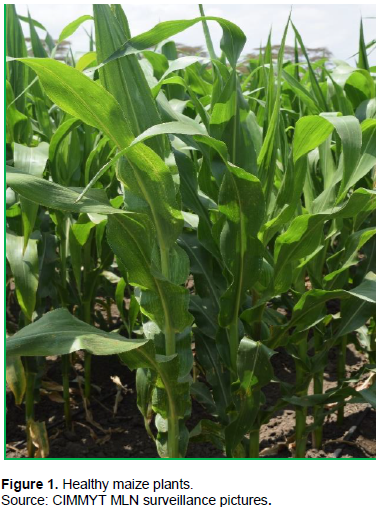
MLN is a relatively new disease in Africa having been first detected in Bomet county of Kenya in 2011 (Wangai et al., 2012). Indeed, MCMV is a new virus to infect maize in Africa in synergy with SCMV to cause MLN (Adams et al., 2013). The possibility of MCMV to combine with other native potyviruses of cereals poses a challenge to maize production in eastern Africa. The synergy between MCMV and SCMV or any other potyviruses can lead to the full development of MLN symptoms (Figures 1 and 2) in the field (Goldberg and Brakke, 1987; Niblett and Claflin, 1978; Uyemoto et al., 1980).

The spread of transboundary pests and diseases has increased significantly in recent years, affecting the food security and livelihoods of several million resource-constrained smallholders, especially in SSA, Asia, and Latin America. Globalization, trade, and climate change, as well as reduced resilience in production systems due to decades of agricultural intensification, have all played a part. Maize lethal necrosis has emerged as one serious transboundary disease in recent times.
It is evident that MLN had a serious impact on maize production and grain yield in eastern Africa from the time it was first reported in Kenya in 2011. Yield losses of between 23-100% were reported in maize growing counties of Kenya in the period 2012-2013 (Prasanna et al., 2020). De Groote et al. (2016) estimated that the aggregate national loss of maize production due to MLN in Kenya alone was about 0.5 million tons with a value of US$180 million. An average yield reduction of 1.4 t/ha was reported in Uganda, estimated at US$ 332 per ha (ASARECA, 2014; Kagoda et al., 2016). An estimated financial loss of KShs. 2 billion (approximately US$23.3 million) to smallholder farmers (Ministry of Agriculture and Livestock Development, Annual Report, 2014).
MCMV has been declared a regulated non-quarantine pest in Kenya and eastern Africa region by all the National Plant Protection Organizations (NPPOs) in countries where the disease has been officially reported (Kenya Plant Health Inspectorate Service Report, 2013). The pathogen is still classified as a quarantine disease in non-endemic countries that are close to the East African region (Malawi, Zambia and Zimbabwe) and have vibrant maize and seed maize industry. Due to a high exchange of seed material and trade in grain between these three countries and the Eastern Africa countries, there is a danger of introducing MCMV in these countries, which will adversely affect their prime maize grain and seed industry. In the same breath, seed producers in the endemic countries need to have internal regulation in place to produce MLN free seed as required by their respective seed certification agencies.
The first step in management of plant diseases is an accurate, affordable and user friendly diagnostics tool. Detection of viruses that cause viral diseases in maize is important to monitor maize diseases through surveillance programs, hence facilitating development and deployment of appropriate management measures (Riley et al., 2002). MCMV has been detected in leaves, pollen, female and male inflorescences, ear husks, cotyledons, and seeds (pericarps, endosperm, cotyledons, and embryo) (Scheets, 2004).
The current methods used for the detection of MCMV in host tissues include ELISA (enzyme-linked immunosorbent assay), Northern blots, and polymerase chain reaction (PCR).
The routine detection of MCMV is conducted by reverse transcriptase polymerase chain reaction (RT-PCR) as a standard molecular based diagnostics tool in countries that have both the infrastructure and expertise for PCR based diagnostics. Loop mediated isothermal amplification (LAMP) for the detection of MCMV has recently been adopted by the Research and Plant Disease Quarantine Laboratories in Kenya. As such, there is a need for a simple, rapid, and cost-effective method with equivalent specificity and sensitivity to PCR or quantitative PCR (qPCR).
A variety of isothermal amplification methods have been recently developed that can be deployed in the field for easier and faster diagnostics solutions (Piepenburg et al., 2006). One example is the ESEQuant Tube Scanner (Qiagen Lake Constance GmbH, Stockach, Germany). This Tube-scanner is a portable, battery operated fluorimeter that enables on-site, real-time detection. It has an advanced fluorescence sensor that slides back and forth under a set of eight tubes, collecting fluorescence signals over time and allowing for real-time documentation of increasing fluorescence signals. The increasing fluorescence in the Tube Scanner can be interpreted as nucleotide amplification by the recombinase polymerase amplification (RPA). RPA is a novel DNA amplification technique that operates at a low isothermal condition (Piepenburg et al., 2006; Mary, 2006). It combines isothermal recombinase-driven primer targeting of template material with strand-displacement DNA synthesis (Piepenburg et al., 2006). It achieves exponential amplification without pre-treatment of DNA sample. The detections are probe-based, and therefore sensitive, specific, and rapid. For RNA-based RPA assays, a reverse transcription (RT) step is required hence RT-RPA. RPA’s technology potentiates highly accessible and sensitive nucleic acid amplification outside of laboratory, and even self-testing (Li et al., 2019; Jia et al., 2019).
There are five main components of RPA: template DNA; a primer–recombinase complex to bind to atemplate and initiate the copying process; nucleotides; a polymerase to synthesize nascent strand; and single-stranded DNA-binding proteins (SSBs) to prevent double stranded deoxyribonucleic acid (dsDNA) to anneal, resulting in an exponential increase in the DNA sample (Mary, 2006). RPA combines with a sequence specific fluorescent probe for real-time detection. Probes are made up of an oligonucleotide backbone that contains an abasic nucleotide analogue, a tetrahydrofuran residue (THF), flanked by a dT-fluorophore and a corresponding dT-quencher group (Euler et al., 2012a, b). The probes are also blocked at the 3’ end by a modification group (such as a C3- spacer). Any fluorescent signal generated by the fluorophore will normally be absorbed by the quencher located 2-6 bases 3’ to the fluorophore. Once the probe has paired with its target sequence, the THF residue presents a target for the DNA repair enzyme, exonuclease III. The exonuclease cleaves the probe at the THF position, separating the fluorophore from the quencher to generate a detectable fluorescent signal (Haberstroh and Reiff, 2007). The separation of the fluorophore from the quencher occurs only when the probe has annealed to its target sequence within the amplification product and is an indication that amplification has occurred. The increase in signal allows real-time monitoring of the reaction.
Apart from the ability of RPA to operate under a low constant temperature, its reagents, and enzymes are lyophilized into individual reaction pellets. They are only activated in the field for reactions by mixing with the relevant rehydration buffer solution. The current documented RT-RPA for MCMV detection (Jiao et al., 2019) employs the end-point analysis of gel electrophoresis which limits it only to laboratory conditions since it is not practical to run gels in the field.
RPA extends the application of DNA amplification in fieldwork and in laboratories where thermocycling instruments are not available. As such, this study aimed at developing a real time recombinase polymerase amplification assay for the detection of MCMV both in the laboratory and in field conditions.
MATERIALS AND METHODS
Ribonucleic acid (RNA) extraction and detection of MCMV
RNA was extracted from several maize plants’ leaf samples infected by inoculation with MCMV grown and maintained in the quarantine screen house in Ohio Agricultural Research Development Center (OARDC), Ohio State University, USA in June - October 2017. RNA from maize leaf samples showing symptoms of MLN was also extracted for subsequent specificity analysis of the assay developed in this study. The ZR RNA MiniPrep™ from Zymo Research was used for RNA extraction from the samples. RNA is isolated from homogenized leaf samples using Fast-Spin column technology. RNA was eluted into volumes of 25 µl or more suitable for use in RT-PCR and other RNA-based procedures. RNA samples were also obtained from maize plants infected with Maize Dwarf Mosaic Virus (MDMV), Sugarcane Mosaic Virus (SCMV), healthy maize plants, and grass samples infected with Panicum Mosaic virus (PMV). The routine testing in the OARDC laboratories for MDMV, SCMV and PMV ensured only positive samples for these viruses were used in evaluating this assay.
The quality and concentration of RNA were tested using a NanoDrop ND-1000 Spectrophotometer (Thermo Fischer Scientific, Wilmington USA). A 2.2M formaldehyde RNA denaturing agarose gel electrophoresis was also performed to determine the integrity of the extracted RNA. RT-PCR was performed on the samples to ascertain the presence of MCMV presence using the primers designed by the USDA – Research Unit at OARDC, Ohio (MCMV F 5' – CCG GTC TAC CCG AGG TAG AAA – 3' and MCMV R 5' – TGG CTC GAA TAG CTC TGG ATT T – 3'). The detection MCMV was done in a two-step RT-PCR. The first step involved cDNA synthesis using the Maxima First Strand cDNA synthesis kit (ThermoFisher, Waltham, MA) for RT-qPCR with dsDNase.
RPA primers and probe design
The AmplifyRP Handbook (AmplifyRP Discovery Kit Assay Design Help Book, Agdia 2014) (https://d163axztg8am2h.cloudfront.net/static/doc/b0/4f/4e8d445a51f202f9f57f24a74c8d.pdf) was used for guidance in designing RPA primers and probes for this assay. The primers were designed using the Primer Quest tool, Integrated DNA Technologies Inc. (https://www.idtdna.com/Primerquest/Home/Index).
The primers and probes were designed targeting position 2765 to 2948 and 1869 to 2067 on the MCMV consensus genome sequence. The MCMV sequences used in the alignment were MCMV Taiwan – KJ782300, MCMV Nebraska USA – EU358605, MCMV Rwanda – KF744396, MCMV China – KF010583 and the Kenyan MCMV sequence, KP798454.1. Alignment was done using the ClustalW alignment algorithm in the MEGA X (MX) Alignment Explorer (Kumar et al., 2018) with the sequences to get consensus regions with conserved sequences for primer and probe design (Figure 3).

The XRT probes were designed manually by getting a reverse complementary sequence in the targeted region and modified with a Fluorophore (dT-Fam), tetrahydrofuran residue (THF, H), and a quencher dye (dT- Q) as per the AmplifyRP guidelines. The probe was designed from the sequences in the amplification region targeted by the forward and reverse primers without overlapping the priming regions denoted by the red colour of the bases (Figure 4). Complementary bases to these chosen regions were generated manually yielding the probe denoted in blue in Figure 2. The dT-Fam, -H and the-dT-Q- were plugged in the probe sequence by replacing the area with TTT (highlighted yellow in Figure 4). The final designed raw and modified probe is shown below:
AGTAACGAGCGCTTTCTTGGACCTCCTGCCATTTGCGATTGGATAGTTTGC
AGTAACGAGCGCTTTCTTGGACCTCCTGCCA--dT-Fam-H-dT-Q-GCGATTGGATAGTTTGC

The probe position in the sequence to be amplified was 2813 – 2863 as shown in Figure 2.
The primers and probes were designed by considering several parameters that are important in designing RPA primers and probes. The size of the amplicons produced should be 200bp or less with the primers typically between 30 to 35 nucleotides long. Shorter oligonucleotides are bound less efficiently by the recombinase. A “G/C clamp” at the 3’ end was adopted. The GC content between 30 and 70% was employed since Tm is not a factor in RPA.
Primer sets with long strings of repeated bases, as well as excessive numbers of short repeats were avoided. As with PCR, sequences that could contribute to primer-primer interactions, hairpins, and secondary structures were also avoided.
Designed RPA primers and probes can be found in Table 1. For each probe, three sets of F and R primers were designed.

The first set of primers and the probe (Table 2) were ordered from Integrated DNA Technologies (IDT, 1710 Commercial Park Coralville, Iowa 52241). The primers and probes were reconstituted upon receipt and a stock of 100 µM was prepared for each primer.

RPA primers and probe evaluation for testing Maize Chlorotic Mottle Virus in maize leaves
The following experiments were designed to evaluate the primers and the probe. The primers and the probe for the first set were ordered and used in assessing the primers amplification efficiency. A primer combination approach was used. Each forward primer (1AF, 1BF and 1CF) was screened against the three reverse primers at a time. The primer combinations were arranged and carried out as shown below.
i. 1AF - 1AR, 1BR, and 1CR
ii. 1BF - 1AR, 1BR, and 1CR
iii. 1CF - 1AR, 1BR, and 1CR
The Agdia XRT-RPA protocol was used to make the reaction master mix for the RT-RPA reactions (AmplifyRP Discovery Help Book (2), 2014). Each primer combination was screened with positive MCMV and water control. The rehydration solution for the RPA experiments was prepared as follows; 14.75 µl rehydration solution, 1.05 µl of the forward and reverse primers, 0.3 µl XRT probe, 0.25 µl of reverse transcriptase (Protoscript II, New England Biolabs (NEB), Ipswich, Massachusetts, United States), 4.37 µl of water and 1.0 µl of the RNA template. The total working volume of the rehydration mixture was 22.75 µl.
For each reaction, 22.75 μl of rehydration mixture was added to the reaction pellet for each sample. This was mixed briefly by pipetting up and down. The reaction tubes were capped immediately, spun briefly, and 1.25 μl of 280 mM magnesium acetate solution was added to start the reaction. The tubes were then vortexed and spun briefly before they were loaded into the fluorimeter (ESEQuant TS95, ID: ESTS10-MB-3020 Serial No.: 0014 from Qiagen). Fluorescence measurements (excitation, 470 nm; detection, 520 nm (FAM channel) were performed at 39°C for 20 min. This reaction temperature was found to yield the best performance in terms of sensitivity in a range tested from 39 to 42°C. The tube scanner software offers threshold validation, that is, evaluation of fluorescence by an increase of fluorescence above three standard deviations over the background determined in minute 1 (adaptable) of the reaction. The reactions progression was monitored on the computer connected to the ESEQuant fluorimeter.
For the first set of experiments, the first forward primer (1AF) was combined with all the reverse primers under the first set (1AR, 1BR and 1CR). These runs were repeated three times to assess the reproducibility of the assay. The same process was done for the evaluation of primer set 2; 1BF-1AR, 1BR -1CR and set 3; 1CF-1AR, and 1BR-1CR.
Determination of specificities for MCMV detection by the RPA primers/probe
The assay was tested for discrimination of the common potyviruses that are known to co-infect maize with MCMV namely Sugarcane mosaic virus (SCMV), Maize dwarf mosaic virus (MDMV) and Wheat streak mosaic virus (WSMV). The closest relative of MCMV, Panicum mosaic virus (PMV) in the family Tombusviridae was also used in evaluating the primers and probe specificity for MCMV amplification only. RNA was extracted from the plants infected by the aforementioned maize infecting viruses in the glasshouse chambers in OARDC, Ohio State University. The primer sets IBF and 1AR were used together with probe 1. These runs were replicated three times for consistency and reproducibility assessment.
Determination of Recombinase Polymerase Amplification sensitivity for Maize Chlorotic Mottle Virus detection
One positive sample from the RT-PCR testing experiment described earlier was selected and quantified with the Nanodrop to be used in the sensitivity analysis. The total plant RNA concentration was found to be 701 ng/µl. This was then standardized to 100 ng/µl; followed by six 10-fold serial dilutions, yielding samples of 100, 10, 1, 0.1, 0.01 and 0.001 ng/µl. These MCMV viral dilutions were analyzed in the RT-RPA assay for MCMV to determine the sensitivity of the assay.
Assessment of Recombinase Polymerase Amplification performance using fresh maize leaf sap extract
Extracts from fresh maize hybrid DEKALB DKC55-84RIB leaf samples from the OARDC Quarantine Screenhouse infected with MCMV, MCMV+SCMV, SCMV, and a healthy control (HC) were obtained using the General Extraction Buffer 3 (GEB3) (Agdia Agdia, Inc. 52642 County Road 1, Elkhart, IN 46514 USA). The GEB3 extraction buffer was reconstituted from the components as per the user guide from Agdia. To make 1000 ml of GEB3 sample extract buffer, smooth slurry was made by adding a small amount of water to 48 g of powder. 20 mls of Tween-20 were added to the slurry while mixing with water. Water was added to bring the final volume to 1 L (1000 mls) and the mixture was stirred for 30 min.
The maize leaf extracts from maize plants infected singly by MCMV, MCMV+SCMV, SCMV, and a healthy control (HC) were evaluated for the RPA assay in this experiment using the XRT RT-RPA protocol aforementioned. RPA primer 1BF and 1AR in combination with probe1 were used in subsequent tests.
RESULTS
Testing primer combinations
Amplification of positive samples occurred between the 6th and 10th minute of the assay for samples with high MCMV viral titers and the 10th and 20th minute for lower titer samples. By the 20th minute, the sample status had been determined as either positive or negative for MCMV. The primer and probe optimization and combinations were evaluated to get the most efficient combinations. The primer combination of 1CF-1BR gave the best amplification with a sharper curve at the 5th minute, showing it was the most efficient in terms of amplification (Figure 2). This was followed by 1AF-BR, 1AF-AR and 1CF-AR primer combinations. All the primer evaluations are shown in Table 3 with each primer combination reaction in a specific tube.

This combination of primers 1CF-1BR was used for subsequent experiments for evaluation on other maize viruses, sensitivity tests, and on maize leaf tissue extracts. However, other primer combinations also showed amplifications but not as efficient and precise as the combinations of 1CF-1BR and 1BF-1AR (Figure 5). These primer combinations were 1AF-1BR and 1AF-1AR. The remaining primer combinations did not yield any amplification whatsoever.

Primer combinations 1CF-1BR, 1BF-1AR, 1AF-1BR and 1AF-1AR amplified the targeted viral RNA whereas the others: 1BF-1BR, 1CF-1AR, 1BF-1CR and 1AF-1CR did not amplify the targeted viral RNA.
Specificity analysis of the Reverse Transcriptase-Recombinase Polymerase Amplification Assay
Evaluation of the specificity of the developed assay was paramount. The samples with MCMV, SCMV, PMV, MDMV, and WSMV were found to be well discriminated by the assay as illustrated in Figure 6. The healthy control (HC) and water were used as negative controls. The primer set, 1CF-1BR was used in this trial as indicated earlier. The assay was found to be highly specific picking up only MCMV present samples with no amplification registered in samples with other maize infecting viruses (Figure 6).

The RPA assay was able to segregate samples without MCMV hence displaying a high level of specificity (Figure 7). Only the three samples with different levels of MCMV were detected with the assay while SCMV was not detected by the assay as demonstrated with other maize infecting viruses.

RT-Recombinase Polymerase Amplification Maize Chlorotic Mottle Virus sensitivity analysis results
The RT-RPA assay for MCMV demonstrated the ability to detect MCMV to the 10-3 dilution with the 100 being 1 ng/µl. All the four dilutions amplified between the 4th and 8th minute of the assay (Figure 8a and b). This is consistent with the initial amplifications recorded in the primer evaluation stage. The healthy control (HC) and water registered no amplification as expected which showed that the assay was successfully executed.

Reverse Transcriptase-Recombinase Polymerase Amplification assay for Maize Chlorotic Mottle Virus using direct fresh leaf extracts
The MCMV RT-RPA assay was able to amplify samples with MCMV from maize hybrid DEKALB DKC55-84RIB leaf samples sap solution after grinding the leaf samples in the GEB extraction buffer. No RNA was extracted from these leaf samples, yet amplification was realized as seen in Figure 9. The assay also illustrated the specificity power by posting amplifications only with samples having MCMV infected samples. Amplification was not realized in samples with SCMV and PMV or HC. Amplifications were delayed by a few minutes in some samples, but early amplifications started at minute 5, which is quite comparable with MCMV RNA samples.

All the fresh leaf samples with MCMV or a combination of MCMV and SCMV were amplified and gave higher fluorescent signals than the controls. This is an indication that indeed the RPA method for the detection of MCMV may work with fresh maize leaf sample extract without isolation of RNA, which is a strenuous and expensive process. This also qualifies the assay suitability for field conditions. The test was run with leaves infected with other maize viruses for segregation. Only samples infected with MCMV tested positive. The samples of MCMV were in different concentrations and one sample had both MCMV and SCMV but the assay was able to amplify only samples with MCMV.
DISCUSSION
Maize Chlorotic Mottle Virus has been declared a regulated non-quarantine pest in Kenya and in eastern Africa where it has been endemic for the last seven years and a quarantine pest in the non-endemic countries in the region. This is due to its serious effect on maize yield in the MLN disease complex and its spread through infected and contaminated seed (Ministry of Agriculture and Livestock Development Kenya Annual Report, 2014; Hugo De Groote et al., 2016; Marenya et al., 2018). Since its first report in Bomet County, Kenya in 2012, the disease spread fast to other countries in the eastern and central Africa region (Wangai et al., 2012). Several prediction models show that the disease may spread to the central and western Africa region in the next five years (Isabirye and Rwomushana, 2016). As such, strengthening of phytosanitary regulatory systems in these countries bordering the MLN endemic countries to prevent the introduction of the disease in both new areas in endemic and non-endemic countries is crucial.
One of the important phytosanitary tools towards this effort is a dependable, efficient, effective, and affordable diagnostic tool for MCMV. The most reliable methods for detecting MCMV in host tissues include the enzyme-linked immunosorbent assay (ELISA), Northern blots, and polymerase chain reactions (PCR) for the detection of virus RNA (Mary, 2006). Immunostrips are also handy for testing MCMV in the field (CIMMYT MLN Diagnostics and Management Report, 2017). Currently, there are Immunostrips for detection of MCMV namely MCMV Agristrips from Bioreba, Switzerland, and MCMV Immunostrips manufactured by Agdia, USA. It is difficult to diagnose MCMV based on symptoms alone for some of its symptoms (stunting and chlorosis) resemble those caused by nutrient deficiency (Marchand et al., 1995). It has also been demonstrated that some infected plants may not exhibit MCMV, or MLN symptoms early enough hence are potential in spreading the disease in a localized manner. Molecular based diagnostic assays have been adopted by several National Plant Protection Organizations (NPPOs) and seed certification agencies for testing MCMV in fresh leaf tissues and seed. This is mostly from surveillance programs, for seed certification and phytosanitary requirements for trade. Of these molecular methods, qRT-PCR has been used due to higher sensitivity levels in comparison with conventional RT-PCR (Mackay et al., 2002). This has led to high testing charges up to USD 35 per sample due to the expensive equipment, maintenance, and reagents for the qRT-PCR MCMV assay. On the other hand, conventional RT-PCR is also used but the laborious process of gel-electrophoresis is a setback. The tests also require highly trained/skilled personnel and take up to 3 h excluding the RNA extraction process or more to generate enough copies of the amplicons for visualization under real-time endpoint analysis. It is also worth noting that the RT-PCR assays are only limited in laboratories hence are not field deployable like the RT-RPA assay developed.
This study developed an RT-RPA diagnostics method for the detection of MCMV. RPA is a rapid isothermal nucleic acid amplification and detection platform that is based on patented RPA technology (Piepenburg et al., 2006). The method achieved amplification within the 4th to the 6th minute from the start of the reactions (Figures 3 and 4) compared to the 20th minute to 1 h for the MCMV Loop-mediated isothermal amplification (LAMP) assays (Zhanmin et al., 2015; Chen et al., 2016; Mwatuni et al., 2020b). This demonstrates how fast the assay is in detecting the virus. The primer combination 1CF-1BR and Probe1 demonstrated its high level of specificity by only targeting the MCMV viral genome. The test discriminated against the viral RNA from PMV, SCMV, MDMV, WSMV, and in samples that had both MCMV and SCMV. This is an important aspect for testing of MCMV since other isothermal assays like LAMP that use color change due to DNA intercalating dyes are prone to cross-contamination leading to false positives (Chen et al., 2016). The LAMP MCMV assays require the opening of the reaction tubes to add the dyes for visualization of the amplification through colour change. False positive results can have devastating economic effects on seed companies, especially when testing seed lots for MCMV in seed certification procedures. In RPA reactions, the reaction tubes are sealed after all the reagents are added and analyzed in real-time due to fluorescence from the probe activity (AmplifyRP Discovery Help Book, 2014; Piepenburg et al., 2006).
The assay was also evaluated for sensitivity which showed detection limits of up to 0.1 ng/µl. The advantage of RPA over qPCR is that it misses the range of equivocal or false positives of the tested real-time PCRs for all the amplifications that are over in 20 min (Euler, 2012b). In this study, AmplifyRP reagents were used to successfully amplify target RNA in a procedure that does not require nucleic acid extraction and purification steps. Amplification was recorded from leaf sap samples extracted using the GB33 extraction buffer (Agdia) (Figure 6), but the differentiation between positive samples and controls was not as sharp as with RNA samples. Many common sample extraction buffers, such as those used in Agdia’s Immunostrips and ELISA test kits are suitable for use with AmplifyXRT format which was used to develop this MCMV RT-RPA assay. Physical extraction of plant tissue or insect vectors can be performed using any common laboratory technique such as macerating with a mortar and pestle and/or mesh extraction bags (AmplifyRP Help Book, 2014).
The rapid real-time RT-RPA MCMV test hereby developed facilitates faster testing periods and is easily deployable in the field. The small fluorimeter which runs on a lithium phosphate battery is well adopted for field conditions. The lyophilized kits contain pre-mixed enzymes and reagents in a pellet form necessary for the amplification. The reagents can be carried around in the field without storage/transport problems and with low risk of degradation. This makes the reaction amenable to field use or any other point of use applications. The cost per sample was evaluated and found to be USD 4 which was quite affordable compared to USD 35 for qRT-PCR and USD7.5 for LAMP (Mwatuni et al., 2020b) as earlier mentioned.
The rapid and highly sensitive isothermal real-time RT- for the detection of MCMV and by extension MLN developed in this study is well adapted for both laboratory and field usage hence quite mobile. This methodology could be utilized by both regulatory agencies and seed maize production entities for surveillance, quality control, regulation, and internal self-regulation.
The MCMV RT-RPA method has been recommended to the Kenya Plant Health Inspectorate Service (KEPHIS) management, other regional Plant Protection, seed certification agencies, and seed companies for adoption due to the short turnaround time, short time in sample preparation and flexibility in point of use (International Phytosanitary Conference Proceedings, KEPHIS, Kenya 2018). However, the direct testing from fresh maize leaves using the direct leaf sample extract needs to be optimized to improve its sensitivity for low sensitivity which was recorded in this study.
CONCLUSION
It is evident that this developed molecular based assay will go a long way in facilitating diagnostics of MCMV both in the laboratories and in the field. The fact that it has impressive limits of detection that is well comparable with qPCR and LAMP; it will be a preferred molecular method due to its relatively low cost, short turnaround time, and ease in carrying out the tests. It also has an extra advantage of testing MCMV without the nucleic acid extraction stage which is laborious with cost implications. It is therefore recommended for adoption for research institutions, phytosanitary regulatory institutions and in MLN surveillance programs.
CONFLICT OF INTERESTS
The authors have not declared any conflict of interests.
ABBREVIATIONS
CIMMYT, International Maize and Wheat Improvement Center; KEPHIS, Kenya Plant Health Inspectorate Service; MCMV, Maize chlorotic mottle virus; MLN, Maize lethal necrosis; NPPOs, National Plant Protection Organizations; SCMV, Sugarcane mosaic virus; OARDC, Ohio Agricultural Research Development Center.
ACKNOWLEDGEMENTS
The authors sincerely thank the US government through the USDA-FAS office in Nairobi for granting Francis Mwatuni a research fellowship under the Norman Borlaug fellowship to carry out this work in the Department of Plant Pathology, Ohio Agriculture and Research Development Center (OARDC) - Wooster, Ohio State University. The authors also recognize the extremely valuable guidance by Dr. Margaret Peg Radinbaugh and Dr. Sally Miller during the entire research study at OARDC. Also highly appreciated are Christine Willey, Xing Ma, Fulya Baysal for their technical backstopping on the bench while in OARDC, Ohio State University. Finally, with the support from BMGF (Investment ID: INV-009934/OPP1134248), the authors were able to evaluate the assay in Kenya.
REFERENCES
|
Adams IP, Miano, DW, Kinyua, ZM, Wangi A, Kimani E, Pahari N, Reeder R, Harju V, Glover R, Hany U, Souza-Richards R, Deb Nat P, Nixon T, Fox A, Barnes A, Smith J, Skelton A, Thwaite's R, Mumford R, Bonham N (2013). Use of next-generation sequencing for the identification and characterization of Maize chlorotic mottle virus and Sugarcane mosaic virus causing maize lethal necrosis in Kenya. Plant Pathology 62(4):741-749. |
|
|
Amplify RP Discovery Kits. Assay Design Help Book (2), (2014). Available at: |
|
|
ASARECA (2014). ASARECA Annual Report, 2014. Available at: |
|
|
Braidwood L, Quito-Avila DF, Cabanas D, Bressan A, Wangai A, Baulcombe DC (2018). Maize chlorotic mottle virus exhibits low divergence between differentiated regional sub-populations. Scientific Reports 8(1):1-9. |
|
|
Chen L, Jiao Z, Liu D, Liu X, Xia Z, Deng C, Zhou T, Fan Z (2016). One-step reverse transcription loop-mediated isothermal amplification for the detection of Maize chlorotic mottle virus in maize. Journal of Virological Methods 240:49-53. |
|
|
International Maize and Wheat Improvement Center (CIMMYT) (2017) CIMMYT MLN Diagnostics and Management Project Report, Available at: View |
|
|
De Groote H, Oloo F, Tongruksawattana S, Biswanath D (2016). Community-survey based assessment of the geographic distribution and impact of maize lethal necrosis (MLN) disease in Kenya 2016.Elsevier Crop Protection 82:30-35. |
|
|
Goldberg KB, Brake MK (1987). Concentration of Maize chlorotic mottle virus increased in mixed infections with Maize dwarf mosaic virus, Strain-B. Phytopathology 77:162-167. |
|
|
Euler M, Wang Y, Nentwich O, Siedenburg O, Hufert FT, Weidmann M (2012a). Recombinase polymerase amplification assay for rapid detection of Rift Valley fever virus. Journal of Clinical Virology 54:308-312. |
|
|
Euler M, Wang Y, Otto P, Tomaso H, Escudero R, Anda P, Hufert FT, Weidmann M (2012b). Recombinase polymerase amplification assay for rapid detection of Francisella tularensis. Journal of Clinical Microbiology 50:2234-2238. |
|
|
Haberstroh K, Reiff EC (2007). Fluoreszenzmessung wird alltagstauglich pp. 29-33. In: Schanz GW (ed), Sensortechnik aktuell. Oldenbourg Industrieverlag GmbH, Munich, Germany. |
|
|
Isabirye B, Rwomushana I (2016). Current and future potential of distribution of Maize chlorotic mottle virus and risk of maize lethal necrosis disease in Africa. Journal of Crop protection 5(2):21-228. |
|
|
Jia L, Macdonald J, Stetten F (2019). Review: a comprehensive summary of a decade development of the recombinase polymerase amplification. Analyst 144(1):31-67. |
|
|
Jiao Y, Jiang J, Ann M, Xia Z, Wu Y (2019). Recombinase polymerase amplification assay for rapid detection of maize chlorotic mottle virus in maize. Archives of Virology 164(10):2581-2584. |
|
|
Kagoda F, Gidoi R, Isabirye BE (2016). Status of maize lethal necrosis in eastern Uganda. African Journal Agricultural Research 11:652-660. |
|
|
Kenya Plant Health Inspectorate Service (KEPHIS) (2013). Annual reports 2013/2014. Available at: View |
|
|
Kenya Plant Health Inspectorate Service (KEPHIS) (2018). International Phytosanitary Conference proceedings, Kenya Available at: |
|
|
Kumar S, Stecher G, Li M, Knyaz C, Tamura K (2018). MEGA X: Molecular Evolutionary Genetics Analysis across computing platforms. Molecular Biology and Evolution 35:547-1549. |
|
|
Li J, Ma B, Fang J, Zhi A, Chen E, Xu Y (2019). Recombinase Polymerase Amplification (RPA) Combined with Lateral Flow Immunoassay for Rapid Detection of Salmonella in Food. Foods 9(1):27-30. |
|
|
Lommel SA, Kendall TL, Xiong Z, Nutter RC (1991a). Identification of the Maize chlorotic mottle virus capsid protein cistron and characterization of its subgenomic messenger RNA. Virology 181(1):382-385. |
|
|
Mackay IM, Arden K, Nitsche A (2002). Real-time PCR in virology. Nucleic Acids Research 30:1292-1305. |
|
|
Marchand JL, Peterschmitt M, Reynaud B, Dintinger J (1995). Maize streak, Maize stripe and Maize mosaic viruses in the tropics. Agriculture et Development Special issue pp. 55-88. |
|
|
Marenya PP, Erenstein O, Prasanna B, Makumbi D, Jumbo M, Beyene Y (2018). Maize lethal necrosis disease: evaluating agronomic and genetic control strategies for Ethiopia and Kenya. Agricultural Systems 162:220-228. |
|
|
Mary H (2006). DNA Amplification and Detection Made Simple (Relatively) PLoS Biology 4(7):e222. |
|
|
Ministry of Agriculture and Livestock Development (2014). Annual report, 2014. Available at: |
|
|
Mwatuni FM, Radinbaugh MP, Miller S, Ma X, Nyende AB, Suresh LM, Obare I (2020b). A two-end point analysis reverse transcription loop-mediated isothermal amplification (RT-LAMP) assay for the detection of maize chlorotic mottle virus (MCMV). Journal of General and Molecular Virology 10(2):9-21. |
|
|
Niblett CL, Claflin LE, (1978). Maize lethal necrosis a new virus disease of maize in Kansas. Plant Disease Report 62(1):15-19. |
|
|
Nutter RC, Scheets K, Panganiban L, Lommel SA (1989). The complete nucleotide sequence of maize chlorotic mottle virus. Nucleic Acids Research 17(8):3163-3177. |
|
|
Piepenburg O, Williams CH, Stemple DL, Armes NA (2006). DNA detection using recombination proteins. PLoS Biology 4(7):1115-1121. |
|
|
Prasanna BM, Suresh LM, Mwatuni F, Beyene Y, Makumbi D, Gowda M, Olsen M, Hodson D, Worku M, Mezzalama M, Molnar T (2020). Maize lethal necrosis (MLN): Efforts toward containing the spread and impact of a devastating transboundary disease in sub-Saharan Africa. Virus Research 282:197943. |
|
|
Pruss G, Ge X, Shi XM, Carrington JC, Bowman-Vance V (1997). Plant viral synergism: the potyviral genome encodes a broad-range pathogenicity enhancer that transactivates replication of heterologous viruses. Plant Cell 9(6):859-868. |
|
|
Riley M, Williamson MR, Maloy O (2002). Plant disease diagnosis. The Plant Health Instructor. |
|
|
Scheets K (2004). Maize chlorotic mottle virus. Viruses and Virus Diseases of Poaceae (Gramineae). Lapierre H and Signoret PA, eds. Institut National de la Rocherche Agronomique, Paris, France pp. 642-644. |
|
|
Stewart LR, Willie K, Wijeratne S, Redinbaugh G, Massawe D (2017). Johnsongrass mosaic virus contributes to maize lethal necrosis in East Africa. Plant Diseases 101(8):1455-1462. |
|
|
Uyemoto JK, Bockelman DL, Claflin LE (1980). Severe outbreak of maize lethal necrosis disease in Kansas. Plant Diseases 64(1):99-100. |
|
|
Wamaitha JM, Deepti N, Solomon M, Francesca S, Anne W, Joyce N, Timothy AH, Bramwel WW, Mark W, Tanui L, Appolinaire D, Hernan G (2018). Metagenomic analysis of viruses associated with maize lethal necrosis in Kenya. Virology Journal 15(1):90. |
|
|
Wangai AW, Redinbaugh MG, Kinyua ZM, Mahuku G, Sheets K, and Jeffers, D (2012). First report of Maize chlorotic mottle virus and maize lethal necrosis in Kenya. Plant Disease 96(10):15-82. |
|
|
Zhanmin I, Xueying X, Cuiyun Y, Junyi H (2015). Colorimetric detection of Maize chlorotic mottle virus by reverse transcription loop-mediated isothermal amplification (RT-LAMP) with hydroxynapthol blue dye. Royal Society of Chemistry advances 6(1):73-78. |
|
Copyright © 2024 Author(s) retain the copyright of this article.
This article is published under the terms of the Creative Commons Attribution License 4.0


