Full Length Research Paper
ABSTRACT
Anaplasma marginale, Babesia bovis and Theileria annulata infections significantly affect the development and improvement of animal husbandry in Africa. In Côte d’Ivoire, molecular studies for the diagnosis of these hemoparasites are lacking. Therefore, the aim of this study was to determine the prevalence and the distribution of A. marginale, B. bovis and T. annulata and to evaluate the risk of coinfection in cattle in Côte d’Ivoire. A subsample of 180 dried blood spots was randomly selected for analysis with conventional PCR, from a total of 895 in six livestock area. Amplification of the MaR1bB2, Bovar2A and cytochrome b1 genes for the detection of A. marginale, B. bovis and T. annulata were performed. Pearson’s Chi-squared test, Fisher exact test and the confident intervals were also performed. The overall prevalence of A. marginale, B. bovis and T. annulata was 68.9, 57.8 and 10%, respectively. A. marginale was determined to be the most infesting species, especially in the central area where it had the highest prevalence (90%). For B. bovis, the southern and central zones were the most infected with this parasite (70%). Out of 180 examined cattle, 152 (84.4) were infected with one or multiple haemoparasites investigated. Mixed infections were observed in 81/180 (45%) of blood samples and the co-infections of A. marginale and B. bovis were more frequent (63/180; 35%). The monoinfection of A. marginale was significantly higher (49/180; 27.2%) and no monoinfections of T. annulata were detected. The results of this study showed a high prevalence and wide distribution of A. marginale and B. bovis, in all six livestock area, in Côte d’Ivoire. These pathogens pose a risk to animal and human health (especially B. bovis) and food safety.
Key words: Anaplasma marginale, Babesia bovis, Theileria annulata, prevalence, co-infection, Côte d’Ivoire.
INTRODUCTION
Ticks are obligate hematophagous arthropods known to be important vectors of a wide variety of protozoa, fungi, bacteria, viruses and filarial worms of medical and veterinary importance (Aydin et al., 2015; Pereira et al., 2016). Tick-borne diseases (TBDs) are responsible for significant losses among cattle and impact the livelihoods of resource-poor communities worldwide, particularly in sub-Saharan Africa (Byaruhanga et al., 2015; Roy et al., 2018). Haemoparasitosis results in mortality, morbidity resulting in abortions, growth retardation, and losses in milk and meat production. In addition, it is of economic importance due to the cost of veterinary diagnosis and control measures (Simuunza et al., 2011). In tropical and subtropical countries, tick-borne haemoparasites are transmitted mainly by ticks of the genus Hyalomma, Rhipicephalus, and Amblyomma (Ziam et al., 2016). In sub-Saharan Africa, the main tick-borne hemoparasitoses of cattle that cause more damage are babesiosis, theileriosis and anasplasmosis (Simuunza et al., 2011).
In West Africa, anaplasmosis, babesiosis and theileriosis are among the most frequently diagnosed tick-borne haemoparasitoses in sheep and cattle (Farougou et al., 2012; Djakaridja et al., 2014; Adjou Moumouni et al., 2018). In Côte d’Ivoire, data on haemoparasites is scanty and limited to a few departments within the various districts (Achi et al., 2012; Djakaridja et al., 2014; Yéo et al., 2017b). Thus, a parasitological study was conducted throughout the country to map the distribution of tick-borne haemoparasites using microscopy. However, the prevalence obtained was relatively low, below 10% for the whole country, despite the emergence of Rhipicephalus microplus, a potential vector of anaplasmosis and babesiosis (Aké-Bogni et al., 2022). However, microscopic diagnostic methods lack sensitivity and require expertise in reading slides for subclinical or/and chronic infections. Parasitemia is often extremely low and tick-borne haemoparasites can be difficult to find in stained blood smears (Ganguly et al., 2020). Therefore, diagnostic methods with higher sensitivity and specificity than routine microscopic examination, such as molecular detection, will be most suitable for detection of infections with low parasitemia (Chauhan et al., 2015).
Thus, the objective of this work is to determine the presence and prevalence of three important haemoparasites of cattle, Anaplasma marginale, Babesia bovis and Theileria annulata, and to evaluate the risk of coinfection in cattle in Côte d’Ivoire.
MATERIALS AND METHODS
Study areas
Côte d’Ivoire is a country in West Africa, located between latitudes 5° and 11° N, and longitudes 3° and 9° W. The climate is hot with average monthly temperature from 24 to 28°C and monthly rainfall from 10 to 230 mm. The North of the country has one short rainy season from the beginning of June to the end of September, with a high precipitation in August. The Central and Southern regions have two rainy and two dry seasons per year. The two rainy seasons in the Central region include the long March to the end of June season, and the short September to October season. In the South, the main rainy season begin from April to the end of July, and the shorter rainy season from the beginning of October to the end of November.
Sampling and data collection
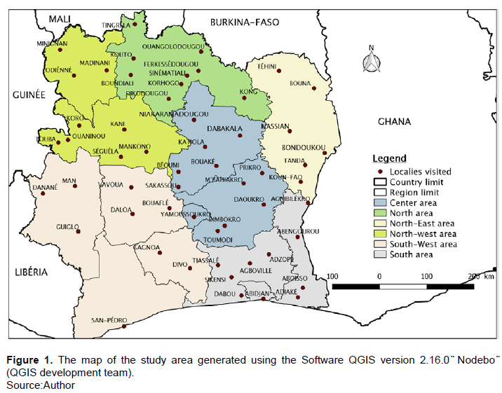
This study was conducted in 59 localities of 54 departments in Côte d’Ivoire (Figure 1), exclusively during the rainy season from April 2014 to May 2015. The study was conducted nationwide according to the administrative division of the Ministry of Animal Resources and Fisheries (MIRAH) which includes 19 regions subdivided into 77 departments. The regions have been grouped into 6 geographical livestock zones (Southeast, Southwest, Central, North, Northeast and Northwest) according to the density of cattle. A total of 150 cattle was sampled in each of the six livestock zone, except in the northeast zone where 145 cattle were sampled. Two or three farms were selected by locality. Blood samples were collected from the auricular vein of at least one-year-old cattle, from 5 individuals per farm. Blood collected in a hematocrit tube was used for preparation of dry blood spots for each cattle. This study follows a parasitological study (microscopy method) carried out on 54 departments of the country. The sample size was calculated according to the following formula (Thrusfield et al., 2018):
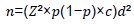
where Z=1,96, d=5% absolute precision, p=77% known prevalence, and c=3 the correction coefficient considering the three main zones of Côte d'Ivoire (North, Center, and South). The total number of cattle in the study was estimated at 895 with n=816 (Aké-Bogni et al., 2022).
For the molecular study, out of a total of 895 cattle, 180 were randomly selected in clusters of 30 cattle per breeding zone.
The farms were georeferenced using a Garmin GPSMAP 64 – Multicolored GPS. The permission was obtained from the owners of the farm to collect blood from cattle.
DNA extraction
DNA was extracted from bovine dried blood spots according to the modified method of Benbouza et al. (2006) using the CTAB lysis buffer. Approximately 1 cm3 of dried blood spot was cut out from filter paper with a sterile pair of scissors. The cut-up blood spot was soaked in PBS for at least one week. Each tube corresponding to an animal was given a specific code so that it can be identified later. Lysis buffer (CTAB) (400 µl) preheated to 65°C, 20 µl proteinase K and 2 µl 2-Mercapto-ethanol were added to the tubes containing blood-PBS solution. The tubes were mixed for 30 s and then incubated in a thermomixer for 1.5 h at 65°C, shaking at 550 rpm. Cold chloroform (400 µl) was added to the tubes and the contents were mixed gently by inversion by hand for 1 min. The tubes were centrifuged for 15 min at 15 000 rpm and the upper aqueous layer was recovered. Cold isopropanol (400 µl) was added to the supernatant and mixed gently by inversion by hand for 1 min. The mixture was stored at -20°C for 1 h to maximize DNA precipitation, then centrifuged for 5 min at 13,000 rpm. The pellet was washed with 70% alcohol and dissolved in 100 µl of 1x TE Buffer.
Polymerase chain reaction (PCR)
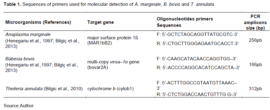
Two types of PCR reactions were performed: a simplex PCR for A. marginale and a duplex PCR for B. bovis and T. annulata, using specific primers and annealing temperatures (Table 1). Each PCR’s reaction was carried out using 1 U Gotaq G2 polymerase Kit (PROMEGA, Madison, WI USA) in a final volume of 50 µl, including 5 µl of genomic DNA and 10 µM primers. All PCR reactions were performed using an automatic thermocycler (Gene Amp PCR Systheme 9700). Thermocycler conditions for the simplex PCR reaction were: 94°C for 3 min, 40 cycles of 94°C for 50 s, 50°C for 50 s, 72°C for 1 min and a final extension of 72°C for 5 min.
The duplex PCR reaction was carried out with similar thermocycler conditions, except for the primer annealing done at 55°C. The PCR products were analyzed in a 1.5% agarose gel (Sigma) SybrGreen incorporated. Amplicon sizes were determined relative to a 250 and 100 bp DNA ladder. All primers were experimentally validated for specificity and only amplified fragments of the desired size.
Sequencing
Fragments corresponding to the predicted amplicons of 265 and 166 bp for A. marginale and B. bovis, respectively, were cut from the gel individually and purified using the QIAquick gel extraction kit (QIAGEN, Germany). After purification of the amplicons, the DNA fragments were analyzed by the Sanger sequencing method using the 3500 xL® Dx genetics analyzer (Applied Biosystems).
Data analysis
Data was analyzed using STATA version 14.2 (StataCorp College Station Texas USA). The prevalence of the identified haemoparasites were calculated and presented in a pie chart. The Pearson’s Chi-squared test and Fisher exact test were also performed for comparing the distribution of haemoparasites throughout the livestock areas. The confident intervals were calculated to compare the prevalence of coinfected cattle. The threshold of significance is 95%. The BLASTn analysis tool was used to compare the sequences obtained in this work with other sequences of the same gene in the GenBank database. Subsequently, sequence alignments and consensus sequences were obtained and edited or trimmed using BioEdit v7.0.1 software. The sequences were then aligned via the ClustalW application and compared with reference sequences of A. marginale and B. bovis from the GenBank, National Center for Biotechnology Information (NCBI), via the BLAST interface.
RESULTS
Prevalence and distribution of tick-borne haemoparasites of cattle determined by conventional PCR
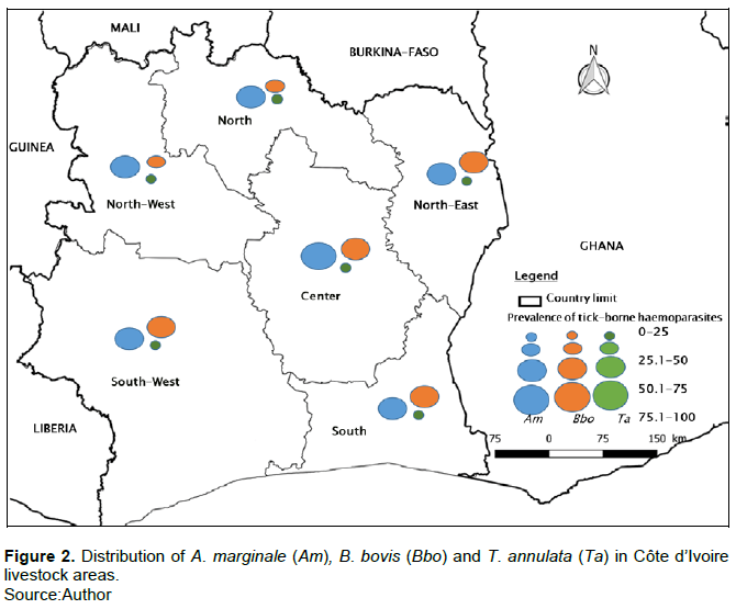
The three haemoparasites investigated were identified in all the livestock areas in Côte d’Ivoire: A. marginale (265 bp), B. bovis (166 bp) and T. annulata (312 bp). The overall prevalence of A. marginale, B. bovis and T. annulata was 68.9, 57.8 and 10%, respectively. A statistical difference between the prevalence and distribution of A. marginale according to the different breeding zones was p=0.038. While, for B. bovis and T. annulata, no statistical difference was observed between prevalences and distributions (p?0.05). A. marginale was the most common haemoparasite found in cattle throughout the country; the central zone was the most infested with a prevalence of 90% (Figure 2, Appendix 1). B. bovis was the second most infesting haemoparasite of the three and the southern and central zones were the most infected with this parasite (70%) (Figure 2, Appendix 1). T. annulata was present at low prevalence in all the livestock areas investigated (>20%). The highest prevalence (16.67%) for this parasite was detected in the northern and north-eastern zone (Figure 2, Appendix 1).
Infection and co-infection of cattle with tick-borne haemoparasites
A total number of 152/180 (84.44%; CI: 79.1-89.7) cattle were infected with one or more tick-borne haemoparasites on conventional PCR (Figure 3 and Appendix 1). The prevalence of co-infected cattle was 45% (81/180) and only 39.4% (71/180; CI: 32.3-46.6) had mono-infections. The number of cattle infected with A. marginale was significantly higher (49/180; 27.2%; CI: 20.7-33.7) than the other two haemoparasites. B. bovis was the second highest monoinfection (22/180; 12.2%; CI: 7.4-17). No T. annulata monoinfections were detected. Regarding co-infections, cattle showed infestation with up to three tick-borne haemoparasites (Figure 3 and Appendix 1).
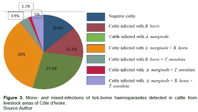
The combination of A. marginale and B. bovis was the most frequent (63/180; 35%; CI: 28-42) and the least frequent was A. marginale and T. annulata (2/180; 1.1%; CI: 0-2.64).
Sequencing of MAR1bB2 and bovar2A amplicons
A total of six PCR amplicons of A. margniale and one of B. bovis were sequenced by Sanger method. Sequences of amplicons detected using primer MAR1bB2 (265 bp) showed 98.8% identity to msp1b gene sequence of A. marginale published in the NCBI database (accession no. MF467524.1) (Appendix 1 and 2). Sequences from the bovar2A PCR products (166 bp) showed 88.43% identity to the B. bovis versa1α gene sequence published in the NCBI database (accession no. XM_00160874.1).
DISCUSSION
Tick-borne haemoparasites are a major obstacle to the development and improvement of cattle breeding worldwide, especially in Africa (Roy et al., 2018). In Côte d’Ivoire, no molecular studies have been conducted up to date to assess the prevalence of important tick-borne haemoparasites of cattle. Thus, our study is the first to provide information on the prevalence of A. marginale, B. bovis and T. annulata in cattle in the country, based on dried blood spots analyzed with conventional PCR methods.
Three haemoparasites were identified in all livestock areas in Côte d’Ivoire, with very high prevalences for A. marginale and B. bovis. However, in a previous, we fund low prevalences for these haemoparasites using ligth microscopy in three livestock areas (below 10%) (Aké-Bogni et al., 2022). These results reflect the low sensitivity of light microscopy compared to PCR (Ganguily et al., 2020).
The abundance of potential vectors such as Rhipicephalus species and Amblyomma variegatum for the transmission of A. marginale, and Rhipicephalus (Boophilus) for the transmission of B. bovis, could explain the high prevalences (>40%) obtained in sampled cattle (Achi et al., 2012; Yéo et al., 2017a). Indeed, the different vector species of these pathogens were identified throughout the Ivorian territory by Boka et al. (2017) with quite high prevalences. The high prevalence of A. marginale was previously reported in the Savannah Region of the country (Soffo, 2010). This high prevalence could be explained by the biological transmission of A. marginale by about 20 species of ticks, including those belonging to the subgenus Rhipicephalus (Boophilus), to the genus Rhipicephalus and Hyalomma found in tropical regions (Amorim et al., 2014; Silaghi et al., 2017).
In addition to the biological route, vertical (or transplacental) transmission and mechanical transmission (biting Diptera, soiled sharp objects) are also implicated. Vertical transmission occurs from cow to fetus during the acute phase of anaplasmosis during gestation or under conditions of constant inoculation in endemic areas (Aktas and Özübek, 2017; Solomon and Tanga, 2020). Vertical transmission of Anaplasma species is partly responsible for their endemic character in a bovine population (Costa et al., 2016; Silvestre et al., 2016). Besides vertical transmission, mechanical transmission through Diptera bites or using soiled objects during the administering of veterinary products against livestock diseases could increase the number of infested animals (Aktas and Özübek, 2017).
Anaplasmosis can be transmitted even through small amounts of blood (Costa et al., 2016; Ringo et al., 2018; Rjeibi et al., 2018). Mirah (2014) and Soffo (2010) attributed the spread of infection to possible poor tick and haemoparasite control practices adopted by lives owners. These situations would cause the maintenance of infected ticks in the different livestock areas and the regular contamination of new animals through the multiple use of needles or soiled tools (Reinbold et al., 2010; Yéo et al. 2017b). Another explanation for the high prevalence of A. marginale and B. bovis would be the self-treatment of cattle by farmers without veterinarian advice, which is the reason for the under-dosing of antibiotics and antiparasitics used in the fight against haemoparasites. This situation would make the treatment against these pathogens (A. marginale and B. bovis) ineffective. To save money and because of ignorance of the seriousness of tick-borne haemoparasites, many owners engage in this practice. This situation is observed in the Poro and Savannah regions of Côte d’Ivoire, where 85.7% of sedentary livestock farmers under-dosing of antibiotics for their cattle to fight tick-borne diseases by over dilution (Yéo et al., 2017b). Finally, there is the problem of illicit veterinary drugs of dubious origin and quality stored in very poor conditions that have flooded rural markets (Soffo, 2010; MIRAH, 2014). In Côte d’Ivoire, the distribution of veterinary drugs is carried out by projects and groups of approved breeders under the direction of a veterinary consultant. However, a large proportion of veterinary drugs, the quantity of which is not known, is smuggled in and ends up on the market (MIRAH, 2014). Also, the quality of veterinary drugs is often questioned. Indeed, studies conducted on the quality of veterinary drugs marketed in sub-Saharan Africa have revealed the existence of poor-quality drugs. Thus, this situation is at the root of the entrenchment of haemoparasitosis in the sub-region (MIRAH, 2014).
The high prevalence of B. bovis and A. marginale could also be explained by the situation of asymptomatic carriers, which is not to be neglected in anaplasmosis and bovine babesiosis. Animals that recover from infection are always low-level carriers of parasites, without apparent clinical signs, with the possibility of relapse under stressful conditions. These relapses are more frequent in the Microbabesia subgenus to which B. bovis belongs. Furthermore, these microbes are more resistant to antiparasitic treatments (Laha et al., 2015). Also, sequential cycles of A. marginale rickettsemias in which new MSP2 variants replicate are the cause of lifelong carriage of the disease in the infected animal, which will however not be clinically affected (De Souza Ramos et al., 2019). Thus, these asymptomatic carrier animals act as a source of infection for vector ticks and would thus contribute to the infection of healthy cattle. This explains the high prevalence of B. bovis and A. marginale in the Ivorian cattle herd (Aktas and Özübek, 2017; De Souza Ramos et al., 2019).
All livestock areas recorded the presence of T. annulata, which could be explained by the numerous movements of animal herds from the North to other parts of the country (transhumance), with all the corollaries of transmission of infectious agents (Capligina et al., 2014). In Côte d’Ivoire, transhumant herds move from the North to the sub-regions in the dry season (January to May) and return at the beginning of the rainy season (Yao et al., 2020; Ouedraogo et al., 2021). Also, the immature stages of some species of ticks of the genus Hyalomma like Hyalomma marginatum rufipes preferentially infest birds and small rodents (Ouedraogo et al., 2021). However, birds have a large range of movement. Thus, this situation contributed to the distribution and discovery of H. m. rufipes in the South of the country (Boka et al., 2017). While although T. annulata was observed in all livestock areas, the prevalences were low (< 20%). This observation could be explained by the low proportion of its vectors (Hyalomma species). Indeed, Hyalomma spp. was identified at 0.49% of all tick species collected by Boka et al. (2017) in Côte d’Ivoire. Tropical theileriosis transmitted by T. annulata is one of the most economically important livestock diseases in North Africa and Asia (Elelu et al., 2016). The presence of this pathogen in West Africa is an additional concern to those that are endemic (Mamman et al., 2021).
A total of 86.1% (155/180) of the cattle were infested by one or more species of haemoparasites. Also, 35% of the infested cattle had a co-infection marked by the combination of A. marginale and B. bovis. The higher prevalence of cattle infested with these pathogens and co-infections could be explained by a favorable environmental climate and the multiplication of vectors of these haemoparasites such as species of the subgenus Rhipicephalus (Boophilus), Hyalomma spp., etc. (Ringo et al., 2018). The main vector of these pathogens widely identified in Côte d’Ivoire is R. (B.) microplus (Boka et al., 2017; Walker et al., 2014); which explains the high prevalence of co-infection in sampled cattle. Also, according to Dib et al. (2008), concurrent infections with tick-borne diseases are common in animals. The association between A. marginale and B. bovis that was very marked in this study was also observed by Farougou et al. (2007) in Benin.
The alignment of the sequences confirmed the presence of the pathogens of interest, A. marginale and B. bovis. Indeed, the msp1β gene sequence is a sensitive target in the genome of A. marginale and specific for the detection of infection in both ticks and cattle (Bilgiç et al., 2013). The same is true for the ves1α gene sequence that is specific for the detection of B. bovis in the blood of cattle in different geographical regions where the disease is endemic (Bilgiç et al., 2013).
Our study has several limitations. Firstly, PCR was not optimal due to the presence of aspecific bands and the deposition of primer residues. However, the target genes were identified. Secondly, we could not have sequenced T. annulata because of technical issue.
CONCLUSION
This work made it possible to determine the actual distribution and prevalence of A. marginale, B. bovis and T. annulata in cattle in Côte d’Ivoire. The study identified the three tick-borne haemoparasites in cattle in all livestock areas and A. marginale was the most detected with a higher prevalence in the central zone (90%). T. annulata was the least represented in all the livestock areas (<20%) and always detected in mixed infections. The prevalence and distribution of A. marginale was observed throughout the territory with a difference depending on livestock areas. Most samples had mixed infection (81/180; 45%) and the infection of A. marginale and B. bovis was the most frequent (63/180; 35%). The high prevalence of the pathogenic A. marginale and B. bovis infections pose a threat to pastoralists and the food security of people living in Côte d’Ivoire. Sanitary authority should implement better disease control measures such as regulating treatment use, especially for tick control. It is also necessary to promote epidemiological surveillance of the Ivorian cattle herd and introduce PCR in routine of analytical laboratories.
CONFLICT OF INTERESTS
The authors have not declared any conflict of interests.
REFERENCES
|
Achi YL, Kone P, Stachurski F, Zinsstag J (2012). Impact des tiques sur des bovins métisses dans le nord de la Cote d'Ivoire. Bulletin de la Santé et de la Production animales en Afrique 60(2):109?118. |
|
|
Adjou Moumouni PF, Aplogan GL, Katahira H, Gao Y, Guo H, Efstratiou A, Jirapattharasate C, Wang G, Liu M, Ringo AE, Umemiya-Shirafuji R, Suzuki H, Xuan X (2018). Prevalence, risk factors, and genetic diversity of veterinary important tick-borne pathogens in cattle from Rhipicephalus microplus-invaded and non-invaded areas of Benin. Ticks and Tick-Borne Diseases 9(3):450?464. |
|
|
Aké-Bogni G, Achi YL, Yao KP, Diaha-Kouamé CA (2022). Distribution of cattle tick-borne haemoparasites in 54 departments of Côte d'Ivoire after the invasion of Rhipicephalus microplus. International Journal of Biosciences 20(1):123-132. |
|
|
Aktas M, Özübek S (2017). Outbreak of anaplasmosis associated with novel genetic variants of Anaplasma marginale in a dairy cattle. Comparative Immunology, Microbiology & Infectious Diseases 54:20?26. |
|
|
Amorim LS, Wenceslau AA, Carvalho FS, Carneiro PLS, Albuquerque GR (2014). Bovine babesiosis and anaplasmosis complex: diagnosis and evaluation of the risk factors from Bahia, Brazil. Revista Brasileira de Parasitologia Veterinária 23(3):328-336. |
|
|
Aydin MF, Aktas M, Dumanli N (2015). Molecular identification of Theileria and Babesia in ticks collected from sheep and goats in the Black Sea region of Turkey. Parasitology Research 114(1):65?69. |
|
|
Bilgiç H.B., Karagenç T., Simuunza M., Shiels B., Tait A., Eren H., Weir W., 2013. Development of a multiplex PCR assay for simultaneous detection of Theileria annulata, Babesia bovis and Anaplasma marginale in cattle. Experimental Parasitology 133(2):222?229. |
|
|
Bilgic HB, Karagenç T, Shiels B, Tait A, Eren H, Weir W (2010). Evaluation of cytochrome b as a sensitive target for PCR based detection of T. annulata carrier animals. Veterinary Parasitology 174(3?4):341?347. |
|
|
Boka OM, Achi L, Adakal H, Azokou A, Yao P, Yapi YG, Kone M, Dagnogo K, Kaboret YY (2017). Review of cattle ticks (Acari, Ixodida) in Ivory Coast and geographic distribution of Rhipicephalus (Boophilus) microplus, an emerging tick in West Africa. Experimental and Applied Acarology 71(4):355-369, |
|
|
Byaruhanga C, Collins NE, Knobel D, Kabasa W, Oosthuizen MC (2015). Endemic status of tick-borne infections and tick species diversity among transhumant zebu cattle in Karamoja Region, Uganda: Support for control approaches. Veterinary Parasitology: Regional Studies and Reports 1:221?230. |
|
|
Capligina V, Salmane I, Keišs O, Vilks K, Japina K, Baumanis V, Ranka R (2014). Prevalence of tick-borne pathogens in ticks collected from migratory birds in Latvia. Ticks and Tick-Borne Diseases 5(1):75?81. |
|
|
Chauhan HC, Patel BK, Bhagat AG, Patel MV, Patel SI, Raval SH, Panchasara HH, Shrimali MD, Patel AC, Chandel BS (2015). Comparison of molecular and microscopic technique for detection of Theileria annulata from the field cases of cattle. Veterinary World 8(11):1370?1374. |
|
|
Costa SCL, De Magalhães VCS, De Oliveira UV, Carvalho FS, De Almeida CP, Machado RZ, Munhoz AD (2016). Transplacental transmission of bovine tick-borne pathogens: Frequency, co-infections and fatal neonatal anaplasmosis in a region of enzootic stability in the northeast of Brazil. Ticks and Tick-Borne Diseases 7(2):270-275. |
|
|
De Souza Ramos IA, Herrera HM, De Fernandes SJ, Do Amaral RB, De Zanatto DCS, Da Silva TMV, Horta BLS, Campos JBV, Alves JVA, De Macedo GC, Machado RZ, André MR (2019). Genetic diversity of Anaplasma marginale in beef cattle in the Brazilian Pantanal. Ticks and Tick-borne Diseases 10(4):805-814. |
|
|
Dib L, Bitam I, Tahri M, Bensouilah M, De Meeûs T (2008). Competitive Exclusion between Piroplasmosis and Anaplasmosis Agents within Cattle. PLoS Pathogens 4(1):1-6. |
|
|
Djakaridja B, Yao KP, Gragnon B, Acapovi-Yao G, Mavoungou JF, N'Goran KE (2014). Situation épidémiologique des hémoparasites des bovins dans deux zones d'élevage de Cote d'Ivoire?: cas des anciennes régions des Savanes et de la vallée du Bandama. Révue de Medecine Véterinaire 165 (9-10):297-303. |
|
|
Elelu N, Ferrolho J, Couto J, Domingos A, Eisler MC (2016). Molecular diagnosis of the tick-borne pathogen Anaplasma marginale in cattle blood samples from Nigeria using qPCR. Experimental & Applied Acarology 70(4):501-510. |
|
|
Farougou S, Adakal H, Biguezoton AS, Boko C (2012). Prévalence de l'infection d'Amblyomma variegatum par Ehrlichia ruminantium dans les élevages extensifs du Bénin. Révue de Medecine Véterinaire 163(5):261?266. |
|
|
Farougou S, Tassou AW, Tchabode DM, Kpodekon M, Boko C, Youssao AKI (2007). Tiques et hémoparasites du bétail dans le nord-Bénin. Révue de Medecine Véterinaire 158 (8-9):463-467. |
|
|
Ganguly A, Maharana BR, Ganguly I (2020). Pentaplex PCR assay for rapid differential detection of Babesia bigemina, Theileria annulata, Anaplasma marginale and Trypanosoma evansi in cattle. Biologicals 63:81?88. |
|
|
Henegariu O, Heerema NA, Dlouhy SR, Vance GH, Vogt PH (1997). Multiplex PCR: critical parameters and step-by-step protocol. BioTechniques 23(3): 504-511. |
|
|
Laha R, Das M, Sen A (2015). Morphology, epidemiology, and phylogeny of Babesia: An overview. Tropical Parasitology 5(2):94-100. |
|
|
Mamman AH, Lorusso V, Adam BM, Dogo GA, Bown KJ, Birtles RJ (2021). First report of Theileria annulata in Nigeria: Findings from cattle ticks in Zamfara and Sokoto States. Parasites and Vectors, 14 (1): 242. |
|
|
MIRAH (2014). Plan stratégique de développement de l'élevage, de la pêche et de l'aquaculture en Côte d'Ivoire (PSDEPA 2014-2020). Ministère des Ressources Animales et Halieutiques, http://extwprlegs1.fao.org/docs/pdf/ivc146471.pdf, consulté le 29/03/2021. |
|
|
Ouedraogo SA, Zannou MO, Biguezoton AS, Yao KP, Belem AMG, Farougou S, Oosthuizen M, Saegerman C, Lempereur L (2021). Cross border transhumance involvement in ticks and tick-borne pathogens dissemination and first evidence of Anaplasma centrale in Burkina Faso. Ticks and Tick-Borne Diseases 12(5):101781. |
|
|
Pereira A, Parreira R, Nunes M, Casadinho A, Vieira ML, Campino L, Maia C (2016). Molecular detection of tick-borne bacteria and protozoa in cervids and wild boars from Portugal. Parasites and Vectors 9(1):251. |
|
|
Reinbold JB, Coetzee JF, Sirigireddy KR, Ganta RR (2010). Detection of Anaplasma marginale and A. phagocytophilum in bovine peripheral blood samples by duplex real-time reverse transcriptase PCR assay. Journal of Clinical Microbiology 48(7):2424-2432. |
|
|
Ringo AE, Adjou Moumouni PF, Lee S-H, Liu M, Khamis YH, Gao Y, Guo H, Zheng W, Efstratiou A, Galon EM, Li J, Tiwananthagorn S, Inoue N, Suzuki H, Thekisoe O, Xuan X (2018). Molecular detection and characterization of tick-borne protozoan and rickettsial pathogens isolated from cattle on Pemba Island, Tanzania. Ticks and Tick-borne Diseases 9(6):1437-1445. |
|
|
Rjeibi MR, Ayadi O, Rekik M, Gharbi M (2018). Molecular survey and genetic characterization of Anaplasma centrale, A. marginale and A. bovis in cattle from Algeria. Transboundary and Emerging Diseases 65(2):456-464. |
|
|
Roy BC, Krücken J, Ahmed JS, Majumder S, Baumann MP, Clausen P-H, Nijhof AM (2018). Molecular identification of tick-borne pathogens infecting cattle in Mymensingh district of Bangladesh reveals emerging species of Anaplasma and Babesia. Transboundary and emerging diseases 65(2):e231-e242. |
|
|
Silaghi C, Santos AS, Gomes J, Christova I, Matei IA, Walder G, Domingos A, Bell-Sakyi L, Sprong H, Von Loewenich FD, Oteo JA, De La Fuente J, Dumler JS (2017). Guidelines for the Direct Detection of Anaplasma spp. in Diagnosis and Epidemiological |
|
|
Studies. Vector-Borne Zoonotic Diseases 17(1):12?22, |
|
|
Silvestre BT, Silveira JAG, Meneses RM, Facury-Filho EJ, Carvalho AU, Ribeiro MFB (2016). Identification of a vertically transmitted strain from Anaplasma marginale (UFMG3): Molecular and phylogenetic characterization, and evaluation of virulence. Ticks and Tick-Borne Diseases 7(1):80?84. |
|
|
Simuunza M, Weir W, Courcier E, Tait A, Shiels B (2011). Epidemiological analysis of tick-borne diseases in Zambia. Veterinary Parasitology 175 (3?4): 331?342. |
|
|
Soffo YVS (2010). Enquête sur les hémoparasitoses et les parasitoses gastro? intestinales des bovins dans la région des savanes en Côte d'Ivoire. Doctorat Véterinaire, Ecole Inter-Etats de Sciences et Medecine Vétérinaire de Dakar, Dakar 158 p. |
|
|
Solomon A, Tanga BM (2020). The first investigation of tick vectors and tick-borne diseases in extensively managed cattle in Alle District, Southwestern Ethiopia. Veterinary Medicine International 2020:1?8. |
|
|
Walker AR, Bouattour A, Camicas JL, Estrada-Peña A, Horak IG, Latif AA, Pegram G, Preston PM (2014). Ticks of domestic animals in Africa: a guide to identification of species, Bioscience Reports, Edinburgh (Scothland) 228 p. |
|
|
Yao KP, Achi YL, Diaha-Kouame CA, Diobo NF (2020). Dynamiques saisonnières des tiques des bovins et de quelques hémoparasites associés le long du couloir de transhumance Est de la Côte d'Ivoire. Science et technique (5):497-511. |
|
|
Yéo N, Karamoko Y, Soro D, Zouh Bi ZF, Traore SI (2017a). Elevages de bétail dans la région du Poro (Côte d'Ivoire)?: Caractérisation et modalités de lutte contre les pathogènes transmis par les tiques. International Journal of Biological and Chemical Sciences 11(1):237-246. |
|
|
Yéo N, Yahaya K, Dofara S, Zahouli FZB, Abou JLO, Biégo GG (2017b). Prevalence of Trypanosoma, Babesia and Anaplasma in cattle reared in the North of Côte d'Ivoire. International Journal of Biosciences 10(2):21?28. |
|
|
Ziam H, Ababou A, Kazadi JM, Harhoura K, Aissi M, Geysen D, Berkvens D (2016). Prévalences et signes cliniques associés des piroplasmoses bovines dans les Wilayates d'Annaba et El Tarf, Algérie. Revue Médecine Vétérinaire 167(9-10):241-249. |
|
Copyright © 2024 Author(s) retain the copyright of this article.
This article is published under the terms of the Creative Commons Attribution License 4.0


