Medical Practice and Reviews

  • Abbreviation: Med. Pract. Rev.
  • Language: English
  • ISSN: 2141-2596
  • DOI: 10.5897/MPR
  • Start Year: 2010
  • Published Articles: 44

Short Communication

Prevalence of ovarian torsion in postmenopausal women in a Nigerian community

Wilson Onuigbo
  • Wilson Onuigbo
  • Department of Pathology, Abia State University Teaching Hospital, Aba, Nigeria.
  • Google Scholar
Paul Femi-Waboso
  • Paul Femi-Waboso
  • Department of Obstetrics and Gynaecology, Abia State University Teaching Hospital, Aba, Nigeria.
  • Google Scholar


  •  Received: 24 November 2014
  •  Accepted: 19 January 2015
  •  Published: 31 March 2016

 ABSTRACT

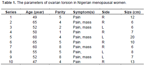
This study aimed at determining the prevalence of ovarian torsion in postmenopausal women in Nigeria. For this purpose, a histopathology data pool was maintained for 30 years among the Igbos, a large ethnic group in Nigeria from where postmenopausal women diagnosed with ovarian torsion were extracted for classification. The data concerned 69 cases. All were presented with acute abdominal pain. None was malignant. The patients were aged between 47 and 65 years (mean 55.5 years); their parity ranged from 1 to 8 (average 5); the size of the specimens ranged from 6 to 20 cm (average 9.5 cm); and the right/left involvement was in the ratio of 3:2. It was concluded that there are well-acknowledged risk factors for ovarian torsion even in postmenopausal women presenting with acute abdominal pain. Consequently, even in a developing country, there is need for clinical awareness of this important entity.

Key words: Ovary, torsion, menopause, Nigeria.


 INTRODUCTION

Although a few studies had reviewed the incidence of adnexal torsion, probably none had for years dwelt specifically on its occurrence in postmenopausal women. Therefore, to remedy this situation, Koonings and Grimes (1989) added to its literature with 19 cases collected from the Women’s Hospital, University of Southern California School of Medicine, Los Angeles. Another US group reported on 2 cases (Shih et al., 1991). The search of this study indicates that it is now being reported from countries such as Taiwan (Peng et al., 2008), Spain (Perez-Lopez et al., 2010), France (Huchon and Fauconnier, 2010), Turkey (Balci et al., 2011), Israel (Eitan et al., 2007), and Tunisia (Bouguizane et al., 2003). Therefore, to present the Nigerian picture, the authors reviewed all cases of this unusual lesion in the files of  a  Reference  Pathology Laboratory that serves the Ibos or Igbos, who constitute one of the three largest ethnic groups in Nigeria (Basden, 1966). Our investigation followed the recommendation of a British group (Macartney et al., 1980) by using a histopathology data pool for epidemiological analysis.


 MATERIALS AND METHODS

The data pool of this study consisted of operation specimens that were submitted by gynecologists working in several hospitals among the Igbos of Nigeria. To obtain large enough materials, the period of study was extended from 20th February, 1970 to 19th February, 2000.
 
The specimens were accompanied by full clinical details. Cases of Igbo postmenopausal women whose  histopathologic  diagnosis was ovarian torsion were manually reviewed. This identified 10 of such patient’s materials. This group constituted 14% of the total 69 patients who were of all ages and presented with ovarian torsion. The results are presented in Table 1.


 RESULTS AND DISCUSSION

Even in a developing country, such as Nigeria, gynecologists will be caring for women who present with acute abdominal pain. Perhaps, the postmenopausal group deserves to be given preferential attention. The fact that this is not being done is exemplified in Ackerman’s Surgical Pathology (Rosai, 1996), which documented not the occurrence of torsion in post-menopausal women per se but in adult women in general.
 
The risk of malignancy in older women with ovarian torsion is not exactly known (Shih et al., 1991). However, as was shown in this series and in a Spanish series (Perez-Lopez et al., 2010), when torsion occurs, the risk of malignancy is likely to be low. Indeed, Lomano et al. (1970) wrote: “the pathologic findings are emphasized as being benign.”
 
Another risk that is either low (McGovern et al., 1999) or not encountered (Bayer and Wiskind, 1994) is that of pulmonary embolism occurring during detorsion of the twisted ovary. Since our patients were postmenopausal, the question of detorsion for ovarian preservation did not arise.
 
The point was made in an Israeli report (Eitan et al., 2007) that under-diagnosis occurred on frozen section analysis. This problem did not apply because our specimens were not obtained with frozen section.
 
Nine of our patients became postmenopausal on account of age and the tenth attained this status because of hysterectomy performed for massive fibroids 2 years before her presentation. In the experience of Houry and Abbott (2001), 15 of their cases were postmenopausal while 7 were posthysterectomy. In contrast, in the experience of Baker and Copas (1995), the most common previous operation was bilateral tubal ligation.
 
Concerning age, the oldest patient was 65 years. This is in line with the age patterns in an undeveloped community (Husain, 2002). This is in contrast with developing Taiwan (Peng et al., 2008) where the oldest patient was 90 years old.
 
The largest size attained by the torsed ovary is of considerable interest (Nicholas and Julian, 1985). Of those who generalized, Houry and Abbott (2001) mentioned it to be equal or more than 5 cm across, while Lomano et al. (1970) have “greater than 8 cm in diameter.” Nichols and Julian (1985) stated that lesions undergoing torsion involve medium-sized ovarian neoplasms up to 10 to 12 cm in diameter and added that “torsion is virtually unknown among the very large ovarian neoplasms because of the limited mobility consequent to their size.” On the other hand, the above 90-year-old Taiwanese exhibited an infarcted ovary measuring 21 × 17 × 6 cm (Peng et al., 2008). Our local cases involved specimens that measured from 6 to 20 cm, averaging 9.5 cm.
 
Parity is another topical aspect. In our series, it ranged from 1 to 8. Bayer and Wiskind (1994) emphasized on its occurrence in the nulliparous state in almost half of their patients.
 
As regards the laterality of involvement, Baker and Copas (1995) found “no dominance in the side involved, as hypothesized by others.” Another group confirmed higher incidence of torsion on the right as being “consistent with the findings of others” (Lomano et al., 1970). In the present series, six were right sided and the remaining four left-sided, that is, R/L of 3/2. Much larger numbers would suffice for a decisive opinion.
 
Of the symptoms, Bayer and Wiskind (1994) noted the order to be pain (83%) and mass (72%). Our comparative figures are 100 and 50%, respectively. Concerning racial considerations, Lomano et al. (1970) mentioned that, among their 44 patients, 36 were white and 8 black. However, our series dealt exclusively with women of Negro stock.
 
The question of detorsion rather than extirpation was widely debated (Descargues et al., 2001; Bider et al., 1991; Oelsner and Shashar, 2006). However, this choice of  treatment  strictly  applies  to   child   bearing   women, whereas old Nigerian patients with average parity of 4.6 do not face this problem.
 
Lastly, a review of the literature has revealed the existence of variable views (Lomano et al., 1970; Gordon et al., 1980; Perez-Lopez et al., 2010). This includes the debate on benignancy and malignancy of torsed ovaries. However, all authors are in agreement that, when a postmenopausal woman presents with sudden abdominal pain in any part of the world, adnexal torsion should be considered strongly in the differential diagnosis and operative treatment undertaken quickly.


 CONFLICT OF INTEREST

The authors have not declared any conflict of interest.


 REFERENCES

Baker TE, Copas PR (1995). Adnexal torsion: A clinical dilemma. J. Reprod. Med. 40(6):447-449.


Balci O, Icen MS, Mahmoud AS, Capar M, Colakoglu MC (2011). Management and outcomes of adnexal torsion: A 5-year experience. Arch. Gynecol. Obstet. 284(3):643-646.
Crossref


Basden GT (1966) (First ed. 1938) Niger Igbos, London Frank Cass


Bayer AI, Wiskind AK (1994). Adnexal torsion: Can the adnexa be saved. Am. J. Obstet. Gynecol. 171(6):1506-1510.
Crossref


Bider D, Mashiach S, Dulitzky M, Kokia E, Lipitz S, Ben-Rafael Z (1991). Clinical, surgical and pathologic findings of adnexal torsion in pregnant and nonpregnant women. Surg. Gynecol. Obstet. 173(5):363-366.


Bouguizane S, Bibi H, Farhat Y, Dhifallah S, Darraji F, Hidar S, Lassoued L, Chaieb A, Khairi H (2003). Adnexal torsion: A report of 135 cases. English Abstract. J. Gynecol. Obstet. Biol. Reprod. 32(6):535-540.


Descargues G, Tinlot-Mauger F, Gravier A, Lemoine JP, Marpeau L (2001). Adnexal torsion: A report on forty-five cases. Eur. J. Obstet. Gynecol. Reprod. Biol. 98(1):91-96.
Crossref


Eitan R, Galoyan N, Zuckerman B, Shaya M, Shen O, Beller U (2007). The risk of malignancy in post-menopausal women presenting with adnexal torsion. Gynecol. Oncol. 106(1):211-214.
Crossref


Gordon A, Rosenshein N, Parmley T, Bhagavan B (1980). Benign cystic teratomas in postmenopausal women. Am. J. Obstet. Gynecol. 138(8):1120-1123.


Houry D, Abbott JT (2001). Ovarian torsion: A fifteen-year review. Ann. Emerg. Med. 38(2):156-159.
Crossref


Huchon C, Fauconnier A (2010). Adnexal torsion: A literature review. Eur. J. Obstet. Gynecol. Reprod. Biol. 150(1):8-12.
Crossref


Husain AR (2002). Life expectancy in developing countries: A cross-section analysis. Bangledesh Inst. Dev. Stud. 28:161-178.


Koonings PP, Grimes DA (1989). Adnexal torsion in postmenopausal women. Obstet. Gynecol. 73(1):11-12.


Lomano JM, Trelford JD, Ullery JC (1970). Torsion of the uterine adnexa causing an acute abdomen. Obstet. Gynecol. 35(2):221-225.


Macartney JC, Rollason TP, Codling BW (1980). Use of a histopathology data pool for epidemiological analysis. J. Clin. Pathol. 33(4):351-355.
Crossref


McGovern PG, Noah R, Koenigsberg R, Little AB (1999). Adnexal torsion and pulmonary embolism: Case report and review of the literature. Obstet. Gynecol. 54(9):601-608.
Crossref


Nichols DH, Julian PJ (1985). Torsion of the adnexa. Clin. Obstet. Gynecol. 28(2):375-380
Crossref


Oelsner G, Shashar D (2006). Adnexal torsion. Clin. Obstet. Gynecol. 49:459-463.
Crossref


Peng YY, Chen CH, Chien SC, Chen JR (2008). Torsion of a huge ovarian cyst in a 90 year-old woman. Taiwan J. Obstet. Gynecol. 47(2):224-225.
Crossref


Perez-Lopez FR, Chedraui P, Troyano-Luque JM (2010). Peri-and post-menopausal incidental adnexal masses and the risk of sporadic ovarian malignancy: New insights and clinical management. Gynecol. Endocrinol. 26(9):631-643.
Crossref


Rosai J (1996). Ackerman's Surgical Pathology. 8th ed. St Louis: Mosby-Year Book Inc, 1996:1450.


Shih S, Vetto JT, Berek JS, Heaps JM, Hiatt JR (1991). Adnexal torsion. An unusual cause of abdominal pain in postmenopausal women. Am. Surg. 57(5):327-329.


          */?>