African Journal of
Agricultural Research

  • Abbreviation: Afr. J. Agric. Res.
  • Language: English
  • ISSN: 1991-637X
  • DOI: 10.5897/AJAR
  • Start Year: 2006
  • Published Articles: 6863

Full Length Research Paper

Sero-epidemiological survey on toxoplasmosis in cattle, sheep and goats in Algeria

Amina Samia Dechicha
  • Amina Samia Dechicha
  • Institute of Veterinary Sciences, University of Blida 1, Algeria.
  • Google Scholar
Fatma Bachi
  • Fatma Bachi
  • Service de Biologie Parasitaire, Institut Pasteur d?Algerie, Algeria.
  • Google Scholar
Ismail Gharbi
  • Ismail Gharbi
  • Institute of Veterinary Sciences, University of Blida 1, Algeria.
  • Google Scholar
Edmee Gourbdji
  • Edmee Gourbdji
  • Service de Biologie Parasitaire, Institut Pasteur d?Algerie, Algeria.
  • Google Scholar
Djamila Baazize-Ammi
  • Djamila Baazize-Ammi
  • Institute of Veterinary Sciences, University of Blida 1, Algeria.
  • Google Scholar
ohamed Brahim-Errahmani
  • ohamed Brahim-Errahmani
  • Faculty of Sciences, University of Blida 1, Algeria.
  • Google Scholar
Djamel Guetarni
  • Djamel Guetarni
  • Department of Biology, Faculty of Nature and Life Sciences, University of Blida 1, Algeria.
  • Google Scholar


  •  Received: 02 February 2015
  •  Accepted: 26 April 2015
  •  Published: 14 May 2015

 ABSTRACT

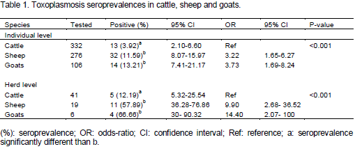
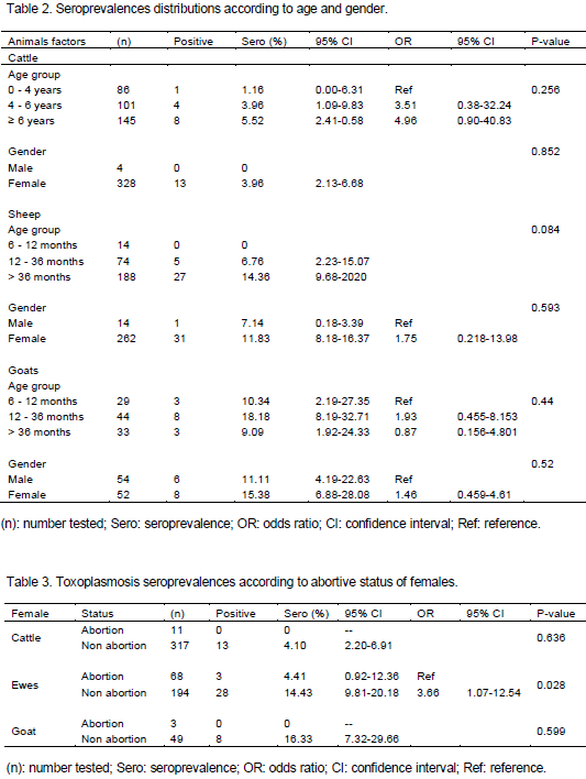
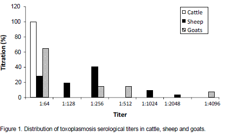
Toxoplasmosis is a parasitic disease with worldwide distribution and a major public health problem. In Algeria, human toxoplasmosis is screened in pregnant women and immunosuppressed persons; however, no information is available on the animal infection and a probable implication of the parasite in abortions occurring in the field. This sero-epidemiological cross-type survey on toxoplasmosis in cattle (332), sheep (276) and goats (106) revealed the presence of anti-Toxoplasma antibodies based on the indirect fluorescent antibody test (IFAT), at the respective rates of 3.92, 11.59 and 13.21%. The likelihood of acquiring Toxoplasma gondii infection was higher in sheep and goats (OR=3.22, 95% confidence interval [CI]: 1.65-6.27 and OR= 3.73, [CI]: 1.69-8.24 respectively) than in cattle (p<0.001). However, the difference between sheep and goats is not significant. At herd level, 5 herds out of 41 (12.19%), 11 herds out of 19 (57.89%) and 4 herds out of 6 (66.66%) showed at least one seropositive case in cattle, sheep and goat herds, respectively. Statistical comparison between genders and age groups showed no significant difference in the three species. The highest serological titers obtained are 1:64, 1:2048 and 1:4096 for cattle, sheep and goats, respectively. Suspicion of the parasite's role in abortions has been investigated, the seroprevalence showed no significant difference between abortive and non abortive females for cattle and goats; however, it was significantly higher in ewes that have not aborted as compared to those having abortions, a high suspicion was done for one abortive ewe whose antibody titer reached 1:1024. The presence of anti-Toxoplasma antibodies has been highlighted for the first time in livestock in Algeria, indicating a contamination with the parasite.


Key words: Toxoplasma gondii, seroprevalence, cattle, sheep, goats, abortion, Algeria.


 INTRODUCTION

Toxoplasmosis occurs worldwide and is present in a benign form in most animals. However, it is being recognized as a major cause of reproductive failure in several countries (Blewett and Watson, 1984; Dubey and Beattie, 1988; Dubey, 2004). Infection with Toxoplasma gondii seems to be the cause of embryonic resorption, mummification, abortions, neonatal deaths or birth of weak and non viable newborns.
 
The protozoan distribution varies widely according to species, farms and countries. Sheep, goats and pigs are the most sensitive species which record the highest seroprevalences and constitute a potential danger to humans (Tenter et al., 2000; Dumetre et al., 2006; Opsteegh et al., 2010). Contamination of human is usually done by ingestion of tissue cysts in undercooked meat (Smith, 1993; Cook et al., 2000) or by oocysts in soil or water that have been contaminated with cat feces (Bahia-Oliveira et al., 2003; Dubey, 2004; Jones et al., 2005; de Moura et al., 2006). Indeed, according to Roberts et al. (1994) and Mead et al. (1999), T. gondii is one of three pathogens (together with Salmonella and Listeria) which account for up to 75% of all deaths due to food borne disease in the USA.
 
Human toxoplasmosis occurs most often asymptomatically, inducing an underestimation of its prevalence. However, the use of serology shows seroprevalences that vary from one country to another; the lowest values are being recorded in the Northern countries, 5 and 10%, respectively in Sweden and Norway (Evengard et al., 2001; Jenum et al., 1998) and those higher in the south, 60 and 74.5% in Ivory coast and Brazil, respectively (Adoubryn et al., 2004; Spalding et al., 2005).
 
In Algeria, human seroprevalence is being estimated at 51.56% and prophylaxis of congenital toxoplasmosis is part of a national surveillance program for pregnant women, with medical care of toxoplasmic seroconversions or evolutive toxoplasmosis (Bachi et al., 2010). By contrast, there is no study available concerning the seroprevalence of animal toxoplasmosis and its implication as the probable origin in the abortions observed on the site, because only Brucellosis is investigated in this case (Dechicha et al., 2010).
 
The aim of this study is to investigate the toxoplasmosis antibodies in cattle, sheep and goats, and to test a probable implication of the parasite in abortions occurring in the field.


 MATERIALS AND METHODS

Study population
 
The sample comprised of three main animal species considered as the primary sources of red meat in Algeria. Cattle (332) originated from 41 farms located in Blida province, whilst the sheep (276) and goats (106) came from 19 and 6 farms, respectively, located in Djelfa province (agro-pastoral region).
 
For each species and from each farm, all females with a history of recent abortion, ten percent of reproductive female and reproductive males were selected. Blood was collected from the jugular vein and the separated serum was stored at -20°C until used. Animals in this study were treated according to the ethical standards of Algerian research centers and universities (MESRS, Algeria). Blood collections were carried out in compliance with EU legislation on research involving animals (EU, 2010).
 
Serological test
 
Toxoplasma antibodies were detected by the indirect fluorescent antibody test (IFAT) according to OIE Terrestrial Manual (2008). Slides were coated with whole tachyzoites of "RH" strain of T. gondii maintained by continuous passages in BALB/c mice and collected intraperitoneally. Test sera deposited on slides were diluted in PBS (0.3 M PBS, pH 7.4) starting from 1:16. Positive and negative control and also a PBS sample were included on each test slide. Species-specific IgG for cattle (Sigma F7887), sheep (Sigma F7634) and goats (Sigma F7363) conjugated to fluorescein isothiocyanate diluted in 0.2% Evans Blue (in PBS), were used appropriately as secondary antibody. The reaction was examined under a fluorescence microscope, and a sample was considered positive if at least 80% of the parasites were surrounded by a continuous peripheral strip of fluorescence at a dilution level of 1:64. Positive sera were subsequently titrated until the maximum positive dilution titer was reached.
 
Statistical analysis
 
Statistical analysis was performed using "Statistica 10.0" of Statsoft Inc., Tulsa, USA and "IBM SPSS Statistics 20.0," IBM Corp., USA. The degree of significance of the link between the seroprevalence and age, sex and the abortive/non abortive status for the females was performed by the χ2 test or Fischer Exact test where values in one or more cells are ≤ 5. These links were considered significant for p < 0.05. The degree of dependence of the disease on various factors was determined by the odds-ratios (with confidence intervals).


 RESULTS

Individual seroprevalences were estimated at 3.92, 11.59 and 13.21% respectively in cattle, sheep and goats. At herd level, 12.19, 57.89 and 66.66% herds showed at least one seropositive case in cattle, sheep and goats, respectively. Within-herd seroprevalence ranged from 11.76 to 30.76% in cattle; 4.54 to 63.63% in sheep and 20 to 42.85% in goat herds.
 
Statistical analysis shows that sheep and goats seroprevalences at individual or herd level are significantly higher than cattle seroprevalence (p<0.001). However, the difference between sheep and goats is not significant (Table 1).
 
 
Statistical comparison between age groups and gender of animals showed no significant difference in the three species (Table 2). The link between the seroprevalence and the abortive/non abortive status of the females showed no significant difference for cattle and goat. Whereas, the seroprevalence of non abortive ewes is significantly higher (p<0.05) than those having abortions (Table 3). Titration of positive sera showed a maximum titer of 1:64 for cattle, 1:2048 for sheep and 1:4096 for goats (Figure 1).


 DISCUSSION

The presence  of  anti-Toxoplasma  antibodies  has  been highlighted for the first time in livestock in Algeria, indicating a contamination with the parasite. In cattle, the seroprevalence estimated  in  the  study  is  relatively  low (3.92%), it is comparable to rates found in Brazil (1.96%), Iran (1.6%) and Poland (3.15%) as reported by Luciano et al. (2011), Raeghi et al. (2011) and Holec-G?sior et  al. (2013), respectively. This seroprevalence and the rate of infected herds (12.82%) may reflect a low distribution of the parasite in the farms of the study area. In this species, natural infection does not appear to cause clinical disease, however the seroprvalences reported in the literature range from 0 to 92% (Tenter et al., 2000). Indeed, cattle are considered a poor host for T. gondii; although they can be successfully infected with oocysts, the parasite is eliminated or reduced to undetectable levels within a few weeks (Dubey, 1986), perhaps due to innate resistance (Dubey and Jones, 2008).
 
Distribution of seroprevalence according to age showed no significant difference even if the odds ratio shows that it is 4.96 times higher in subjects aging ≥ 6 years. Rozette et al. (2005) report a similar finding in contrast to those of El fahal et al. (2013) who found a higher seroprevalence in cattle aging less than 1 year, thing that could be explained by the capacity of this species to eliminate the parasite with age. The distribution of seroprevalence according to gender showed no significant difference what is in agreement with the reports of Klun et al. (2006) and Garcia et al. (1999); however, those of Nematollahi and Moghddam (2008) showed that seropositivity is higher among males. The low antibody titer of 1:64 suggests that toxoplasmosis spreads in these farms in a latent form; it is similar to the findings of Santos et al. (2013). However, the low titer obtained is adequate with the hypothesis regarding the rapid removal of cysts by cattle.
 
In sheep, seroprevalence at individual level (11.59%) and at herd level (57.89%) reflects a strong environmental contamination and a wide distribution of the parasite in these farms. According to Ruiz and Frenkel (1980) and Dubey et al. (1996), the ground of farms can be strongly contaminated with oocysts as demonstrated in numerous studies. Indeed, animals kept on pastures with an increased pressure of infection due to contamination of the  environment  with  oocysts  show high seroprevalences in many areas of the world (Tenter et al., 2000).
 
The difference between age groups was not significant and is in agreement with reports of Sevgili et al. (2005). However, other studies reported a significant increase of the prevalence with the age of animals which could be explained by a greater possibility for the older subjects to be exposed to environment oocysts (Clementino et al., 2007; Vesco et al., 2007). In fact, according to Dubey and Kirkbride (1989), most of sheep acquire the infection before the age of 4 years; however, only one third of the older females remain seronegative in heavily contaminated farms. As well, we found no significant difference between sexes; a similar observation was reported by Khezri et al. (2012) and Gebremedhin et al. (2014), unlike Clementino et al. (2007) who reported a higher seroprevalence in females.
 
The antibody titers obtained are higher as compared to cattle, with a maximum titer of 1:2048; a similar titer was reported by Rossi et al. (2011) and higher titers reaching 1:65536 were reported by Garcia et al. (1999). A high antibody level would favor an acute toxoplasmosis or a reactivation of latent toxoplasmosis after an immunosuppressed conditions.
 
For goats, the seroprevalence of 13.21% is similar to rates reported in China (14.1%), Saudi Arabia (12%) and Ethiopia (19.7%) respectively by Zhao et al. (2011), Al Mohammed (2011) and Zewdu et al. (2013). At herd level, two thirds of the farms (66.66%) presented at least one seropositive case, which also goes in favor of a strong environmental contamination. In fact, this large distribution of the parasite among sheep and goat herds could be attributed to cats residing on site and the access of animals to contaminated feed and water. Indeed, according to Dubey and Frenkel (1972), cats can shed as many as 500 million oocysts after ingesting one T. gondii infected mouse under laboratory conditions. Furthermore, while only a few cats may be shedding T. gondii oocysts at any given time, the enormous numbers produced and their resistance to destruction assures widespread contamination (Dubey, 2004).
 
The difference between age groups was not significant in contrast to studies which showed a significant increase in seroprevalence with age as in the case of sheep (Figueiredo et al., 2001; Kamani et al., 2010; Zewdu et al., 2013). We observed no significant difference between genders as found by Bisson et al. (2000) and Carneiro et al. (2009), while the report of Swai and Kaaya (2012) showed that females are more infected than males. The majority of subjects have shown a titer ranging from 1:64 to 1:512, indicating a latent form of toxoplasmosis, the highest titer recorded is of 1:4096 expressing an acute form of toxoplasmosis. In fact, much higher titers reaching 1:8192 and 1:16384 were reported in other studies by Figueiredo et al. (2001) and Neto et al. (2008), respectively.
 
The  statistical  comparison  between  species  showed that cattle are the least infected species as compared to sheep and goats (p<0.001); this situation has already been described worldwide (Pita Gondim et al., 1999; Sharif et al., 2007). However, the comparison between sheep and goats showed no significant difference in contrast to Mahboub et al. (2013) whom observed a higher rate in sheep that could be explained by the variability of the species sensitivity and methods of farming.
 
Whatever the seroprevalence obtained, a particular attention should be given to the risk of human infection through consumption of undercooked meat (Halos et al., 2010; Boughattas et al., 2014) or raw goat and sheep milk (Camossi et al., 2011; Lafi et al., 2014). In fact for cattle, ingestion of beef or dairy products is not considered important in the epidemiology of T. gondii because cattle are not a good host for this parasite. However, we cannot be sure that beef does not play a role in T. gondii transmission as only relatively small amounts of beef have been tested for viable parasites (Dubey and Jones, 2008).
 
Furthermore, according to Smith (1993), meat from infected sheep and goat milk are shown to be primary sources of infection for men. In Algeria, the consumption of undercooked meat appeared as a major risk factor for toxoplasmosis infection in pregnant women (Messerer et al., 2014). Therefore, investigation of T. gondii in livestock is required in order to detect potential risk for human infection, especially where consumption of undercooked meat and raw milk is a popular tradition. To date, no protective measures have been taken by the farmers for herds because the sanitary and economic repercussions of this disease are unknown.
 
For cattle, no female that abort was seropositive; in fact, T. gondii is not recognized as an abortive agent in cattle (Dubey, 1986). Contrarily, for sheep and paradoxically to what has been reported in the literature, where toxoplasmosis is presented among the leading causes of abortion in many countries (Dubey and Kirkbride, 1989; Jackson and Hutchison, 1989) the seroprevalence of non-abortive ewes is significantly higher than of the abortive ones (14.43% vs. 4.41%). Hamzy El Idrissi et al. (1995) reported a seroprevalence rates that are equal between the abortive females (8.8%) and the normal lambing females (9.7%), while Benkirane et al. (1990) found a significantly higher rate in the abortive ewes (21%) as compared to normal lambing females (8%). The highest seroprevalence obtained in non-abortive ewes could be explained by the fact that the female is able of giving birth to a viable but infected lamb if it contracted the parasite in late gestation, and it would abort if it contracted the parasite for the first time during mid-gestation. Moreover, whether pregnant or not, it would develop an immunity that will prevent it from miscarrying during subsequent pregnancies. Probable hypothesis is that our non-abortive females have contracted the parasite  during  a  previous  gestation, or outside a gestation period, as they are all multiparous and aged between 3 and 6 years (Data not shown). As concerns the abortive and seropositive ewes, we cannot blame T. gondii as the unique responsible agent for these abortions, because the serology of the fetal fluids and further examination of aborted fetuses were not practiced, nevertheless the suspicion is high for one of the abortive ewes whose antibody titer reached 1:1024.
 
In goats, the disease is a common cause of abortion and neonatal mortality (Tenter et al., 2000; Lindsay and Dubey, 2007). However, in the present study, abortive females are seronegative; abortion may be due to another cause, whereas the seropositivity of the non-abortive females (16.33%) would have the same arguments as those given for the sheep. According to Ndiaye et al. (1996), toxoplasmosis in goats contributes more to neonatal deaths (82%) than to other types of loss, namely: abortions (44%) and weakness (56%).


 CONCLUSION

In Algeria, anti-Toxoplasma antibodies were revealed for the first time, in cattle, sheep and goats proving thereby the farm contamination by oocysts. The parasite has never been incriminated before because no research has been carried out, it could be responsible for one part of reproductive disorders, such as embryonic mortality and observed abortions. Further studies are needed to assess its involvement as an abortive agent other than Brucella.
 
A positive serology of livestock would favor contamination of their products and the high human seroprevalence recorded in Algeria could be partly explained by the consumption of contaminated animal products. 


 CONFLICT OF INTEREST

The authors have not declared any conflict of interest.


 REFERENCES

 
Al-Mohammed HI (2011). Seroprevalence of Toxoplasma gondii Infection in Cats, Dogs and Ruminant Animals in Al-Ahsa Area in Saudi Arabia. Res. J. Med. Sci. 5:190-192.
Crossref
 
Bachi F, Gourbdji E, Taourirt L, Boudhane L, Lazizi L (2010). Toxoplasmose congénitale: bilan du centre national de référence (Algérie). Archives de l'institut Pasteur de Tunis. Tome 87:3-4.
 
Bahia-Oliveira LM, Jones JL, Azevedo-Silva J, Alves CC, Oréfice F, Addiss DG (2003). Highly endemic, waterborne toxoplasmosis in north Rio de Janeiro state, Brazil. Emerg. Infect. Dis. 9:55-62
Crossref
 
Benkirane NJ, Rodolakis A (1990). Fréquence d'avortement et séroprévalence des principales maladies infectieuses abortives ovines dans la région de Rabat (Maroc). Ann. Rech. Vet. 21:267-273.
 
Bisson A, Maley S, Rubaire-Akiiki CM, Wastling JM (2000). The seroprevalence of antibodies to Toxoplasma gondii in domestic goats in Uganda. Acta Tropica. 76:33-38.
Crossref
 
Blewett DA, Watson WA (1984). The epidemiology of ovine toxoplasmosis. III. Observations on outbreaks of clinical toxoplasmosis in relation to possible mechanisms of transmission. Br. Vet. J. 140:54–63.
Crossref
 
Boughattas S, Ayari K, Sa T, Aoun K, Bouratbine A (2014). Survey of the Parasite Toxoplasma gondii in Human Consumed Ovine Meat in Tunis City. PLoS One 9(1):1-5.
Crossref
 
Camossi LG, Greca-Júnior H, Corrêa AP, Richini-Pereira VB, Silva RC, Da Silva AV, Langoni H (2011). Detection of Toxoplasma gondii DNA in the milk of naturally infected ewes. Vet. Parasitol. 177:256-261.
Crossref
 
Carneiro ACAV, Carneiro M, Gouveia AMG, Vilas-Boas LS, Vitor RWA (2009). Seroprevalence and risk factors of sheep toxoplasmosis in Minas Gerais, Brazil. Revue Méd. Vét. 160(11):527-531.
 
Clementino MM, Souza MF, Andrade Neto VF (2007). Seroprevalence and Toxoplasma gondii-IgG avidity in sheep from Lajes, Brazil. Vet. Parasitol.146:199-203.
Crossref
 
Cook AJC, Gilbert RE, Buffolano W, Zufferey J, Petersen E, Jenum PA, Foulon W, Semprini AE, Dunn DT (2000). Sources of Toxoplasma infection in pregnant women: european multicentre case-control study. Br. Med. J. 321:142–147.
Crossref
 
de Moura L, Bahia-Oliveira LM, Wada MY, Jones JL, Tuboi SH, Carmo EH, Ramalho WM, Camargo NJ, Trevisan R, Graca RM, da Silva AJ, Moura I, Dubey JP, Garrett DO (2006).Waterborne toxoplasmosis, Brazil, from field to gene. Emerg. Infect. Dis. 12:326–329.
Crossref
 
Dechicha A, Gharbi S, Kebbal S, Chatagnon G, Tainturier D, Ouzrout R, Guetarni D (2010). Serological survey of etiological agents associated with abortion in two Algerian dairy cattle breeding farms. J. Vet. Med. Anim. Health 2(1):001-005.
 
Dubey JP, Frenkel JK (1972). Cyst-induced toxoplasmosis in cats. J. Protozool. 19:155–177.
Crossref
 
Dubey JP (1986). A review of toxoplasmosis in cattle. Vet. Parasitol. 22: 177-202.
Crossref
 
Dubey JP, Beattie CP (1988). Toxoplasmosis of Animals and Man. CRC Press, Boca Raton, FL. P. 200.
 
Dubey JP, Kirkbride CA (1989). Enzootic toxoplasmosis in sheep in North-Central United-States. J. Parasitol. 75:673-676.
Crossref
 
Dubey JP, Lunney JK, Shen SK, Kwok OCH, Ashford DA, Thulliez P (1996). Infectivity of low numbers of Toxoplasma gondii oocysts to pigs. J. Parasitol. 82:438–443.
 
Dubey JP (2004) Toxoplasmosis – a waterborne zoonosis. Vet. Parasitol.126:57–72.
Crossref
 
Dubey JP, Jones JL (2008). Toxoplasma gondii infection in humans and animals in the United States. Int. J. Parasitol. 38:1257–127.
Crossref
 
Dumetre A, Ajzenberg D, Rozette L, Mercier A, Dardé ML (2006). Toxoplasma gondii infection in sheep from Haute-Vienne, France: Seroprevalence and isolate genotyping by microsatellite analysis. Vet. Parasitol. 142:376-379.
Crossref
 
El fahal AM, Elhassan AM, Hussien MO, Enan KA, Musa AB, El Hussein AM (2013). Seroprevalence of Toxoplasma gondii in Dairy Cattle with Reproductive Problems in Sudan. ISRN Vet. Sci. 1-4.
Crossref
 
EU (2010). Directive on the protection of animals used for scientific purposes. 
 
Evengard B, Petersson K, Engman ML, Wiklund S, Ivarsson SA, Tear-Fahnehjelm K, Forsgren M, Gilbert R, Malm G (2001). Low incidence of toxoplasma infection during pregnancy and in newborns in Sweden. Epidemiol Infect.127: 121-127.
Crossref
 
Figueiredo JF, Silva DAO, Cabral DD, Mineo JR (2001). Seroprevalence of Toxoplasma gondii Infection in Goats by the Indirect Haemagglutination, Immunofluorescence and mmunoenzymatic Tests in the Region of Uberlândia, Brazil. Mem Inst Oswaldo Cruz, Rio de Janeiro. 96(5):687-692.
 
Garcia JL, Navarro IT, Ogawa L, Claret de Oliveira R (1999). Seroprevalência do toxoplasma gondii, em suínos, bovinos, ovinos e eqüinos, e sua correlação com humanos, felinos e caninos, oriundos de propriedades rurais do norte do paraná-brasil. Ciência Rural, Santa Maria. 29(1):91-97.
Crossref
 
Gebremedhin EZ, Abdurahaman M, Hadush T, Tessema TS (2014). Seroprevalence and risk factors of Toxoplasma gondii infection in sheep and goats slaughtered for human consumption in Central Ethiopia. BMC Research Notes. 7:696.
Crossref
 
Halos L, Thébault A, Aubert D, Thomas M, Perret C, Geers R, Alliot A, Escotte-Binet S, Ajzenberg D, Dardé ML, Durand B, Boireau P, Villen I (2010). An innovative survey underlining the significant level of contamination by Toxoplasma gondii of ovine meat consumed in France. Int. J. Parasitol. 40:193-200.
Crossref
 
Hamzy El Idrissi A, Manyari A, Benkirane A (1995). Fréquence des avortements infectieux des ovins au Maroc (régions des Zaer et du Moyen Atlas). Actes Inst. Agron. Veto (Maroc). 15(4):11-14.
 
Holec-GÄ…sior L, DrapaÅ‚a D, Dominiak-Górski B, Kur J (2013). Epidemiological study of Toxoplasma gondii infection among cattle in Northern Poland. Ann. Agric. Environ. Med. 20(4|):653–656.
 
Jackson MH, Hutchison WM (1989). The prevalence and source of Toxoplasma infection in the environment. Adv. Parasitol.28:55-105.
Crossref
 
Jenum PA, Kapperud G, Stray PB, Melby KK, Eskild A, Eng J (1998). Prevalence of Toxoplasma gondii specific immunoglobulin G antibodies among pregnant women in Norway. Epidemiol. Infect.120:8792.
Crossref
 
Jones JL, Lopez B, Alvarez Mury M, Wilson M, Klein R, Luby S, Maguire JH (2005). Toxoplasma gondii infection in rural Guatemalan children. Am. J. Trop. Med. Hyg. 72:295–300.
PMid:15772325
 
Kamani J, Mani AU, Egwu GO (2010). Seroprevalence of Toxoplasma gondii infection in domestic sheep and goats in Borno state, Nigeria. Trop Anim Health Prod. 42(4):793-797.
Crossref
 
Khezri M, Mohammadian B, Esmailnia K, Khezri O (2012). Toxoplasmosis in sheep from Kurdistan province, Iran. Afr. J. Microbiol. Res. 6(18):3989-3992.
Crossref
 
Klun I, Djurkovic´-Djakovic´ O, Katic´-Radivojevic´ S, Nikolic´A (2006). Cross-sectional survey on Toxoplasma gondii infection in cattle, sheep and pigs in Serbia: Seroprevalence and risk factors. Vet. Parasitol. 135:121-131.
Crossref
 
Lafi SQ, Giadinis ND, Papadopoulos E, Filioussis G, Koutsoumpas A (2014). Ovine and caprine toxoplasmosis: experimental study. Pak. Vet J. 34(1): 50-53.
 
Lindsay DS, Dubey JP (2007). Toxoplasmosis in wild and domestic animals L.M. Weiss, K. Kami (Eds.), Toxoplasma gondii. The Model Apicomplexan: Perspectives and Methods, Academic Press, London, UK. pp. 133–152.
 
Luciano DM, Menezes RC, Ferreira LC, Nicolau JL, Das Neves LB, Luciano RM, Dahroug MAA, Amendoeira MRR (2011). Occurrence of anti-Toxoplasma gondii antibodies in cattle and pigs slaughtered, State of Rio de Janeiro. Rev. Bras. Parasitol. Vet. Jaboticabal. 20(4):351-353.
Crossref
 
Mahboub HD, Helal MA, Abd Eldaim MA, Abd El-Razek EM, Elsify AM (2013). Seroprevalence of abortion causing agents in egyptian sheep and goat breeds and their effects on the animal's performance. J. Agric. Sci. 5(9):92-101.
Crossref
 
Mead PS, Slutsker L, Dietz V, McCaig LF, Bresee JS, Shapiro C, Griffin PM, Tauxe RV (1999) Food-related illness and death in the United States. Emerg. Infect. Dis. 5:607–624.
 
Messerer L, Bouzbid S, Gourbdji E, Mansouri R, Bachi F (2014). Séroprévalence de la toxoplasmose chez les femmes enceintes dans la wilaya d'Annaba, Algérie Revue d'Épidémiologie et de Santé Publique. 62(2):160–165.
Crossref
 
Nematollahi A, Moghddam G (2008). Survey on Seroprevalence of Anti-Toxoplasma gondii Antibodies in Cattle in Tabriz (Iran) by IFAT. Am. J. Anim. Vet. Sci. 3(1):40-42.
Crossref
 
Neto JOA, Azevedo SS, Gennari SM, Funada MR, Pena HFJ, Arau’jo ARCP, Batista BSA, Silva MLCR, Gomes AAB, Piatti RM, Alves CJ (2008). Prevalence and risk factors for anti-Toxoplasma gondii antibodies in goats of the Serido´ Oriental microregion, Rio Grande do Norte state, Northeast region of Brazil. Vet. Parasitol. 156:329-332.
Crossref
 
OIE (2008). Manuel terrestre de l'OIE. 
 
Opsteegh M, Teunis P, Mensink M, Zuchner L, Titilincu A, Langelaar M,van der Giessen J (2010). Evaluation of ELISA test characteristics and estimation of Toxoplasma gondii seroprevalence in Dutch sheep using mixture models. Prev. Vet. Med. 96:232–240.
Crossref
 
Pita Gondim LF, Barbosa Jr HV, Ribeiro Filho CHA, Saeki H (1999). Serological survey of antibodies to Toxoplasma gondii in goats, sheep, cattle and water buffaloes in Bahia State, Brazil. Vet. Parasitol. 82:273–276.
Crossref
 
Raeghi S, Akaberi A, Sedeghi S (2011). Seroprevalence of Toxoplasma gondii in Sheep, Cattle and Horses in Urmia North-West of Iran. Iran J. Parasitol. 6(4):90-94.
 
Roberts T, Murrell KD, Marks S (1994). Economic losses caused by foodborne parasitic diseases. Parasitol. Today. 10:419–423.
Crossref
 
Rossi GF, Cabral DD, Ribeiro DP, Pajuaba AC, Corrêa RR, Moreira RQ, Mineo TW, Mineo JR, Silva DA (2011). Evaluation of Toxoplasma gondii and Neospora caninum infections in sheep from Uberlândia, Minas Gerais State, Brazil, by different serological methods. Vet Parasitol. 75:252-259.
Crossref
 
Rozette L, Dumètre A, Couquet CY, Dardé ML (2005). Séroprevalence de la toxoplasmose chez des ovins et des bovins en Haute-Vienne. Epidémiol. Santé Anim. 48:97-99.
 
Ruiz A, Frenkel JK (1980). Toxoplasma gondii in Costa Rican cats. Am. J. Trop. Med. Hyg. 29: 1150–1160. PMID: 7446806.
 
Santos LM, Damé MC, Cademartori BG, da Cunha Filho NA, Farias NA, Ruas JL (2013). Occurrence of antibodies to Toxoplasma gondii in water buffaloes and meat cattle in Rio Grande do Sul State, southern Brazil. Acta Parasitol. 58(3):334-336.
Crossref
 
Sevgili M, Babur C, Nalbantoglu S, Karas G, Vatansever D (2005). Determination of Seropositivity for Toxoplasma gondii in Sheep in Sanliurfa Province. Turk. J. Vet. Anim. Sci. 29:107-111.
 
Sharif M, Gholami SH, Ziaei H, Daryani A, Laktarashi B, Ziapour SP, Rafiei A, Vahedi M (2007). Seroprevalence of Toxoplasma gondii in cattle, sheep and goats slaughtered for food in Mazandaran province, Iran, during 2005. Vet. J. 174:422-424.
Crossref
 
Smith JL (1993). Documented outbreaks of toxoplasmosis. Transmission of Toxoplasma gondii to humans. J. Food Protect. 56:630-639.
 
Spalding SM, Amendoeira MRR, Klein CH, Ribeiro LC (2005). Serological screening and toxoplasmosis exposure factors among pregnant women in South of Brazil. Rev. Soc. Bras. Med. Trop. 38(2):173-177.
Crossref
 
Swai ES, Kaaya JE (2012). A survey of Toxoplasma gondii antibodies by latex agglutination assay in dairy goats in Northern Tanzania. Trop. Anim. Health Prod. 45(1):211-217.
Crossref
 
Tenter AM, Heckeroth AR, Weiss LM (2000). Toxoplasma gondii: from animals to humans. Int. J. Parasitol. 30:1217–1258.
Crossref
 
Vesco G, Buffolano W, La-Chiusa S, Mancuso G, Caracappa S, Chianca A, Villari S, Curro V, Liga F, Petersen E (2007). Toxoplasma gondii infections in sheep in Sicily, Southern Italy. Vet. Parasitol. 146:199-203.
Crossref
 
Zewdu E, Agonafir A, Tessema TS, Tilahun G, Medhin G, Vitale M, Di Marco V, Cox E, Vercruysse J, Dorny P (2013). Seroepidemiological study of caprine toxoplasmosis in East and West Shewa Zones, Oromia Regional State, Central Ethiopia. Res. Vet. Sci. 94(1):43-48.
Crossref
 
Zhao GH, Zhang MT, Lei LH, Shang CC, Cao DY, Tian TT, Li J, Xu JY, Yao YL, Chen DK, Zhu XQ (2011). Seroprevalence of Toxoplasma gondii infection in dairy goats in Shaanxi Province, Northwestern China. Parasites Vectors. 4:47.
Crossref


          */?>