Full Length Research Paper

ABSTRACT
Synthesis of nanoparticles was done by green method. Aqueous extracts of Vernonia amygdalina, Telferia occidentalis and Lasianthera africana were used as the model vegetables. Silver nitrate was used as the silver precursor, while the plant extracts served as the reducing agents and Xanthan gum (0.25, 0.5, 0.75 and 1.00% w/v) was introduced as stabilizing agents. Twelve batches of silver nanoparticles were synthesized: 0.25% V. amygdalina, 0.50% V. amygdalina, 0.75% V. amygdalina, 1.00% V. amygdalina, 0.25% T. occidentalis, 0.5% T. occidentalis, 0.75% T. occidentalis, 1.00% T. occidentalis, 0.25% L. africana, 0.50% L. africana, 0.75% L. africana, and 1.00% L. africana. The nanoparticle formation was confirmed with the visible colour change from colourless to characteristic reddish brown and the plasmon resonance peak ranges from 350 to 500 nm. The surface plasmon resonance (SPR) characteristic peak for synthesized nanoparticles gave values from 371 to 452 nm. Nanoparticles synthesized from V. amygdalina and T. occidentalis had similar (p > 0.05) peaks for the surface plasmon resonance. Nanoparticles synthesized from L. africana had the least SPR (371 nm). After 9 months of storage, SPR and colour of nanoparticles remain unchanged. The antimicrobial activities of these nanoparticles were studied against Escherichia coli, Staphylococcus aureus, Pseudomonas aeruginosa, and Bacillus subtilis. They had satisfactory inhibitions against the four test microorganisms. Among the different vegetables used in the study, L. africana had the highest sensitivity.
Key words: Silver nanoparticles, green method, characterisation, antimicrobial.
INTRODUCTION
The use of plants for the preparation of nanoparticles has gained more relevance in the last decade as the technique is simple and involves the use of plants extracts which contain biomolecules of medicinal value (Roy and Das, 2015). Extensive researches have been carried out on silver nanoparticles as a major group of nanomaterials.
They have attracted a great deal of attention due to their peculiar physico-chemical, optical and biological properties (Rao and Tang, 2017). Nano silver has immense applications in the field of detection, diagnostics, therapeutics, and antimicrobial activity (Sachin et al., 2012).
Various chemical and physical methods have been developed to prepare silver nanoparticles (AgNPs). Among them, the chemical reduction is the most widely used. These approaches are usually associated with the use of hazardous chemicals such as reducing agent, stabilizers, and organic solvents. This may also involve special requirements for the employed techniques such as high energy radiation and microwave irradiation (Rao and Tang, 2017). Some physical and chemical techniques used in the preparation of AgNPs have also been reported (Yang et al., 2011; Korbekandi and Abbasi, 2009). Many of these methods are either expensive or involve the use of harmful chemicals. Therefore, there is an increasing need to develop the eco-friendly, nontoxic and cost-effective methods for the preparation of AgNPs without the application of toxic chemicals and special equipments. In recent years, the biological approaches using microorganisms and plant extracts have become valuable alternatives to chemical synthesis.
The synthesis of silver nanoparticles has been carried out using fruit rind extract of Citrullus lanatus (Ndikau et al., 2017). Synthesis of silver nanoparticles have also been done using Murraya koenigii (Jackson et al., 2016), Pulicaria glutinosa (Khan et al., 2013), Eriobotrya japonica leaf extract (Rao and Tang, 2017), and apple extract (Ali et al., 2016). The preparation of silver nanoparticles using Cinnamomum camphora (Xin et al., 2010) and Nicotiana tobaccum (Kumar et al., 2011) has been found in literature. Seaweed (Ganesan et al., 2013a) and enzymes (Ganesan et al., 2013b) have also been employed in silver nanoparticle preparation.
In spite of all these researches, there is dearth of information on the synthesis of silver nanoparticles using xanthan in addition to edible plants, hence, the need for this study with the following objectives; to prepare aqueous extract of some edible plants (Vernonia amygdalina, Telfaria occidentalis, and Lasianthera africana) and use the extracts as reducing agents in the synthesis of silver nanoparticles. The antimicrobial properties of the nanoparticles were also determined.
MATERIALS AND METHODS
Xanthan gum (Sigma Aldrich, USA), silver nitrate (BDH Chemicals, England), and plant extract (T. occidentalis, V. amygdalina, and L. africana) were the materials used. Other chemicals and reagents were of laboratory grade.
Preparation of plant extract
Fresh leaves of V. amygdalina (bitter leaf), L. africana (editan leaf) and T. occidentalis (pumpkin leaf) were collected from a farm in Uyo and washed several times with water to remove the dust particles. They were sun dried to remove the residual moisture and ground to form powder. Then plant extract was prepared by mixing 1% of plant extract in deionized water in a 250 ml of conical flask. Then, the extract was centrifuged for 30 min at 5000 rpm. The supernatant was separated with Whatman filter paper. Then the resultant solution was used for the reduction of silver ions (Ag+) to silver nanoparticles (Ag°).
Synthesis of silver nanoparticles
Preparation of 1% w/v of silver nitrate solution
Silver nitrate solution (1% w/v) was prepared by dissolving 5.0 g of silver nitrate in distilled water and making the volume to 500 ml.
Preparation of polymer solution
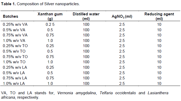
Xanthan gum suspension (0.25, 0.50, 0.75 and 1.0% w/v) was prepared by dissolving 0.25, 0.5, 0.75, and 1.0 g of Xanthan gum in 100 mL of distilled water. These served as stabilizing agents in the nanoparticle synthesis.
Preparation of silver nanoparticles
To each of the polymer suspension, 2.5 ml of silver nitrate solution was added in drops over a period of 30 s under constant stirring using a magnetic stirrer assembly. This was followed by incorporation of 10 ml of reducing agent (freshly prepared edible plant extract). The composition of the silver nanoparticles is shown in Table 1.
UV Vis spectroscopy
The optical property of AgNPs was determined by UV-Vis spectrophotometer, UNICO 2100, China. UV-Vis spectrophotometer allows identification, characterization and analysis of metallic nanoparticles. In general, 200 to 800 nm light wavelength is used for the characterization.
Antimicrobial studies
Agar dilution method was used to determine the antimicrobial activities of the silver nanoparticle against S. aures, Bacillus subtilis, Pseudomonas aeruginosa, and Escherichia coli. Microbes were pipetted and dropped on 3 batches of petri dishes (12 plates) already labeled with respect to the nanoparticles and their concentrations. 20 ml of Mackonkey agar (HiMedia) was swirled with the cultures. Sterile Cork borer of 4 mm diameter was used to bore four holes in each plate (three holes for nanoparticles’ concentrations 0.05, 0.10, and 0.15% w/v and one hole for the control of which an antibacterial standard drug-Erythromycin were used). The silver nanoparticles were placed in each hole of the agar plate and incubated at 37°C for 24 h. Based on inhibition zones around the hole, the antimicrobial activities were measured saturated with plant extract synthesized silver nanoparticle. The results are shown in Table 3.
RESULTS AND DISCUSSION
The results for the characterization of synthesized nanoparticles are shown in Table 2. The results for the determination of optimized batches of antimicrobial activities of synthesized silver nanoparticles are shown in Table 3.
Antimicrobial studies
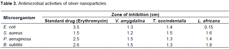
The antimicrobial activity of silver nanoparticles was carried out against both Gram positive and Gram negative bacteria. The synthesized silver nanoparticles exhibited good antibacterial activity against both Gram negative and Gram positive bacteria. Based on the zones of inhibition produced (Table 3), E. coli was most sensitive to L. Africana (inhibition zone diameter = 1.5 cm) and least sensitive to T. occidentalis (inhibition zone diameter = 1.3 cm). B. subtilis was also least sensitive to T. occidentalis (inhibition zone diameter = 1.3 cm) and most sensitive to L. africana (inhibition zone diameter = 1.8 cm). A similar trend of sensitivity was observed with P. aeruginosa. However, with Staphylococcus aureus, the sensitivity pattern was slightly different. The microorganism was most sensitive to L. africana (inhibition zone diameter = 1.6 cm) but least sensitive to V. amygdalina (inhibition zone diameter = 1.2 cm). Among the different vegetables used in the study, L. africana appeared to have the broadest antimicrobial activity.
Silver nanoparticles are widely known for their antimicrobial properties. The widespread cases of multidrug resistant bacteria against the standard antibiotics have led scientists to potentially incorporate AgNPs and other nanomaterials as ingredient to enhance the antibiotic efficacy (Ali et al., 2016). In some cases, all the microbes were all eliminated (Sathishkumar et al., 2009). There have been several proposed mechanisms on how AgNPs work as antibacterial, although the exact mechanism is still unknown. Several reports including Kumar and Münstedt (2005) suggested that the AgNPs could produce Ag ions which will damage the cell membrane, interrupt the metabolic activity, and subsequently lead to denaturation of protein and finally cell death. AgNPs could also produce reactive oxygen species (ROS) such as singlet oxygen 1O2, hydroxyl radical ∙OH, and peroxide radical R-O-O• which are harmful to the bacteria (Carlson et al., 2008).
Colour change
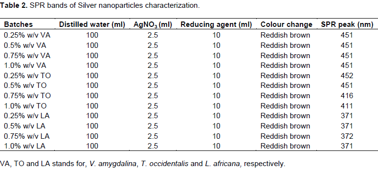
Reduction of silver ions into silver nanoparticles during exposure to plant extracts was observed as a result of the colour change. The colour change was due to the surface plasmon resonance (SPR) phenomenon. The metal nanoparticles have free electrons, giving the SPR absorption band, due to the combined vibration of electrons of metal nanoparticles in resonance with light wave. Silver nanoparticles synthesized from plants extracts have been observed to exhibit brownish colour in aqueous solution due to SPR (Logeswari et al., 2015).
UV Vis spectroscopy
The sharp bands of silver nanoparticles were observed around 451 nm in case of V. amygdalina for all four concentrations, whereas the bands for L. africana were observed around 371 nm (0.25 g), 371 nm (0.75 g), and 371 nm (1.0 g). For T. occidentalis, the bands were observed at 452 nm (0.25 g), 451 nm (0.5 g), 416 nm (0.75 g), and 411 nm (1.0 g). From different literature, it was found that the silver nanoparticles show SPR peak at around 420 to 450 nm (Rao and Tang, 2017; Ali et al., 2016). From these studies, the SPR peak for T. occidentalis was found at 451 nm and the SPR peak for V. amygdalina was found at 451 nm, whereas for L. africana, it was found at 371 nm. These results are similar to the SPR peaks observed by Jackson et al. (2016). T. occidentalis and V. amygdalina leaf extracts seem to have more potential to reduce Ag ions into Ag nanoparticles than L. africana. The intensity of absorption peak increases with time. This characteristic colour variation is due to the excitation of the SPR in the metal nanoparticles. The reduction of the metal ions occur fairly rapidly; more than 90% of reduction of Ag+ ions was complete within 4 h after addition of the metal ions to the plant extract. The metal particles were observed to be stable in solution even 4 weeks after their synthesis. There was no visible change in colour and the UV-Vis peaks of the nanoparticle solutions with time.
CONCLUSION
The rapid biological synthesis of silver nanoparticles using bitter leaf (V. amygdalina), editan leaf (L. africana), and pumpkin leaf (T. occidentalis) extract provides environmental friendly, simple and efficient route for synthesis. The change in colour from yellow to reddish brown is the characteristic of silver nanoparticles. SPR characteristic peak for the synthesized nanoparticles gave values from 371 to 452 nm which confirmed the formation of silver nanoparticles. Nanoparticles synthesized from V. amygdalina and T. occidentalis have similar (P<0.05) peaks for the SPR. Nanoparticles synthesized from L. africana had at least SPR (371 nm). After nine months of storage at room temperature and at 4°C, the SPR peak and colour of nanoparticles remain unchanged.
These edible plants used in the synthesis are readily available, affordable, non-toxic and biocompatible. They are generally regarded as safe (GRAS). The antimicrobial activity of these nanoparticles was studied against E. coli, S. aureus, P. aeruginosa and B. subtilis. They have the satisfactory inhibitions against the four mentioned microorganisms. Among the different vegetables used in the study, nanoparticles synthesized from L. africana appeared to have the highest antimicrobial activity.
CONFLICT OF INTERESTS
The authors have not declared any conflict of interests.
ACKNOWLEDGEMENT
The authors are grateful to Tertiary education Trust Fund ( TETFund), Nigeria for supporting the work..
REFERENCES
|
Ali ZA, Yahya IR, Sekaran SD, Puteh R (2016). Green Synthesis of Silver Nanoparticles Using Apple Extract and Its Antibacterial Properties. Advances in Materials Science and Engineering Volume 2016, Article ID 4102196, 6 pages. |
|
|
Carlson C, Hussein SM, Schrand AM (2008). Unique cellular interaction of silver nanoparticles: size-dependent generation of reactive oxygen species. The journal of physical chemistry B 112(43):13608-13619. |
|
|
Ganesan V, ArunaDevi J, Astalakshmi A, Nima P, Thangaraja A (2013a). Eco-friendly synthesis of silver nanoparticles using a sea weed, Kappaphycus alavarezii. International Journal of Nanotechnology 2(5):559-563. |
|
|
Ganesan V, Astalalshmi A, Nima P, Arunkumar C (2013b). Synthesis and characterization of silver nanoparticles using Merremia tridentate (L). Hall, F. International Journal of Current Science 6:87-93. |
|
|
Jackson TC, Agboke A, Jackson I, Ekpuk E (2016). Biosynthesis of Silver nanoparticles using Murraya Koenigii and Acacia Gum. International Journal of Research in Pharmacy and Biosciences 3(2):29-32. |
|
|
Khan M, Khan M, Adil SF, Tahir MN, Tremel W, Alkhathlan HZ, Al-Warthan A, Siddiqui MRH (2013). Green synthesis of silver nanoparticles mediated by Pulicaria glutinosa extract. International journal of nanomedicine 8:1507-1516. |
|
|
Korbekandi HIS, Abbasi S (2009). Production of nanoparticles using organisms. Critical Reviews in Biotechnology 29:279-306. |
|
|
Kumar P, Suranjit P, Darshit P, Patel A, Dalwadi P, Prasad R, Patel P, Selvaraj K, Prasad S (2011). Biogenic synthesis of silver nanoparticles using Nicotiana tobaccum leaf extract and study of their antibacterial effect. African Journal of Biotechnology 10(41):8122-8130. |
|
|
Kumar R, Münstedt H (2005). Silver ion release from antimicrobial polyamide/silver composites. Biomaterials 26(14):2081-2088. |
|
|
Logeswari P, Silambarasan S, Abraham J (2015). Synthesis of Silver nanoparticles using plants extracts and analysis of their antimicrobial property. Journal of Saudi Chemical Society 19:311-317. |
|
|
Ndikau M, Noah NM, Andala DM, Masika E (2017). Green Synthesis and Characterization of Silver Nanoparticles Using Citrullus lanatus Fruit Rind Extract. International journal of analytical chemistry Article ID 8108504, 9 pages. |
|
|
Rao B, Tang RC (2017). Green synthesis of silver nanoparticles with antibacterial activities using aqueous Eriobotrya japonica leaf extract. Advances in Natural Sciences: Nanoscience and Nanotechnology 8(1):015014. |
|
|
Roy S, Das TK (2015). Plant mediated green synthesis of Silver nanoparticles. A Review. International Journal of Plant Biology and Research. 3(3):1044-1055. |
|
|
Sachin S, Anupama P, Meenal K (2012). Biosynthesis of silver nanoparticles by marine bacterium, Idiomarine Sp. PR – 58-5. Bulletin of Materials Science 35(7):1201-1205. |
|
|
Sathishkumar M, Sneha K, Won SW, Cho CW, Kim S, Yun YS (2009). Cinnamon zeylanicum bark extract and powder mediated green synthesis of nano-crystalline silver particles and its bactericidal activity. Colloids and Surfaces B: Biointerfaces 73(2):332-338. |
|
|
Xin Y, Qingbiao LI, Huixuan W, Jiale H, Liqin L, Wenta W, Daohua S, Yuanbo S, James B, Luwei H, Yuanpeg W, Ning H, Lishan J (2010). Green synthesis of palladium nanoparticles using broth of Cinnamomum camphora leaf. Journal of Nanoparticle Research 12:1589-1598. |
|
|
Yang WT, Li H, Gong Y, Chen WY, Gaidau C (2011). Preparation of silver nanoparticles of enhanced antibacterial effect with benzalkonium bromide. Journal of Optoelectronics and Advanced Materials 13(6):661-665. |
|
Copyright © 2024 Author(s) retain the copyright of this article.
This article is published under the terms of the Creative Commons Attribution License 4.0


