ABSTRACT
Jatropha curcas L. (Jatropha) has gained popularity for its potential use in biodiesel production in arid regions, and its yield improvement by molecular breeding has been anticipated. However, Jatropha is known for its recalcitrance in the extraction of pure genomic DNAs. In this study, four DNA extraction methods were comparatively examined for their efficiency and quality of genomic DNA isolations from Jatropha. Consequently, one method, designated as nuclear isolation and purification using SDS/urea (NIPSU) method was found to be the most effective for rapid preparation of high yield and quality genomic DNA from Jatropha leaves. NIPSU method allowed extraction of genomic DNAs from leaves, stems, and roots tissues of Jatropha in high yield (6.8 to 24.5 µg gFW-1), with spectroscopically high purity (A260/A280 and A260/A230 >1.8). NIPSU method was also applicable to the dehydrated leaves for genomic DNA preparations. The isolated genomic DNA was amenable for downstream molecular procedures such as polymerase chain reaction, restriction enzyme cutting and Southern blotting analyses.
Key words: Jatropha curcas L., arid region, genomic DNA isolation, nuclear isolation, polymerase chain reaction, Southern blotting analysis.
Jatropha (Jatropha curcas L.), a perennial shrub belonging to the Euphorbiaceae family, originated in Central America (Heller, 1996), but is now distributed widely over tropical and subtropical areas including Africa (Silitonga et al., 2013). Jatropha has been traditionally used as a hedge and a medicinal material. Recently, increasing concern regarding the global climate change and the depletion of fossil fuel reserves have been raised and Jatropha has received a considerable attention as a feedstock for renewable biodiesel fuel production. This species shows a number of advantageous traits, including its high yield of seed oil and resistance to adverse growing conditions such as drought, high salinity, and high temperature (Openshaw, 2000; Pandey et al., 2012). Elite varieties of Jatropha will be required for large-scale commercial production. However, Jatropha has a short history of breeding and is still essentially a wild plant; therefore, improved cultivars with stable yields in fluctuating environments remain to be developed. With the development of molecular breeding technology, various kinds of PCR-based molecular markers have been established for Jatropha, such as random amplified polymorphic DNA (RAPD), simple sequence repeat (SSR), single nucleotide polymorphism (SNP), inter simple sequence repeat (ISSR), and amplified fragment length polymorphism (AFLP) (Basha and Sujatha, 2007; Sudheer Pamidimarri et al., 2009b; Wang et al., 2011; Kanchanaketu et al., 2012; Sillma et al., 2016). These technologies have the potential to assist in rapid breeding advances in Jatropha. However, isolation of high-purity genomic DNA from Jatropha is a challenging task because of the presence of a range of contaminants in the extracts, such as polyphenolics, polysaccharides and latex, which tend to be co-purified with DNAs during the conventional isolation procedures, and interfere with the reactions of DNA-modifying enzymes such as restriction enzymes and DNA polymerases (Kumar et al., 2003). Genomic DNA extraction from Jatropha tissues using commercially available kits often leads to low yield. Moreover, the cost for purchasing commercial kits should not be ignored. Cetyltrimethylammonium bromide (CTAB) is a cationic detergent that has a useful property of forming complexes with proteins and polysaccharides but not with nucleic acids at high ionic strength, and the CTAB-based methods have often been used to obtain genomic DNA from Jatropha tissues (Dhakshanamoorthy and Selvaraj, 2009; Sudheer Pamidimarri et al., 2009a; Mastan et al., 2012). However, the conventional CTAB-based methods require a time-consuming 30 to 90 min incubation step, which are accompanied by the labor-intensive manual procedures such as repeated phenol/chloroform extraction.
On the other hand, Liu et al. (1995) developed an isolation buffer containing SDS/urea and successfully applied to the extraction of genomic DNA from Arabidopsis. In this method, a strong anionic detergent SDS and a denaturant urea in the isolation buffer was allowed to extract genomic DNA without an incubation step, and the procedure was simplified by combining the procedures of DNA extraction and phenol/chloroform purification into single step. In other study, a one-step nuclear isolation procedure was successfully introduced to remove impurities from DNA-rich nuclear fraction in the plant extracts, using a nuclear isolation solution containing polyvinylpyrrolidone (PVP), polyethylene glycol (PEG), and β-mercaptoethanol (Wagner et al., 1987). However, to the best of the authors’ knowledge, effects of the nuclear isolation procedure and the SDS/urea isolation buffer on Jatropha DNA isolation has not been examined so far. Therefore, the objective of this study was to comparatively examine the efficiency of these procedures on DNA extraction from Jatropha tissues, and to optimize a rapid and efficient method for the DNA extraction for practical use in molecular analyses. The study also examined whether the optimized DNA extraction procedure could be applicable to ethanol-treated dehydrated tissue samples (Sharma et al., 2003), which has been developed for stabilizing tissues for longer storage periods without cooling condition in large-scale field experiments.
Reagents
Ethylenediamine-N,N,N',N'-tetraacetic acid disodium salt (EDTA) was purchased from Dojindo Laboratories (Kumamoto, Japan). Chloroform, ethanol, and sodium chloride were from Wako Pure Chemical Industries (Osaka, Japan). Polyethylene glycol #6000 (average molecular weight: 7,400 to 10,200) and other reagents were from Nacalai Tesque (Kyoto, Japan).
Plant materials
Jatropha curcas L. variety IP-3P was obtained from Pakuwon Station, Indonesian Center for Estate Crop Research and Development, Sukabumi, West Java, Republic of Indonesia. For evaluation of various DNA extraction methods, fully-expanded fresh leaves collected from Jatropha trees of 1 to 2 m height grown in a greenhouse were used. For comparison of DNA extraction efficiency in leaf, stem, and root tissues, juvenile Jatropha seedlings at a height of 20 to 25 cm were used, which were grown in a growth room at 25 to 28°C with a 14 h/10 h (light/dark) photoperiod using white fluorescent lamps (150 to 170 µmol photons m-2 s-1, Plantlux, Toshiba Lightning and Technology, Tokyo, Japan).
Dehydration of leaf tissues was performed by detaching a leaf from the Jatropha plants grown in the greenhouse, submerged immediately in 40 mL of absolute ethanol in a 50 mL tube, and incubated at room temperature for 30 to 60 min. The ethanol-treated leaf samples were then allowed to air dry. Dehydrated leaves were kept at room temperature for short-term storage, or at -30°C for longer preservation.
Evaluation of DNA extraction methods
To compare the methods for DNA extraction from Jatropha, the following four methods were examined. In each method, a 50 to 80 mg sample of fresh tissue was ground to a fine powder using a mortar and pestle under liquid nitrogen, then used for the genomic DNA extractions. In each method, the extractions were repeated four times using four different Jatropha samples.
Method I
This method was a CTAB-based protocol essentially in accordance with the report by Murray and Thompson (1980), with an attached modification related to the DNA precipitation procedure. After the CTAB-DNA precipitation, the pellet was dissolved in 400 µL of 1.0 M CsCl. The 800 µL of absolute ethanol was added and mixed by gentle inverting, then incubated at -20°C for 20 min. The precipitate was recovered by centrifugation at 15,000 × g for 10 min at 4°C. After discarding the supernatant, the pellet was washed with 70% ethanol, then dissolved in TE buffer containing 10 mM Tris-HCl (pH 8.0) and 1 mM EDTA.
Method II
The method was a SDS/urea-based protocol essentially in accordance with the report by Liu et al. (1995), with a minor modification to the extraction buffer, which was composed of 50 mM Tris-HCl (pH 7.5), 20 mM EDTA, 0.3 M NaCl, 0.5% SDS, 5 M urea, 5% (v/v) TE-saturated phenol, and 10 mM β-mercaptoethanol.
Method III
This method was a combination of a procedure of nuclear isolation followed by the method I described previously. For the nuclear isolation procedure, 1 mL of ice-cold nuclear isolation buffer containing 10% PEG #6000, 5% PVP K-30 (average molecular weight 40,000), 0.35 M sorbitol, 100 mM Tris-HCl (pH 8.0), 1 mM EDTA, and 0.5% β-mercaptoethanol was added to the ground tissues and vortexed vigorously. The suspension was centrifuged at 15,000 × g for 10 min at 4°C. After discarding the supernatant, genomic DNA was extracted from the pellet using the procedure in the method I described previously.
Method IV (NIPSU: nuclear isolation and purification using SDS/urea)
This method was a combination of the nuclear isolation procedure described in the method III, followed by the modified method II described previously. After the nuclear isolation treatment described in the method III, the pellet was dissolved in 500 µL of extraction buffer (described in the method II) and vortexed vigorously. After centrifuging at 15,000 × g for 10 min at room temperature (20 to 25°C), the supernatant was transferred to a new tube and centrifuged again using same condition to remove insoluble impurities. The supernatant was transferred to a new tube, and one volume (that is, 500 µL) of TE-saturated phenol: chloroform: isoamyl alcohol=25:24:1 (PCI) was added and mixed gently by inverting the tube. After centrifuging at 15,000 × g for 5 min at room temperature, the aqueous phase was transferred to a new tube. Two volumes of ethanol (that is, 1 mL) was added to the supernatant and centrifuged at 15,000 × g for 5 min at 4°C. The pellet was washed with 1 mL of 70% ethanol and repeated the same centrifugation step. DNA was dissolved in TE buffer.
Analysis of the spectroscopic property and quantity of genomic DNA
The ratios of optical absorbance at 260 and 280 nm (A260/A280), and of 260 and 230 nm (A260/A230) were used to assess the purity of genomic DNAs using a spectrophotometer (WPA Biowave S2100, Biochrom, Cambridge, UK). Quantity of genomic DNA was estimated by gel electrophoresis analysis with GelAnalyzer 2010 software (http://www.gelanalyzer.com), using a dilution series of StyI-digested lambda DNA with known concentration as the calibration standards.
Polymerase chain reaction
Primers specific for the actin gene (Nanasato et al., 2015, 5'-AGACCTCCAAAACCAGCTCA-3' and 5'-TTGATTTTCATGCTGCTTGG-3'), 620 bp of the curcin promoter region (GenBank accession number AF469003, 5'-CTCGAGAATATTGGAATAGAAGACTTTG-3' and 5'-TCTAGACAAATATCATTATACGAATACG-3', XhoI and XbaI sites are indicated as underlines, respectively), and 5,956 bp of JcDGAT1 (contig number Jcr4S00005, Jatropha genome database; Sato et al., 2011), 5'-ATGACGATTTTGGAGACCACTACTA-3' and 5'-TCATCTTAATTCAGCATTGCCTTTC-3') were used for amplification in 10 µL reaction volumes containing Jatropha genomic DNA as the template. PCR amplification was performed using ExTaq DNA polymerase (Takara Bio, Shiga, Japan) using the following conditions: 94°C for 2 min; 30 cycles of 94°C for 30 s, 55°C (JcDGAT1), 58°C (actin) or 63°C (curcin promoter) for 30 s, and 72°C for 1 min (actin and curcin promoter) or 6 min (JcDGAT1); and a final extension of 72°C for 7 min. PCR products were separated on a 0.8% agarose gel, and visualized by GelGreen Nucleic Acid Gel Stain (Biotium, Fremont, CA).
Southern blotting analysis
Genomic DNA (5 µg) was digested with EcoRV or SacI, separated on a 0.8% agarose gel, then transferred to a positively charged nylon membranes (Pall, Port Washington, NY) with 20× saline sodium citrate buffer as described previously (Sambrook and Russell, 2001). A digoxigenin (DIG)-labeled DNA probe specific for the JcDGAT1 coding sequence was prepared by amplifying 328 bp of JcDGAT cDNA fragment from a cDNA clone of JcDGAT by PCR using a pair of primers (5’-ATGACGATTTTGGAGACCACTACTA-3’ and 5’-CCCTGAGCGCTCGATGAG-3’), and used as a template for the probe synthesis using a DIG-High Prime DNA Labeling and Detection Starter Kit II (Roche, Penzberg, Germany), and hybridized bands were then detected using X-ray film (Hyperfilm ECL, GE Healthcare, Buckinghamshire, UK).
Statistical analysis
One-way analysis of variance (ANOVA) was adopted for the data analysis. Means were separated by Tukey's honest significant difference (HSD) to identify significant differences (P < 0.05) between the samples.
Evaluation of different DNA extraction methods for Jatropha leaves
Efficiency of four DNA extraction methods, that is, methods I to IV, was evaluated using fresh leaves of Jatropha. Agarose gel electrophoresis showed multiple band signals form the extracted DNAs, which were composed of a single band corresponded to the genomic DNAs which migrated at a similar speed with 19.3-kbp DNA size marker, and three bands migrated with 0.42 to 1.88 kbp markers which showed a characteristic pattern for ribosomal and transfer RNAs (Figure 1a). Quantification of the genomic DNAs by image analysis showed that the method I, which was a conventional CTAB method, gave a lowest average yield of 1.12 ± 0.16 µg g FW-1 among the four methods tested in this study (Table 1). DNA yield of method II, a SDS/urea-based method, was 7.22 ± 4.67 µg g FW-1, but the yields were highly variable among four independent extractions as suggested by the large standard deviation. A brownish color was observed in the DNA solution obtained by the method II (Figure 1b), showing the presence of impurities in this preparation. This observation suggested that themethod II was not effective for eliminating inhibitory compounds for downstream molecular analyses. Introducing the nuclear isolation procedure in methods III and IV contributed greatly to the removal of impurities, and drastically improved the extraction efficiency. The amount of genomic DNAs obtained by the methods III and IV were 9.32 ± 2.69 and 7.93 ± 0.86 µg g FW-1, respectively, and were significantly higher than that in the method I. Cumulative total time required for the incubation and centrifugation steps was 140 and 50 min for the method III and IV, respectively, showing that the method III was more time-consuming than the method IV. Therefore, it is judged that the method IV, hereinafter designated as nuclear isolation and purification using SDS/urea (NIPSU) method, was the most convenient protocol for extracting genomic DNA from fresh Jatropha leaves. The whole scheme of the NIPSU method is shown in Figure 2. From this point onward, only the NIPSU method was used for further analyses in this study.


Evaluation of the NIPSU method in various tissues and dehydrated leaf
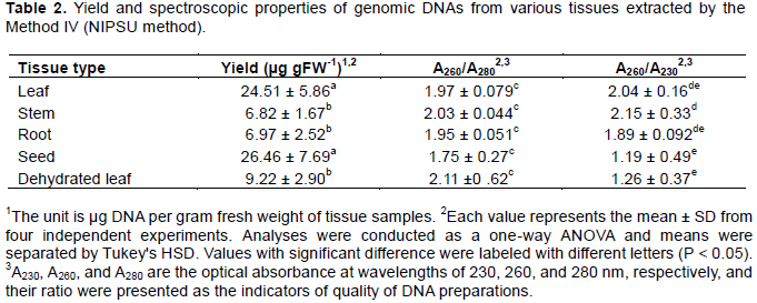
Subsequently, whether the NIPSU method was effective to non-leaf tissues in Jatropha was examined. Consequently, agarose gel electrophoresis analysis showed that genomic DNA preparations extracted from stem, roots, and seeds gave a band signal corresponding to the high molecular weight DNAs (Figure 1c), suggesting that the NIPSU method was also applicable to range 6.82 to 26.46 µg g FW-1 (Table 2). The yields from stem and root tissues were lower than that from leaves, which was consistent with previous report (Sudheer Pamidimarri et al., 2009a). Spectrophotometer analysis non-leaf tissues. Yields of genomic DNAs were in the revealed that the spectroscopic indicators of DNA purity, A260/A280 and A260/A230 ratios, of genomic DNAs extracted from leaf, stem, and roots were in the range of 1.89 to 2.15, suggesting that these DNA preparations were highly pure (Table 2). Relatively low A260/A230 value of 1.19 ± 0.49 was recorded for genomic DNA from seeds, which might be due to the high abundance of organic contaminants such as carbohydrates in this tissue.

The NIPSU method was then applied to dehydrated Jatropha leaves. Dehydration treatment of the leaves using ethanol resulted in the significant decrease in the sample weight, and the ratio of dry weight/fresh weight was 0.19 ± 0.066 (n = 4). This value was similar to that in previous reports (Patel et al., 2010; Nahar and Hoque, 2013). From the dehydrated leaves, 9.22 ± 2.90 µg g FW-1 of genomic DNA was obtained with spectroscopic properties (2.11 ± 0.62 and 1.26 ± 0.37 for A260/A280 and A260/A230 ratios, respectively; Table 2), and the DNA intactness was confirmed by agarose gel electrophoresis (Figure 1c), suggesting that the NIPSU method was applicable to dehydrated leaf tissues.
Molecular analyses using genomic DNAs extracted by the NIPUS method
To examine the behavior of DNAs extracted by the NIPUS method in the molecular analyses, PCR and restriction assays were performed. In the PCR experiments, three representative DNA fragments, that is, actin coding sequence (301 bp), curcin promoter (632 bp), and JcDGAT1 coding sequence (5,956 bp) were amplified from the DNA isolated from either fresh or dehydrated leaves. Gel electrophoresis analysis showed clear, sharp single bands with expected sizes, demonstrating that all PCR reactions were successful in amplifying the target gene fragments (Figure 3a). The suitability of the isolated genomic DNA for downstream enzymatic procedures was also determined by Southern blotting analysis using a probe for a single gene JcDGAT1 (Figure 3b). Consequently, a single band corresponding to JcDGAT1 gene was clearly detected from only 5 µg of EcoRV- and HindIII-digested genomic DNA. These results show that genomic DNAs extracted by the NIPSU method was compatible to PCR amplification and restriction analyses, and can be useful for various molecular analyses in the future, for example, RFLP, RAPD and SSR analyses.

Four methods were comparatively evaluated for the efficiency of DNA extraction from fresh leaves of Jatropha, and one of these methods, NIPSU (nuclear isolation and purification using SDS/urea), was found to be the most effective and fast method for extraction of genomic DNA from Jatropha. This method was applicable to a wide range of Jatropha tissues and dehydrated leaves, and amenable for downstream molecular analyses such as PCR and restriction analysis.
The authors did not declare any conflict of interest.
This work was supported by the Science and Technology Research Partnership for Sustainable Development (SATREPS) program from the Japan International Cooperation Agency (JICA) and the Japan Science and Technology Agency (JST), and the New Energy and Industrial Technology Development Organization (NEDO). The authors acknowledge funding from the Grant-in-Aid for Scientific Research (17K07755) from JSPS, and by the Joint Research Program and the Project Marginal Region Agriculture, the Arid Land Research Center, Tottori University, and the International Platform for Dryland Research and Education (IPDRE) Program, Tottori University.
REFERENCES
|
Basha SD, Sujatha M (2007). Inter and intra-population variability of Jatropha curcas (L.) characterized by RAPD and ISSR markers and development of population-specific SCAR markers. Euphytica 156:375–386.
Crossref
|
|
|
|
Dhakshanamoorthy D, Selvaraj R (2009). Extraction of genomic DNA from Jatropha sp. using modified CTAB method. Romanian Journal of Biology Plant Biology 54:117-125
|
|
|
|
|
Heller J (1996). Physic nut. Jatropha curcas L. Promoting the conservation and use of underutilized and neglected crops. 1. Institute of Plant Genetics and Crop Plant Research, Gatersleben/ International Plant Genetic Resources Institute, Rome. ISBN: 92-9043-278-0
|
|
|
|
|
Kanchanaketu T, Sangduen N, Toojinda T, Hongtrakul V (2012). Genetic diversity analysis of Jatropha curcas L. (Euphorbiaceae) based on methylation-sensitive amplification polymorphism. Genet. Mol. Res. 11:944-955.
Crossref
|
|
|
|
|
Kumar A, Pushpangadan P, Mehrotra S (2003). Extraction of high-molecular-weight DNA from dry root tissue of Berberis lycium suitable for RAPD. Plant Molecular Biology Reporter. 21:309a-309d.
Crossref
|
|
|
|
|
Liu Y-G, Mitsukawa N, Oosumi T, Whittier RF (1995). Efficient isolation and mapping of Arabidopsis thaliana T-DNA insert junctions by thermal asymmetric interlaced PCR. Plant Journal 8:457-463.
Crossref
|
|
|
|
|
Mastan SG, Sudheer PDVN, Rahman H (2012). Molecular characterization of intra-population variability of Jatropha curcas L. using DNA based molecular markers. Molecular Biology Reports 39:4383-4390.
Crossref
|
|
|
|
|
Murray MG, Thompson WF (1980). Rapid isolation of high molecular weight plant DNA. Nucleic Acids Research 8:4321-4326.
Crossref
|
|
|
|
|
Nahar K, Hoque S (2013). A Morphological and Physiological Study of Jatropha curcas Linn . Propagated from Seeds in Bangladesh.
|
|
|
|
|
Nanasato Y, Kido M, Kato A (2015). Efficient genetic transformation of Jatropha curcas L. by means of vacuum infiltration combined with filter-paper wicks. In Vitro Cellular & Developmental Biology-Plant 51:399-406.
Crossref
|
|
|
|
|
Openshaw K (2000). A review of Jatropha curcas: An oil plant of unfulfilled promise. Biomass Bioenergy 19:1-15.
Crossref
|
|
|
|
|
Pandey VC, Singh K, Singh JS, et al (2012). Jatropha curcas: A potential biofuel plant for sustainable environmental development. Renewable. Sustainable. Energy Reviews 16:2870-2883.
Crossref
|
|
|
|
|
Patel AD, Panchal NS, Pandey IB, Pandey AN (2010). Growth, water status and nutrient accumulation of seedlings of Jatropha curcas L. (Euphorbiaceae) in response to soil salinity. An. Biology 32:59-71
|
|
|
|
|
Sambrook J, Russell DW (2001). Molecular Cloning: A Laboratory Manual. Cold Spring Harb Lab Press Cold Spring Harb NY P999. ISBN: 0879695773
|
|
|
|
|
Sato S, Hirakawa H, Isobe S (2011). Sequence analysis of the genome of an oil-bearing tree, Jatropha curcas L. DNA Research 18:65-76.
Crossref
|
|
|
|
|
Sharma R, Mahla HR, Mohapatra T, (2003). Isolating plant genomic DNA without liquid nitrogen. Plant Molecular Biology Reporter 21:43–50.
Crossref
|
|
|
|
|
Silitonga AS, Masjuki HH, Mahlia TMI (2013). A global comparative review of biodiesel production from Jatropha curcas using different homogeneous acid and alkaline catalysts: Study of physical and chemical properties. Renew. Sustain. Energy Reviews 24:514-533.
Crossref
|
|
|
|
|
Sillma R, Daneshwar P, Subhasisa B, Rajesh J (2016). Application of rep-PCR as a molecular tool for the genetic diversity assessment of Jatropha curcas. African Journal of Biotechnology 15:172-179.
Crossref
|
|
|
|
|
Sudheer Pamidimarr DV, Boricha G, Reddy MP (2009a). A simplified method for extraction of high quality genomic DNA from Jatropha curcas for genetic diversity and molecular marker studies. Indian Journal of Biotechnology 8:187-192
|
|
|
|
|
Sudheer Pamidimarri DV, Singh S, Mastan SG, Patel J, Reddy MP (2009b). Molecular characterization and identification of markers for toxic and non-toxic varieties of Jatropha curcas L. using RAPD, AFLP and SSR markers. Molecular Biology Reports 36:1357-1364.
Crossref
|
|
|
|
|
Wagner DB, Furnier GR, Saghai-Maroof MA (1987). Chloroplast DNA polymorphisms in lodgepole and jack pines and their hybrids. Proceedings of the National Academy of Sciences USA 84:2097–2100.
Crossref
|
|
|
|
|
Wang CM, Liu P, Yi C(2011). A first generation microsatellite- and SNP-based linkage map of Jatropha. PLoS One 6:4-11.
Crossref
|
|