African Journal of
Biotechnology

  • Abbreviation: Afr. J. Biotechnol.
  • Language: English
  • ISSN: 1684-5315
  • DOI: 10.5897/AJB
  • Start Year: 2002
  • Published Articles: 12487

Full Length Research Paper

Genetic diversity among Fusarium species associated with sorghum stalk rot in Southern Ethiopia

Alemayehu Chala
  • Alemayehu Chala
  • School of Plant and Horticultural Sciences, College of Agriculture, Hawassa University, P. O. Box 5, Hawassa, Ethiopia.
  • Google Scholar


  •  Received: 06 December 2018
  •  Accepted: 13 March 2019
  •  Published: 03 April 2019

 ABSTRACT

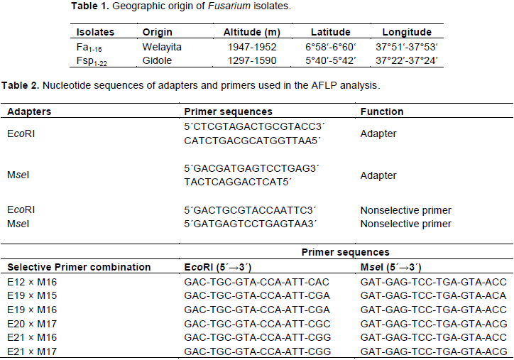
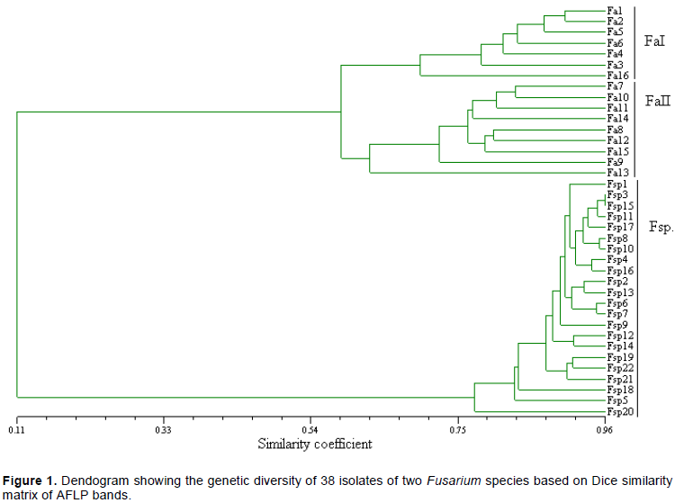
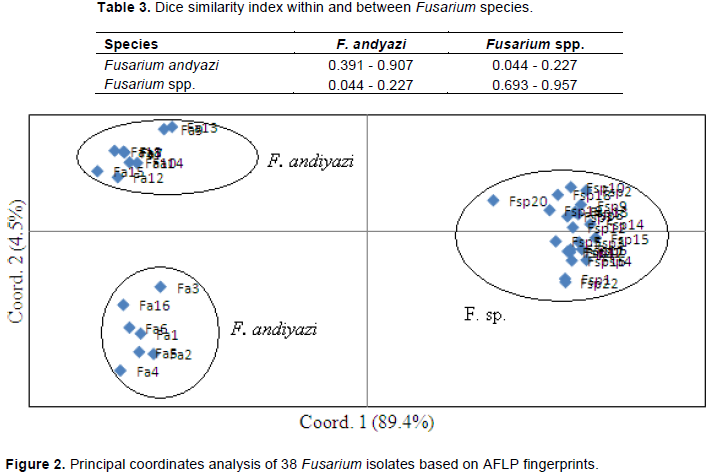
Fusaria are very diverse and destructive pathogens affecting different crops. However, their identity and diversity are unresolved in countries like Ethiopia, where various crop species are grown under differing environmental conditions. The objectives of this paper were to identify Fusarium spp. associated with sorghum stalk rot in Southern Ethiopia, and elucidate the genetic diversity within and between the species. For this purpose, Fusaria associated with sorghum from two locations in Southern Ethiopia were isolated. Sequencing of the elongation factor 1-alpha gene (EF-1α) was used for species identification. In addition, AFLP analysis was employed for further diversity studies within and between the Fusarium spp. Sequence analyses revealed the presence of two Fusarium spp. The first was identified as Fusarium andiyazi, while the identity of the second remains to be solved. AFLP analysis clustered the isolates into two major groups. The Dice similarity coefficients ranged from 0.39 to 0.91 for isolates of F. andiyazi while isolates within the new Fusarium spp. had a Dice similarity coefficient varying between 0.69 and 0.96. Cluster analysis and principal coordinate analysis clearly indicated a genetic separation between the two species. Both groups were pathogenic to mature sorghum plants following a toothpick inoculation test. More researches are required to identify the new species and elucidate the pathogenicity of the isolates.


Key words: EF-1α, Fusarium andiyazi, genetic similarity, sequence analysis, Sorghum bicolor.


 INTRODUCTION

Sorghum (Sorghum bicolor, (L.) Moench) is the fifth most important cereal accounting for more than 65 million tons of annual production on over 45 million ha of land worldwide (FAO, 2017). The bulk of sorghum is produced in less developed nations (Berenji and Dahlberg, 2004), however, USA is the leading producer with more than 12 million tons of production (FAO, 2017). In Ethiopia, the crop is grown on more than 2 million ha of land making it one of the three most important crops both in terms of area coverage and total production (5 million tons) (CSA, 2018).
 
Sorghum is grown for its various purposes in different parts of the world. It serves as a major source of food and is also used as feed source for livestock especially in developed nations. In addition, sorghum is used as raw material for industries and for the production of bio-fuel. Despite its versatile use and ability to withstand adverse environmental conditions including moisture stress and high temperatures, sorghum production is hampered by various biotic stresses among which diseases caused by different pathogens are one (Thakur and Mathur, 2000; Chala et al., 2011; 2012; Eshte et al., 2015). Fusarium species that cause stalk rot, ear rots and grain mold are among the major pathogens that infect sorghum (Frederiksen and Odvody, 2000; Leslie et al., 2005). The fungus Fusarium belongs to the most harmful pathogens of cultivated crops all over the world (Antonia, 1995; Ramdial et al., 2017).
 
Once they occur in the field or storage, Fusaium spp. are known to cause significant qualitative and quantitative yield reduction (Parry et al., 1995; Brandfass and Karlovsky, 2008). In addition, they produce mycotoxins that pose serious health risks to humans and animals that feed on contaminated grains (D'Mello et al., 1999; Pestka and Smolinsky, 2005; Antonissen et al., 2014; Wu et al., 2014 and Duan et al., 2016). Despite continued efforts to manage diseases caused by Fusaria, they still pose serious threats to grain producers across the world (Brandfass and Karlovsky, 2008). Since the early reports by Wollenweber and Reinking (1935), lots of researches have been conducted on the taxonomy/genetic diversity of the genus Fusarium (Summerell et al., 2011; O’Donnell et al., 2015; Laurence et al., 2016; Moussa et al., 2017 and Valente et al., 2017). However, research on Fusarium spp. from sorghum has been given only peripheral importance (Leslie et al., 2005). The only exceptions, in this regard, are earlier reports by Claflin (2000) and Leslie (2000; 2002), which identified more than 10 Fusarium species from sorghum, with many of them known to infect the stalk and grain. On the other hand, the identity and diversity of Fusarium species infecting sorghum in Africa, particularly in Ethiopia remains unresolved. In Ethiopia, there are limited reports (Ayalew, 2002; Ayalew et al., 2006; Chala et al., 2014; Taye et al., 2016; 2018) on Fusarium spp. and associated mycotoxins from sorghum even though the country is one of the primary centers of origin and diversity for the crop. The objectives of this work were: i) to identify Fusarium spp. associated with sorghum stalk rot in Southern Ethiopia; and ii) to elucidate the genetic diversity within and between the species.


 MATERIALS AND METHODS

Isolate collection
 
Twenty sorghum stalks with visible rotting were randomly collected from sorghum fields in Southern Ethiopia during a routine field survey. The stalk samples were stored in paper bags at room temperature until isolation. Geographic description of the locations is given in Table 1.
 
Isolation, identification and storage of the isolates
 
Infected stalks were cut into pieces, surface sterilized using 0.5% sodium hypochlorite  (NaOCl)  solution  for  90 s,  and  rinsed  three times in sterile, distilled water. The cut and surface-sterilized stalks were placed on potato dextrose agar (PDA) and incubated at 25°C under continuous fluorescent light for 10 days. After 10 days of incubation, sporulation was observed in the PDA plates. Isolates were identified as Fusaria based on pigmentation and conidial morphology. On these bases, a total of 37 single spore isolates were transferred to new PDA plates. The isolates were grouped into two groups based on pigmentation and form of conidia. Pure cultures were maintained on potato dextrose agar (PDA) amended with 50 ppm of streptomycin, and stored at 4°C as stock cultures.
 
Molecular characterization
 
DNA extraction
 
Approximately 100 mg of fresh mycelium per isolate was crushed in liquid nitrogen using mortar and pestle. Fine powdered mycelium was transferred to a 2 ml microcentrifuge tube and genomic DNA was extracted using the DNeasy Plant Mini Kit (Qiagen Inc., Valencia, CA) following the manufacturer’s instructions. The quality of the extracted DNA was controlled on 0.8% agarose gels and the DNA was stored at -20°C.
Species identification
 
Elongation factor 1-alpha genes of seven isolates, randomly selected from the two morphological groups, were partially sequenced using the EF-1α primers that is, EF-728F: 5´-CATCGAGAAGTTCGAGAAGG-3´ and EF-986R: 5´-TACTTGAAGGAACCCTTACC-3´ (Carbone and Kohn, 1999). The resulting sequences were BLAST searched with the NCBI nucleotide database for molecular species identification.
 
AFLP analysis
 
AFLP analysis was conducted following the method developed by Vos et al. (1995) with modifications that include the use of fluorescent labeled primers instead of radioactive labeled isotopes. Six combinations of MseI and EcoRI primers were used for selective amplification (Table 2). The primers differ by two selective nucleotides at their 3´ ends and the EcoRI primers were labeled with the fluorescent dye FAM (6-carboxyfluorescein). The selective amplification reaction mix contained 1.6 µl dNTP (2.5 mM), 2 µl of 10× polymerase chain reaction (PCR) buffer, 0.08 µl of Taq DNA polymerase (5 U/µl), and 5 µl MseI (6 ng/µl) and 1 µl EcoRI (1 pmol) primers to which 5 µl of 10 fold diluted preamplification PCR product was added as a template. The PCR amplification conditions were as follows: 1 cycle of 94°C for 30 s, 65°C for 30 s and 72°C for 60 s; 12 cycles where the annealing temperature was lowered by 0.7°C for each cycle; 23 cycles at 94°C for 30 s, 56°C for 30 s and 72°C for 60 s; finally 72°C for 7 min. The accuracy of the analysis was checked by running a randomly selected sample in duplicates.


Data scoring and analysis
 
Amplification products were separated in an ABI3730 DNA analyzer (Applied Biosystems Inc., Foster City, California) following the manufacturer’s protocol and using GeneScan-1200 LIZ size standard (Applied Biosystems). The presence (1) and absence (0) of peaks were scored using Gene-Mapper software version 4.0 (Applied Biosystems Inc., Foster City, California), checked manually, and only clear and unambiguous peaks with fluorescence greater than or equal to 100 arbitrary units were entered into a binary  data  matrix  for  further  analysis. The  binary matrices were then used to calculate genetic similarities between the isolates based on the Dice similarity coefficient (Dice, 1945), and the unweighted pair-group method with arithmetic average (UPGMA) was used to construct a genetic similarity tree with the help of the NTSYS-pc software, version 2.0 (Exeter Biological Software, Setauket, NY). To further elucidate the genetic relationship among the tested isolates, principal coordinate analysis was conducted using the software GenAlEx6 (Peakall and Smouse, 2006).
 
In-vitro growth rate of Fusarium isolates
 
To study the phenotypic characters (growth rate and colony morphology) of the Fusarium isolates, 5 mm portions of the 38 single spore isolates were transferred from the stock cultures and cultivated on PDA at 25°C in the dark. After five days of incubation, 3 mm mycelia plugs were taken from the actively growing edges of each isolate, transferred to the centre of four replicate PDA plates, and incubated in the dark at 25±2 and 30±2°C. For each isolate, radial growth was recorded at 24 h intervals for 7 days.


 RESULTS

Species identification and characterization
 
Sequence analysis
 
Based on sequencing of the elongation factor 1-alpha gene (EF-1α) of isolates, the Fusaria were categorized into two groups. The first group of isolates were identified as F. andiyazi.  The sequence of the second group of isolates did not match with the sequence of Fusarium species deposited in NCBI, and hence their identity remains to be resolved.
 
Morphological and cultural characterization
 
F. andiyazi isolates produced both macro- and micro- conidia and had a white mycelium that become some what pale through time. Isolates belonging to the unidentified Fusarium species also produced both micro- and macro- conidia on PDA while their mycelium consistently appeared white with a mass of orange colored spores. Isolates of similar morphological appearance were also obtained from sorghum and finger millet grains collected from different locations in Ethiopia (data not shown).
 
Isolates belonging to the two species also varied in terms of radial growth rate. The growth rate of isolates belonging to F. andiyazi ranged from 8 to 14 mm/day and 8 to 12 mm/day at 25±2 and 30±2°C, respectively. On the other hand, isolates belonging to the new species grew considerably faster (10-17 mm/day) at 25°C than at 30°C (7-12 mm/day). When five isolates representing F. andiyazi and 10 isolates from the new Fusarium species were used to inoculate the stalks of mature sorghum plants using the toothpick inoculation method (Cumagun et al., 2009), all of them produced typical lesions that were absent in the control plants. This suggested the pathogenicity of both species to sorghum and proved that they were responsible for the stalk rot of sorghum.
 
AFLP analysis
 
AFLP analysis of 38 isolates clustered them into two major groups (Figure 1). The first major group consists of 16 isolates identified as F. andiyazi by sequence analyses. Dice similarity coefficient for isolates belonging to this major group varied from 0.39 to 0.91 (Table 3). Results differentiated isolates of F. andiyazi into two sub-groups. The first sub-group consists of 7 isolates while the second sub-group is made of the remaining 9 isolates. The second major group consists of 22 isolates belonging to the unidentified Fusarium species and hence named as Fusarium spp. Isolates within this group had a Dice similarity coefficient ranging between 0.69 and 0.96, and hence they were considered as genetically more similar with one another than those within F. andiyazi, and likely represent a single species. One isolate within this group had 69% similarity while the rest had at least 70% similarty between each other. The six primer combinations used in this study generated a total of 200 clearly scorable bands. Of these, 71 (35.5%) were unique to the new Fusarium species while 70 bands (35%) were unique to F. andiyazi isolates. The remaining 59 bands (29.5%) were shared across the species. Of the 71 bands unique to Fusarium spp., 31 (56%) were polymorphic while 60 (86%) bands unique to F. andiyazi were also polymorphic.
 
Principal coordinates analysis (PCO) also revealed the population subdivision within and between the two Fusarium species. Accordingly, the isolates were categorized into three groups (Figure 2). The first three principal coordinates accounted for 89.4, 4.5 and 2.3% of the total variation, respectively. PCO grouped 16 of the F. andiyazi isolates that formed the first two clusters of the UPGMA tree into two groups. The first group was made of 9  isolates  while the second PCO group consisted of 7 isolates. The 22 remaining isolates belonging to the new Fusarium species were aggregated within a single PCO group with only 1 isolate barely separated from the rest. These results are in line with the cluster analysis of UPGMA.


 DISCUSSION


The Fusarium isolates included in the current study fullfilled the morphological characteristics of Fusarium as described in Leslie and Summerell (2006). However, sequence analysis revealed the presence of two Fusarium species associated with stalk rot in Southern Ethiopia. The current results are in line with previous works that reported the co-occurence of different Fusarium spp. on the same plant (Summerell et al., 2011; Ramdial et al., 2017; Minnaar-Ontong et al., 2017). Results from sequencing confirmed the first species as F. andiyazi, a species which was first described by Marasas et al. (2001). This species was subsequently reported to be present in different parts of the world including Australia, Ethiopia, Nigeria, South Africa and United States (Marasas et al., 2001; Marley et al., 2004; Leslie et al., 2005; Leslie and Summerell, 2006 and Summerell et al., 2011). Nevertheless, except for initial reports, no
further work has been done on this particular species in Ethiopia to the best of the author’s knowledge. As a result, the diversity of this pathogen remains largely unknown to date.
 
Isolates belonging to the two Fusarium species varied not only in terms of their morphology and sequence but they also differed in growth rate, when incubated at 25 and 30°C. Isolates of F. andiyazi grew slower than those of the newly recovered Fusarium species at both temperatures. Besides, the growth rate of F. andiyazi isolates was also consistent across temperatures. Isolates of the same species showed similar growth rate at both of these temperatures in a previous study (Leslie et al., 2005). Isolates of Fusarium species on the other hand did not grow consistently across temperatures.
 
Although cultural/morphological characterizations provide a basis for both inter- and intra-species diversity studies; as suggested in other pathosystems, they may be unstable, highly influenced by the growth environments and rather change with the age of the colonies (Browning et al., 1999; Crouch et al., 2006 and Rivera-Vargas et al., 2006). As a result, such taxonomic features need to be supplemented with other characters like molecular markers that differentiate biological entities at the genetic level. Currently there is a growing interest to assess the genetic diversity of fungi including Fusaria based on sequence analysis (McDonald et al., 2012; Leavitt et al., 2013; Maphosa et al., 2016; Laraba et al., 2017).
 
In accordance with sequence analysis and morpho-cultural characterization, AFLP analysis also showed the presence of at least two genetically distinct Fusarium populations associated with sorghum stalk rot in Southern Ethiopia. The two Fusarium species were not only genetically but also geographically separated as there is more than 50 km distance between the districts from where they were obtained. Backhouse et al. (2001) and Saremi et al., (1999) have reported climatic preferences among Fusarium species from both natural and agricultural ecosystems. This report was also
supported by Vigier et al., (1997); De Wolf et al., (2003) and Moschini et al. (2004) that ascertained the influence of climate and local weather variations on the recovery of Fusarium species. Both UPGMA and PCO analyses of AFLP bands suggested greater variation within F. andiyazi than within Fusarium species. Leslie et al. (2005) proposed a 40% similarity cut-off to identify strains into a single species. As the most distantly related isolates within this species had a 39% Dice similarity, which is just close to the 40% boundary, it is better not to reach a conclusion that F. andiyazi isolates belong to different species.
 
In the current study, all isolates belonging to F. andiyazi were isolated from sorghum stalks collected from the district of Welayita, with elevation ranging from 1947 to 1952 m above sea level (masl), while those belonging to the new Fusarium species were isolated from the Gidole district, with elevation of 1297-1590 masl. Based on eleven years weather data from the National Meteorological Agency, Welayita district has a total annual rainfall of 1262 mm, and temperature of 13.6 - 23.4°C (18.5°C average). There is no reliable weather data for Gidole district. However, this relatively low lying district is known to have a more warm and humid weather than Welayita. Preparations are now underway to work on the speciation of isolates belonging to the new species, and to further characterize them on the basis of mycotoxin profiling, mating types and other characteristic features. F. andyiazi is not a known mycotoxin producer (Leslie et al., 2005). However, the toxin production potential of isolates belonging to the new Fusarium species need to be ascertained especially in light of their isolation from cereal grains. This is of paramount importance as several Fusarium spp. are known producers of mycotoxins that pose health risks to consumers of contaminated plant products (Antonissen et al., 2014; Wu et al., 2014; Van der Lee et al., 2015; Duan et al., 2016).


 CONFLICT OF INTERESTS

The author has not declared any conflict of interests.


 ACKNOWLEDGEMENT

The author apprieciates the Norwegian Agency for Development Cooperation (NORAD) for sponsoring the research through the CSA-NORHED project.


 REFERENCES

Antonia S (1995). The occurrence and biology of some Fusarium spp., on wheat in Slovakia. Institute of Experimental Phytopathology and Entomology. Slovak Academy of Sciences, Ivanka pri Dunaji, Slovakia. 119 p.


Antonissen G, Martel An, Pasmans F, Ducatelle R, Verbrugghe E, Vandenbroucke V, Li S, Haesebrouck F, Immerseel FV, Croubels S (2014). The impact of Fusarium mycotoxins on human and animal host susceptibility to infectious diseases. Toxins 6:430-52.
Crossref


Ayalew A (2002). Mycoflora and mycotoxins of major cereal grains and antifungal effects of selected medicinal plants from Ethiopia. Ph.D. Dissertation]. Goettingen, Germany: Georg August University.


Ayalew A, Fehrmann H, Lepschy J, Beck R, Abate D (2006). Natural occurrence of mycotoxins in staple cereals from Ethiopia. Mycopathologia 162:57-63.
Crossref


Backhouse D, Burgess LW, Summerell BA (2001). Biogeography of Fusarium, p. 122-137. In: Summerell BA, Leslie JF, Backhouse D, Bryden WL, Burgess LW. (Eds.), Fusarium: Paul E. Nelson Memorial Symposium. APS Press, St. Paul, Minnesota.


Berenji J, Dahlberg J (2004). Perspectives of sorghum in Europe. Journal of Agronomy and Crop Science 190(5):332-338.
Crossref


Brandfass C, Karlovsky P (2008). Upscaled CTAB-Based DNA Extraction and Real-Time PCR Assays for Fusarium culmorum and F. graminearum DNA in Plant Material with Reduced Sampling Error. International Journal of Molecular Sciences 9(11):2306-2321.
Crossref


Browning M, Rowley LV, Zang P, Chandlee JM, Jackson N (1999). Morphological, pathogenic and genetic comparisons of Colletotrichum graminicola isolates from Poaceae. Plant Disease 83(3):286-92.
Crossref


Chala A, Tronsmo AM, Brurberg MB (2011). Genetic differentiation and gene flow in Colletotrichum sublineolum in Ethiopia, the centre of origin and diversity of sorghum, as revealed by AFLP analysis. Plant Pathology 60(3):474-482.
Crossref


Chala A, Tronsmo AM (2012). Evaluation of Ethiopian sorghum accessions for resistance against Colletotrichum sublineolum. European Journal of Plant Pathology 132(2):179-189.
Crossref


Chala A, Taye W, Ayalew A, Krska R, Sulyok M, Logrieco A (2014). Multimycotoxin analysis of sorghum (Sorghum bicolor L. Moench) and finger millet (Eleusine coracana L. Garten) from Ethiopia. Food Control 45:29-35.
Crossref


Claflin LE (2000). Fusarium root and stalk rot. In: Frederiksen RA, Odvody GN (eds.). Compendium of Sorghum Diseases. The American Phytopathology Society. St. Paula MN USA. pp 29-30.


Crouch JA, Clarke BB and Hillman BI (2006). Unraveling evolutionary relationships among divergent lineages of Colletotrichum causing anthracnose disease in turfgrass and corn. Phytopathology 96(1):46-60.
Crossref


Central Statistical Agency (CSA) (2018). Agricultural Sampling Survey. Report on area and production of crops. http://www.csa.gov.et. Accessed February 2019.


Cumagun CJR, Ramos JS, Dimaano AO, Munaut F, Van Hove F (2009). Genetic characterization of Fusarium verticilloides from corn in the Philippines. Journal of General Plant Pathology 75:405-412.
Crossref


Dice LR (1945). Measures of the amount of ecologic association between species. Ecology 26(3):297-302.
Crossref


D'Mello JPF, Placinta CM, Macdonald AMC (1999). Fusarium mycotoxins: A review of global implications for animal health, welfare and productivity. Animal Feed Science and Technology 80(3-4):183-205.
Crossref


De Wolf ED, Madden LV, Lipps PE (2003). Risk assessment models for wheat Fusarium head blight epidemics based on within-season weather data. Phytopathology 93(4):428-435.
Crossref


Duan C, Qin Z, Yang Z, Li W, Sun S, Zhu Z, Wang X (2016). Identification of pathogenic Fusarium spp. causing maize ear rot and potential mycotoxin production in China. Toxins 8(6):186. 
Crossref


Eshte Y, Mitiku M, Shiferaw W (2015). Assessment of important plant disease of major crops (Sorghum, Maize, Common Bean, Coffee, Mung bean, Cowpea) in south Omo and Segen Peoples Zone of Ethiopia. Current Agriculture Research Journal 3(1):75-79.
Crossref


Food and Agriculture Organization of the United Nations (FAO) (2017). FAOSTAT-Crop production data. http://faostat.fao.org. Accessed February 2019.


Frederiksen RA, Odvody GN (2000). Compendium of Sorghum Diseases. 2nd ed. The American Phytopathological Society, St. Paul, MN.


Laurence MH, Walsh JL, Shuttleworth LA, Robinson DM, Johansen RM, Petrovic T, Vu TTH, Burgess LW, Summerell BA, Liew ECY (2016). Six novel species of Fusarium from natural ecosystems in Australia. Fungal Diversity77(1):349-66.
Crossref


Laraba I, Boureghda H, Abdallah A, Bouaicha O, Obanor F, Moretti A, Geiser DM, Kim HS, McCormick SP, Proctor RH, Kelly AC, Ward TJ, O'Donnell K (2017). Population genetic structure and mycotoxin potential of the wheat crown rot and head blight pathogen Fusarium culmorum in Algeria.  Fungal Genetics and Biology 103:34-41.
Crossref


Leavitt SD, Esslinger TL, Spribille T, Divakar PK, Thorsten LH (2013). Multilocus phylogeny of the lichen-forming fungal genus Melanohalea (Parmeliaceae, Ascomycota): insights on diversity, distributions, and a comparison of species tree and concatenated topologies. Molecular Phylogenetics and Evolution 66(1):138-152.
Crossref


Leslie JF, Zeller KA, Lamprecht SC, Rheeder JP, Marasas WFO (2005). Toxicity, pathogenicity, and genetic differentiation of five species of Fusarium from sorghum and millet. Phytopathology 95(3):275-283.
Crossref


Leslie JF (2000). Fusarium species associated with sorghum. In: Frederiksen RA, Odvody GN. (eds.). Compendium of Sorghum Diseases. The American Phytopathology Society. St. Paula. MN. USA P 30.


Leslie JF (2002). Sorghum and pearl millet diseases in the horn of Africa. In: Leslie JF (ed.). Sorghum and millet diseases. Blackwell publishing. Iowa State Press pp. 383-387.


Leslie JF, Summerell BA (2006). The Fusarium laboratory manual. Blackwell, IA, P 388.
Crossref


Maphosa MN, Steenkamp ET, Wingfield BD (2016). Genome-based selection and characterization of Fusarium circinatum-specific sequences. G3 (Bethesda) 6(3):631-639.
Crossref


Marasas WFO, Rheeder JP, Lamprecht SC, Zeller KA, Leslie JF (2001). Fusarium andiyazi sp. nov., a new species from sorghum. Mycologia 93(6):1203-1210.
Crossref


Marley P, Marasas WFO, Hester V (2004). Occurrence of Fusarium andiyazi associated with sorghum in Nigeria. Archives of Phytopathology and Plant Protection 37(3):177-181.
Crossref


McDonald MC, Razavi M, Friesen TL, Brunner PC, McDonald BA (2012). Phylogenetic and population genetic analyses of Phaeosphaeria nodorum and its close relatives indicate cryptic species and an origin in the fertile crescent. Fungal Genetics and Biology 49(11):882-895.
Crossref


Minnaar-Ontong A, Herselman L, Kriel WM, Leslie JF (2017). Morphological characterization and trichothecene genotype analysis of a Fusarium Head Blight population in South Africa. European Journal of Plant Pathology 148(2):261-269.
Crossref


Moschini RC, Carranza MR, Carmona MA (2004). Meteorological-based predictions of wheat head blight epidemics in the Southern Argentinian Pampas region. Cereal Research Communications 32(1):45-52.


Moussa TAA, Al-Zahrani HS, Kadasa NMS, Ahmed SA, de Hoog GS, AlHatmi AMS (2017). Two new species of the Fusarium fujikuroi species complex isolated from the natural environment Antonie van Leeuwenhoek 110(6):819-832.
Crossref


O'Donnell K, Ward TJ, Robert VARG, Crous PW, Geiser DM, Seogchan K (2015). DNA sequence-based identification of Fusarium: current status and future directions. Phytoparasitica 43(5):583-595.
Crossref


Parry DW, Jenkinson P, Mc Leod L (1995). Fusarium ear blight (scab) in small grain cereals – a review. Plant Pathology 44(2):207-238.
Crossref


Peakall R, Smouse PE (2006). GENALEX 6: genetic analysis in Excel. Population genetic software for teaching and research. Molecular Ecology Notes 6(1):288-295.
Crossref


Pestka JJ, Smolinski AT (2005). Deoxynivalenol: Toxicology and potential effects on humans. Journal of Toxicology and Environmental Health, Part B 8(1):39-69.


Ramdial H, Latchoo RK, Hosein FN, Rampersad SN (2017). Phylogeny and haplotype analysis of fungi within the Fusarium incarnatum-equiseti species complex. Phytopathology 107(1):109-20.
Crossref


Rivera-Vargas LI, Lugo-Noel Y, McGovern RJ, Seijo T, Davis MJ (2006). Occurrence and distribution of Colletotrichum spp. on mango (Mangifera indica L.) in Puerto Rico and Florida, USA. Plant Pathology Journal 5(2):191-98.
Crossref


Saremi H, Burgess LW, Backhouse D (1999). Temperature effects on the relative abundance of Fusarium species in a model plant-soil ecosystem. Soil Biology and Biochemistry 31(7):941-47.
Crossref


Summerell BA, Leslie JF, Liew ECY, Laurence M, Bullock S, Petrovic T, Bentley AR, Howard CG, Peterson SA, Walsh JL, Burgess LW (2011). Fusarium species associated with plants in Australia. Fungal Diversity 46(1):1-27.
Crossref


Taye W, Ayalew A, Chala A, Dejene M (2016). Aflatoxin B1 and total fumonisin contamination and their producing fungi in fresh and stored sorghum grain in East Hararghe, Ethiopia. Food Additives and Contaminants: Part B Surveillance 9(4):237-245.
Crossref


Taye W, Ayalew A, Dejene M, Chala A (2018). Fungal invasion and mycotoxin contamination of stored sorghum grain as influenced by threshing methods. International Journal of Pest Management 64(1):66-76.
Crossref


Thakur RP, Mathur K (2000). Anthracnose: Compendium of sorghum diseases. In Frederiksen RA, Odvody GN (Eds.). The American Phytopathological Society, St. Paul MN. pp. 10-12.


Valente MT, Desideriob F, Infantinoa A, Vale G, Abbruscatod P, Aragona M (2017). Genetic variability of Fusarium fujikuroi populations associated with bakanae of rice in Italy. Plant Pathology 66(3):469-79.
Crossref


Van der Lee T, Zhang H, Van Diepeningen A, Waalwijk C (2015). Biogeography of Fusarium graminearum species complex and chemotypes: a review. Food additives and contaminants. Part A. Chemistry, analysis, control, exposure & risk assessment 32(4):453-460.
Crossref


Vigier B, Reid LM, Seifert KA, Stewart DW (1997). Distribution and prediction of Fusarium species associated with maize ear rot in Ontario. Canadian Journal of Plant Pathology 19(1):60-65.
Crossref


Vos P, Hogers R, Bleeker M, Rijans M, Van de Lee T, Hornes M, Frijters A, Pot J, Kuiper M, Zebeau M (1995). AFLP: A new technique for DNA fingerprinting. Nucleic Acids Research 23(21):4407-4414.
Crossref


Wollenweber HW, Reinking OA (1935). Die Fusarien, ihre Beschreibung, Schadwirkung und Bekämpfung. Paul Parey, Berlin, Germany.


Wu F, Groopman JD, Pestka JJ (2014). Public health impacts of foodborne mycotoxins. Annual Review of Food Science and Technology 5:351-372.
Crossref


          */?>