ABSTRACT
There has been a growing interest in medicinal plants by the locals of the Pacific Islands since it is readily available. Annona muricata, commonly called soursop is one of such plants which is popular because it is being used to treat initial stages of cancer due to its antioxidant properties. This study investigated various extraction methods which could be used to compare and see the effectiveness of each on the chromosomes. The cytotoxicity and genotoxicity effect were studied using Allium cepa root tip cells. Eight onion bulbs were subjected to the treatment groups which were made using methanol, water and hexane extracts of A. muricata. Three different concentrations from each extraction solvent were used. The A. cepa root tip cells were subjected for 24 h to three different leaf extractions after left to grow in water. Concentrations of 12.5, 25 and 50 g/L were used. After the treatment, onion bulbs were put back in water to recover. Hence, 5 root tips from each bulb were removed (before treatment, treatment and recovery) with cells undergoing mitosis counted under 400x of a compound microscope. All studied concentrations of A. muricata methanol extract showed significant (p≤0.05) drop in the mean mitotic index when compared with control (mean MI reduced by 9.9, 44.4 and 54.2% for 12.5, 25 and 50 g/L respectively). A. muricata water extract also showed significant differences at 12.5 g/L where mitotic index (MI) was reduced by 8.6%. At 25 g/L, the reduction was by 35.3% and by 42.8% at 50 g/L. Drastic effect (p≤0.05) was observed with the soursop-hexane extract that reduced the MI significantly in 12.5 g/L by 5.0% and by 29.7% at 25 g/L. A huge reduction in MI was observed at 50 g/L where MI declined by 72.8%. Chromosomal aberrations were also observed which indicates the genotoxicity of the extracts. All the extraction methods contributed significantly towards the drop in total MI. Compounds in the extracts is mitodepressive.
Key words: Allium cepa, Annona muricata, chromosome aberration, cytotoxicity, mitosis, mitotic index.
The Melanesian countries have more diverse flora among all the countries in the South Pacific. For example in Fiji, there are nearly 2500 reported vascular plants, 20% of which is used for medicinal purposes (Han, 1998). Even
the medicinal system was enriched upon the arrival of Indians to Fiji since they introduced the herbal system that was being used in the Indian subcontinent (Han, 1998). Hence, there is an urgent need to study the benefits of these plants before the knowledge on its usage dies out (Mahmoud and Gairola, 2013).
Many remedies are common and are being used in all the tropical regions. For instance, the use of immature guava leaves is being used to treat diarrhoea (Han, 1998). Likewise, most of the common plants in the Pacific are being used to treat similar ailments. The popular herbal medicines are used as dressing on wounds or ointments to cure skin problems. Despite the influence of Western methods to solve health problems, the Islanders still have faith in herbal methods of curing by the healers of the natives (Han, 1998). However, recent civilization which brought about changes in lifestyle and developmental programs has decreased the ethno pharmacological information along with the tribal cultures (Kumar et al., 2015).
Moreover, epidemiological studies have suggested that the usual uptake of specific phyto-chemicals can lower the occurrence of certain types of cancer (Rahman and Khan, 2013). Medicinal plants richly contain herbal properties which have contributed in the development of new drugs including cancer with no adverse effects. Moreover, the consumption of fruits also prevents and slows down the onset of chronic degenerative diseases like cancer, hypertension and cardiovascular diseases (Okullo et al., 2014).
According to Raina et al. (2014), treatment of cancer by traditional medicine would be attaining a significant level to cancer research. The reason as to why medicinal plants have come under spotlight is due to the fact that it contains good immunomodulatory and antioxidant properties enabling these plants to be considered as anticancer drugs.
One common medicinal plant which have been associated with treatment of cancer is Annona muricata (soursop) (Biba et al., 2014). It is commonly referred to as seremaia, sarifa (Fiji), and apele (Tonga) (Han, 1998). A. muricata has been gaining popularity as an anti-cancer treatment due to reports being made of its cytotoxic activity (George et al., 2012).
The objectives were to determine: i) Which extraction method being methanol, hot water and hexane affected mitosis the most. Since plants contain both polar and non-polar compounds, and it is these compounds that play a major role in controlling cell division, it was necessary to use both types of solvents in order to compare which one would be more effective to extract the compounds dictating cell division strongly. Methanol and hot water (50°C) which are polar solvents and hexane being non-polar were used to extract compounds from the leaves of both plants; ii) Which concentrations of leaf extracts being either 12.5, 25 or 50 under different extraction methods reduced the mitotic index the most.
This gives a breakthrough as to which concentration starts to affect MI significantly.
Hence, three different extracts were used to investigate the effectiveness of each on reducing the cell division and the cytotoxic and genotoxic effect on the cells. It is also stated that the Allium test results are similar to mammalian tests (El-Shahaby et al., 2003).
Preparation of the aqueous extracts
The plant leaves were collected locally around the Laucala Campus of The University of the South Pacific, Fiji with reference to Smith (1981)and a research officer from Koronivia Research Station, washed and dried at 50°C. Once the leaves were dry, it was pounded using mortar and pestle and stored in an air tight bag till further use (Bhat et al., 2013).
500 g of the dried powdered material was then soaked in 1000 ml of methanol (0.5 g/ml) for 7 days for extraction (Ping et al., 2012). The same procedure was repeated using water and hexane. The solvent was then evaporated to dryness using rotary evaporator after filtration. For methanol, temperature of 40ºC was used (Tagne et al., 2014)since it evaporates well at this temperature, whereas for the hot water extraction, 60ºC was used and 68ºC for hexane. Stock solution was diluted with distilled water to 12.5, 25 and 50 mg/ml concentrations. Similar concentrations were used by Ratanavalachai et al. (2010). Magnetic stirrer was used to mix the extract homogenously with water.
Allium cepa root cap cells preparation
Onion bulbs (2n=16) were obtained from a local supermarket. The outer scales and the dry bottom plates of the bulbs were removed without destroying the root primordia. For each separate extract with certain concentration, a series of eight bulbs were placed in distilled water for 48 h to germinate. These pyrex glass beakers were covered with a black plastic to keep in dark (Akinboro et al., 2011)and placed in a room at 22°C.
After the newly emerged roots were 1-2 cm in length, the onion roots were treated with the specific leaf extracts for 24 h before it was returned to distilled water to recover for another 24 h.
This makes exposure of root tips to distilled water as negative control. Treatment is when after exposure to negative control root tips are put in extracts for 24 h and recovery is when after this treatment, the root tips are put back into distilled water for another 24 h.
Genotoxic effect
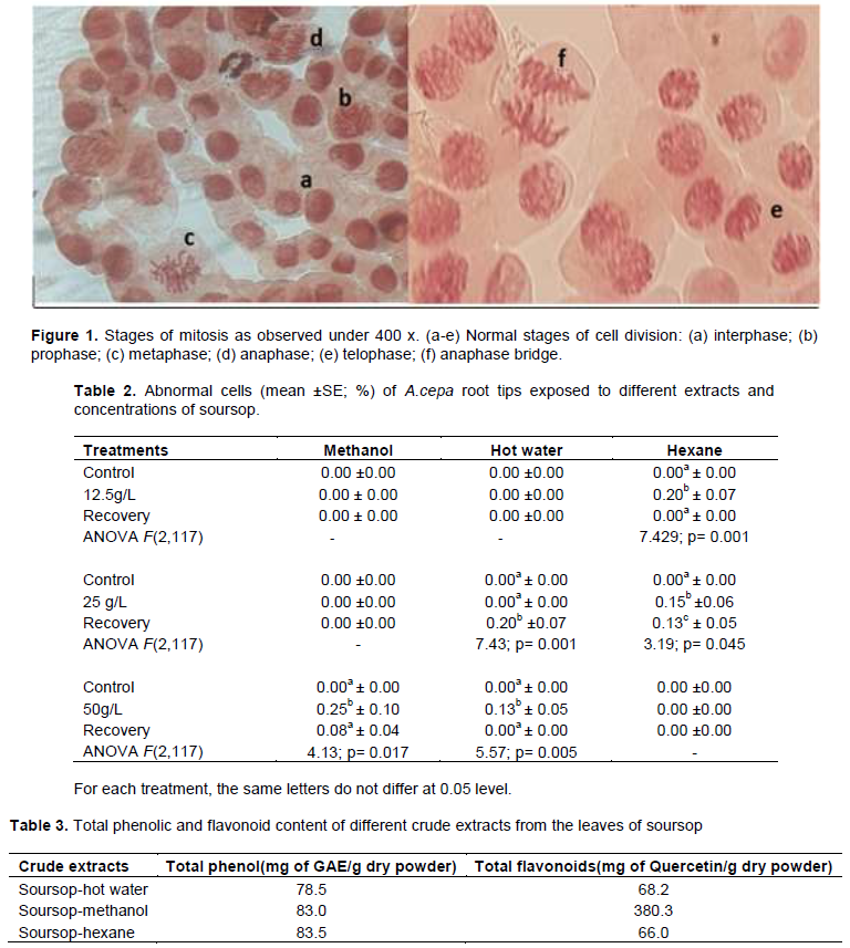
After 24 h under each exposure, 5 root tips were cut from each bulb (8 bulbs for each treatment method which makes it up to 40 root tips for each exposure), and returned for next step. The root tips were fixed in 3:1 (v/v) ethanol: glacial acetic acid and stored overnight at 4°C. They were placed in 70% (v/v) aqueous alcohol the next day and refrigerated until used. Five slides on average were made for each bulb for each treatment using root tips that were hydrolyzed in 1N HCl for 3 min. Stained root tips were squashed in acetocarmine stain. Each slide was viewed at 400x using a compound microscope to determine the MI, while photomicrographs were taken at x1000 for chromosomal aberration under oil immersion.
Analysis of cytotoxicity and genotoxicity
(i) The mitotic index (MI) was calculated per 1000 cells by using the ratio between the number of mitotic cells and the total number of cells scored and expressed as percentage (Balog, 1982; Sehgal et al., 2006; Al-Ahmadi, 2013).
(ii) Cytogenic effects were evaluated by considering chromatin aberrations like anaphase bridges, sticky metaphase and breaks of chromosomes (Akinsemolu et al., 2015).
Determination of total phenolic and flavonoid contents
The total phenolic content of the plant extracts was determined by the Folin-Ciocalteu method (Meda et al., 2005). According to Nassr-Allah et al. (2009), the method similar to the one that follows was used for seven medicinal plants of Egypt since medicinal plants which has antioxidant properties contains phenolic and flavonoid compounds.
Lyophilized powder of 0.1 g of the plant samples were dissolved in 1 ml of deionized water. 0.1 ml of this solution was mixed with 2.8 ml of deionized water, 2 ml of 2% (w/v) sodium carbonate, and 0.1 ml of 50% (v/v) Folin-Ciocalteu phenol reagent. After incubation for 30 min at room temperature, the relative absorbance of the reaction mixture was compared to deionized water with a UV spectrophotometer. Gallic acid was used as a standard phenolic to construct a seven point standard curve (0 to 200 mg/L), the total phenolic contents in plant extracts was determined in triplicate.
The aluminium chloride colorimetric method was used to determine the total flavonoid contents. Aliquots of 0.1 g of plant extracts were dissolved in 1 ml deionized water. 0.5 ml of this solution was mixed with 1.5 ml of 95% (v/v) alcohol, 0.1 ml of 10% (w/v) aluminium chloride hexahydrate, 0.1 ml of 1 M potassium acetate and 2.8 ml of deionized water. The absorbance was measured at 415 nm after incubation at room temperature for 40 min and compared to deionized water with a UV spectrophotometer. In order to construct a seven point standard curve, quercetin was chosen. Triplicated assays were used to determine the total flavonoid contents. The result was expressed as milligram quercetin equivalents (QE)/g dry weight of lyophilized powder.
Statistical analysis
MSTATc statistical software was used for analysis. Analysis of variance was used to compare the data on mitotic and phase indices to reassure the viability of the data and validity of results. Differences between the individual dosage group and the control of each extract was analyzed by means of the post hoc Tukey’s HSD test of significance at p ≤ 0.05 level.
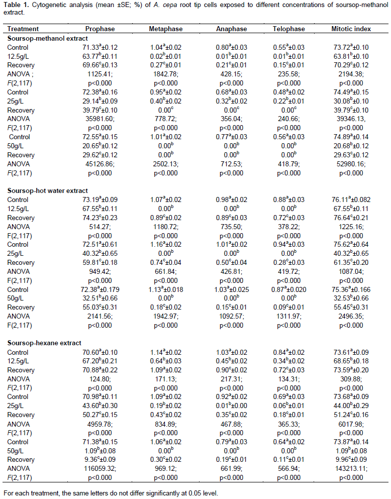
Analyses of variance of the mitotic indices indicated significant differences (p≤0.05) between concentrations of soursop-methanol extract as far as inhibition effect and MI was concerned (Table 1). Overall decrease in MI was contributed to by all the stages of mitosis. MI was above 70% in control groups which decreased to 20.7% with 50 g/L soursop-methanol extract exposure. The recovery rate declined as the concentration of the extract increased.
A significant (p≤0.05) inhibition effect of all three concentrations of soursop-hot water extract on MI (Table 1) was observed. All phase of cell cycles contributed to the overall decrease in MI. In control group, the MI was above 70% which fell significantly to as low as 32.5% with 50 g/L soursop- hot water extract exposure. The cells gained the potential to divide when allowed to recovery in water; however, the potential decreased as the concentration increased.
Similarly, the study showed a significant (p≤0.05) reduction in the mitotic index of all three concentrations of soursop- hexane extract (Table 1). The reduction in the MI was significantly increased with increasing concentration of the extract. The overall decrease in MI was contributed to by all phases of cell cycles. The MI was above 70% in control group which dropped significantly (p≤0.05) to as low as 1% with 50 g/L soursop-hexane extract exposure. The recovery rate had relatively decreased as the concentration of the extract increased.
Cells showing chromosome laggards, bi-nucleated cell, prolonged bacilliform nucleus, prolonged nucleus, multi-polar anaphase, spindle disturbance in metaphase and anaphase bridge (Figure 1) were counted for each treatment group and mean percentage of cells are presented in Table 2.
The data as tabulated in Table 3 shows that phenolic content was highest in soursop-hexane extract (83.5 mg GAE/g dry weight). This was followed in descending order by extracts of soursop-methanol and soursop- hot water. The highest amount of flavonoid was extracted by soursop-methanol extract, that is, 380.3 mg of Quercetin/ g dry powder.
Comparable results were obtained for the soursop-methanol extract (Table 1). There was significant difference in the mitotic index between the control and the different concentrations used since p≤0.05. Mitotic index dropped by 9.9% in 12.5 g/L. There was a huge reduction observed in 25 and 50 g/L where MI decreased by 44.4 and 54.2% respectively. According to Panda and Sahu (1985), a drop below 50% is called cytotoxic limit value and usually has sub lethal effects on the organism. This means that there had been a notable drop in the mitotic index and that the extract from soursop had interfered with the proteins in the cells hindering cell division.
As the concentration of soursop- methanol extract increased to 50 g/L, the ability of the cells to divide dropped drastically to 20.7%. This showed that at this concentration, the extract becomes cytotoxic. Similarly, a cytotoxicity study was conducted on MCF-7 cell line and its effect on expression of bcl-2 also using soursop- methanol extract leaves by Rachmani et al. (2012). As the concentration of the test material increased, the percentage of mean number of living cells MCF-7 cells was reducing. At the least concentration of 31.25 mg/mL, the average percentage of cell survival was 75.3, whereas 5.3% was the cell survival rate at 50 mg/ml. Hence they concluded that soursop- methanol leaf extract shows cytotoxicity. Results obtained by Raybaudi-Massilia et al. (2015) also demonstrated that methanol extract of soursop seeds had significant cytotoxic effect (p ≤ 0.05) on two human tumor cell lines it being HeLa and PC3.
For soursop-hot water extract, the mitotic index was significantly different from control, 12.5 g/L and recovery. This means that even at a lower concentration, this extract was able to reduce the mitotic index significantly by 8.6%. Once put back in distilled water to recover, mitosis rate increased with a significant difference at p≤0.05. Moreover, there was a significant (p≤ 0.05) drop by 5.6, 1.07, 0.98 and 0.88% for individual stages like prophase, metaphase, anaphase and telophase respectively. All these stages contributed towards an overall drop in the MI since division of the cells was slowed down and also hindered.
When the concentration was increased to 25 g/L, there was a significant drop in mitotic index by 35.3%. Mitotic stages like prophase, metaphase, anaphase and telophase recorded a significant (p≤0.05) drop of 32.2, 1.16, 1.01 and 0.94% respectively. When allowed to recover in water, it showed a marked increase in the mitotic index although not being able to divide fast enough to match the control’s mitotic index. This means that at higher concentrations, recovery period of more than 24 h would be needed in order for the cells to reach its full capability of cell division.
The highest concentration (50 g/L) recorded the lowest mitotic index. Division of cells dropped significantly (p≤0.05) by 42.8% (Table 1). This could be the most toxic dose concentration since it hugely affected the division of cells. Recovery with water was possible; however, more than 24 h is essential for the cells to return to its full potential of dividing. Prophase decreased by 39.9%, metaphase by 1.13%, anaphase by 1.03% and telophase by 0.87% significantly (p≤0.05).
It has also been proven that the in vitro activity of antioxidant by soursop-water extract has significant antioxidant activity, hence can manage diseases by reducing oxidative stress (Gavamukulya et al., 2014).
The results obtained from this experiment were similar to the previous studies on A. muricata by Diaz et al. (2016). A. cepa cells that were treated with 1, 3, 5 and 6 and 7% of A. muricata -hot water extract showed differences in the mitotic index obtained. Out of the five extracts used, 1% extract showed the highest MI of 85.59% whereas 7% concentration showed the lowest index of 7.82%. This decrease in the MI as concentration increased is due to the cytotoxic compounds.
Soursop-hexane extract showed a marked potential in reducing MI especially at higher concentration. (Table 1). When the bulbs were exposed to 12.5 g/L, there was a difference by 5.0% in the reduction of division of cells from the control. This showed that even at the lowest concentration, soursop-hexane extract does have the potential in affecting the proteins in the cells. However, when the root tips were put back in water to recovery, it recovered by 4.9% and reached to a level within 24 h where there was no significant difference between the control and recovery (p≤0.05). Prophase, metaphase, anaphase and telophase stages dropped significantly by 3.4, 0.5, 0.58 and 0.5%, respectively.
As the concentration was increased to 25 g/L, there was more hindrance to mitosis by 29.7%. A drop in the percentage of prophase was by 27.3%, metaphase by 0.9%, anaphase by 0.91% and telophase was by 0.63%. Recovery was significantly high when compared to the treatment group (increased by 7.24%). However, since the root tips were exposed to a higher concentration, recovery was not able to reach the fullest since the MI was significantly different and lower in the recovery group to the control.
Further increase in concentration to 50 g/L caused a drastic effect on the cells. It reduced the mitotic index by 72.8% which could have been the most toxic dose for the cells. There was cessation of all the mitotic stages except for prophase. When allowed to recover, there was significant (p≤0.05) difference recorded for cell division; however, it was still away by 63.9% to reach the full potential of cell division and may have needed more than 48 h to reach normality (Table 1). There was a drop in prophase by 70.30%, metaphase by 1.06%, anaphase by 0.79% and telophase by 0.64%. All stages of mitosis contributed significantly towards the total MI.
The ability of the soursop- hexane extract to inhibit mitosis was due to the fact that it contains compounds restricting the cells to divide. A research study conducted by Rosdi et al. (2015)found out that A. muricata –hexane leaves extract consist of flavonoids which played a role in decreasing cell viability in pancreatic cancer cells of humans (Capan-1) in vitro. Hence they concluded that this leaf might have anti-cancer agent. It has also been reported by Haro et al. (2014)after the characterisation and phytochemical screening of soursop leaves that it contains tannins, saponins, glycosides, alkaloids, flavonoids and steroids/triterpenoids.
Alongside this, it was also found out by Ningsih et al. (2015)that as the concentration of A. muricata–hexane extract increased, the inhibition zone diameter against P. acnes got bigger. Similarly, antibacterial activity decreased with decreasing concentration. Hence it was concluded that as concentration of the extract increased, its inhibition ability also increased.
Soursop crude extracts also have different levels of cytotoxicity towards breast cancer cell lines. The tumor’s size and weight was reduced proving that soursop indeed plays a significant role in inhibiting cancer (Najmuddin et al., 2016).
The order of total phenol content was soursop- hexane > soursop -methanol > soursop-hot water. The highest flavonoid was detected in soursop-methanol extract (380.3 mg of Quercetin/g dry powder) and the least amount was found to be in soursop- hexane extract (66.0 mg of Quercetin/g of dry powder). The order of total flavonoids are soursop- methanol > soursop- hot water > soursop-hexane (Table 3). Soursop-methanol extract did reduce the MI the most indicating the presence of most flavonoids which was the possible cause of hindrance to cell division.
Similar results were reported by Rantam et al. (2014)where they found that soursop leaves extract contained flavonoid from flavonol group which improved the activation and propagation of T-cell. Their result from immunocytochemistry for expression on rabbit PBMC culture showed that even the lowest dose of 5 μg of soursop leave extract was able to arouse the expression of T-cell.
Cancer research regarding soursop has focused mainly on the phytochemicals known as Annonaceous acetogenins. These natural compounds are produced in the leaf, stem, bark and fruit seeds. From 37 species, 370 Annonaceous acetogenins have been obtained out of which 50% of over 80 of them were found to be significantly bioactive and could be fractionated. A rapidly growing class of compounds whose genuine anticancer ability as ATP inhibitors is currently being researched on. One of the very potent acetogenins is bullatacin which proves to be effective in vivo models (Kedari and Khan, 2014).
Moreover, few chromosomal aberrations were seen at all the three types of extracts (soursop-hexane extract at 12.5 and 25 g/L, soursop-methanol extract at 50 g/L and soursop-hot water extract at 50 g/L (Table 2). This could be due to chromosomal aberrations being observed mostly after prophase stage since A. muricata restricted most of the cells to proceed beyond prophase. Hence, only interphase and prophase stages were seen.
Hence, it can be said that A. muricata has compounds which affect the proteins in the cells and hinders cell division. The most hindrance was caused by soursop-hexane extract at the highest concentration which could be having the most compounds with antioxidant properties. The leaf extracts did contain significant amount of phenols and flavonoids. Further studies could be carried out using mammalian cancer cells to encourage its consumption as a medicine.
The authors have not declared any conflict of interests.
REFERENCES
|
Akinboro A, Mohamed KB, Asmawi MZ, Sofiman, OA (2011). Mutagenic and antimutagenic potentials of fruit juices of five medicinal plants in Allium cepa L.: Possible influence of DPPH free radical scavengers. African Journal of Biotechnology 10(50):10248-10257.
|
|
|
|
Akinsemolu AA, Nwangburuka CC, Ogunwenmo KO (2015). Evaluation of tobacco industrial wastewater for genotoxic characteristics on Allium cepa L. root cell mitosis. Journal of Advances in Biology and Biotechnology 2(3):165-173.
Crossref
|
|
|
|
|
Al-Ahmadi MS (2013). Effects of organic insecticides, Kingbo and Azdar 10 EC, on mitotic chromosomes in root tip cells of Allium cepa. International Journal of Genetics and Molecular Biology 5(5):64-70.
Crossref
|
|
|
|
|
Balog C (1982). The mitotic index in diploid and triploid Allium roots. Cytologia, 47(3/4): 689-697.
Crossref
|
|
|
|
|
Bhat SK, Singhal K, Shruti P, Pal S (2013). Evaluation of antimitotic effect of Calotropis Procera L on Allium cepa L. International Journal of Pharmacy 3(2):356-359.
|
|
|
|
|
Biba VS, Amily A, Sangeetha S, Remani P (2014). Anticancer, antioxidant and antimicrobial activity of Annonaceae family. World Journal of Pharmacy and Pharmaceutical Sciences 3(3):1595-1604.
|
|
|
|
|
Diaz JC, Jimenez JJL, Pagoso EJA, Tomas JV, Dulay RMR (2016). Cytogenotoxic assessment of aqueous extract of Annona muricata Linn. (Annonaceae) leaves on Allium cepa L. root-tip meristem cells. Der Pharmacia Sinica 7(3):1-5.
|
|
|
|
|
El- Shahaby OA, Migid HM, Soliman MI, Mashaly IA (2003). Genotoxicity screening of industrial wastewater using the Allium cepa chromosome aberration assay. Pakistan Journal of Biological Sciences 6(1):23-28.
Crossref
|
|
|
|
|
Gavamukulya Y, Abou-Elella F, Wamunyokoli F, AEl-Shemy H (2014). Phytochemical screening, anti-oxidant activity and in vitro anticancer potential of ethanolic and water leaves extracts of Annona muricata (Graviola). Asian Pacific Journal of Tropical Medicine 7(14):S355-S363.
Crossref
|
|
|
|
|
George VC, Kumar DR, Rajkumar V, Suresh PK, Kumar RA (2012). Quantitative assessment of the relative antineoplastic potential of the n-butanolic leaf extract of Annona muricata Linn. in normal and immortalized human cell lines. Asian Pacific Journal of Cancer Prevention 13(2):699-704.
Crossref
|
|
|
|
|
Han S (1998). Medicinal plants in the South Pacific. World Health Organization (WHO) Regional Publications, Western Pacific Series 19:1-151.
|
|
|
|
|
Haro G, Utami NP, Sitompul E (2014). Study of the antibacterial activities of Soursop (Annona muricata L.) leaves. International Journal of PharmTech Research 6(2):575-581.
|
|
|
|
|
Kedari TS, Khan AA (2014). Guyabano (Annona muricata): A review of its traditional uses phytochemistry and pharmacology. American Journal of Research Communication 2(10):247-268.
|
|
|
|
|
Kumar MS, Ankit S, Gautam DNS, Kumar SA (2015). Biodiversity and indigenous uses of medicinal plant in the Chandra Prabha wildlife sanctuary, Chandauli district, Uttar Pradesh. International Journal of Biodiversity 2015(394307):1-11.
|
|
|
|
|
Mahmoud T, Gairola S (2013). Traditional knowledge and use of medicinal plants in the Eastern Desert of Egypt: a case study from Wadi El-Gemal National Park. Journal of Medicinal Plants 1(6):10-17.
|
|
|
|
|
Meda A, Lamien CE, Romito M, Millogo J, Nacoulma, OG (2005). Determination of the total phenolic,flavonoid and proline contents in Burkina Fasan honey, as well as their radical scavenging activity. Food chemistry 91(3):571-577.
Crossref
|
|
|
|
|
Najmuddin SUFS, Romli MF, Hamid M, Alitheen NB, Rahman NMANA (2016). Anti-cancer effect of Annona muricata Linn leaves crude extract (AMCE) on breast cancer cell line. BMC Complementary and Alternative Medicine 16(311):1-20.
Crossref
|
|
|
|
|
Nassr-Allah AA, Aboul-Enein AM, Aboul-Enein KM, Lightfoot DA, Cocchetto A, El-Shemy HA (2009). Anti-cancer and anti-oxidant activity of some Egyptian medicinal plants. Journal of Medicinal Plants Research 3(10):799-808.
|
|
|
|
|
Ningsih DR, Zusfahair, Kartika D (2015). In vitro antibacterial test of Soursop (Annona muricata Linn) n-hexane extract leaves on Propionibacterium Acnes. Proceeding of The 1st - University of Muhammadiyah Purwokerto - Pharmacy International Conference pp. 63-68.
|
|
|
|
|
Okullo J, Omujal F, Bigirimana C, Isubikalu P, Malinga M, Bizuru E, Namutebi A, Obaa B, Agea J (2014). Ethno-medicinal uses of selected indigenous fruit trees from the lake Victoria Basin districts in Uganda. Journal of Medicinal Plants 2(1):78-88.
|
|
|
|
|
Panda B, Sahu U (1985). Induction of abnormal spindle function and cytokinesis inhibition in mitotic cells of Allium cepa by the organophosphorus insecticide fensulfothion. Cytobios 42(167-
|
|
|
|
|
Ping K, Darah I, Yusuf UK, Sasidharan S (2012). Genotoxicity of Euphorbia hirta on Allium cepa assay. Molecules 17(7):7782-7791.
Crossref
|
|
|
|
|
Rachmani EP, Suhesti, TS, Widiastuti AR, Aditiyono A (2012). The breast of anticancer from leaf extract of Annona muricata against cell line in T47D. International Journal of Applied Science and Technology 2(1):157-164.
|
|
|
|
|
Rahman MM, Khan MA (2013). Anti-cancer potential of South Asian plants. Natural Products and Bioprospecting 3(3):74-88.
Crossref
|
|
|
|
|
Raina H, Soni G, Jauhari N, Sharma N, Bharadvaja N (2014). Phytochemical importance of medicinal plants as potential sources of anticancer agents. Turkish Journal of Botany 38(6):1027-1035.
Crossref
|
|
|
|
|
Rantam FA, Suwanti LT, Erina A (2014). The potential of soursop (Annona muricata Linn.) leaves extract as immunostimulantin rabbit PBMC. Veterinaria Medika 7(3):278-285.
|
|
|
|
|
Ratanavalachai T, Thitiorul S, Nandhasri P, Tanuchit S, Jansom C (2010). Cytotoxic and genotoxic activities of an aqueous extract from Thai noni leaves in human lymphocytes in vitro. Sonklanakarin Journal of Science and Technology 32(1):37-42.
|
|
|
|
|
Raybaudi-Massilia R, Suárez AI, Arvelo F, Sojo F, Mosqueda-Melgar J, Zambrano A, Calderón-Gabaldón MI (2015). An analysis in-vitro of the cytotoxic, antioxidant and antimicrobial activity of aqueous and alcoholic extracts of Annona muricata L. seed and pulp. British Journal of Applied Science and Technology 5(4):333-341.
Crossref
|
|
|
|
|
Rosdi MNM, Daud NNNNM, Zulkifli RM, Ya'akob H (2015). Cytotoxic effect of Annona muricata Linn leaves extract on Capan-1 cells. Journal of Applied Pharmaceutical Science 5(5):45-48.
Crossref
|
|
|
|
|
Sehgal R, Roy S, Kumar VL (2006). Evaluation of cytotoxic potential of latex of Calotropis procera and Podophyllotoxin in Allium cepa root model [Abstract]. Biocell 30(1):9-13.
|
|
|
|
|
Smith A (1981). Flora Vitiensis Nova. Pacific Tropical Botanical Garden 2:44-116.
|
|
|
|
|
Tagne RS, Telefo BP, Nyemb JN, Yemele DM, Njina SN, Goka SM C, Lienou LL, Kamdje AHN, Moundipa PF, Farooq AD (2014). Anticancer and antioxidant activities of methanol extracts and fractions of some Cameroonian medicinal plants. Asian Pacific Journal of Tropical Medicine 7:S442-S447.
Crossref
|
|