African Journal of
Biotechnology

  • Abbreviation: Afr. J. Biotechnol.
  • Language: English
  • ISSN: 1684-5315
  • DOI: 10.5897/AJB
  • Start Year: 2002
  • Published Articles: 12487

Full Length Research Paper

Antimicrobial activity of metabolites extracted from Zanthoxylum gilletii, Markhamia lutea and their endophytic fungi against common bean bacterial pathogens

Lucy Aketch Wanga
  • Lucy Aketch Wanga
  • Department of Biochemistry and Molecular Biology, Faculty of Science, Egerton University, Kenya.
  • Google Scholar
Isabel Nyokabi Wagara
  • Isabel Nyokabi Wagara
  • Department of Biological Sciences, Faculty of Science, Egerton University, Kenya.
  • Google Scholar
Ramadhan Mwakubambanya
  • Ramadhan Mwakubambanya
  • Department of Biochemistry and Molecular Biology, Faculty of Science, Egerton University, Kenya.
  • Google Scholar
Josphat Clement Matasyoh
  • Josphat Clement Matasyoh
  • Department of Chemistry, Faculty of Science, Egerton University, Kenya.
  • Google Scholar


  •  Received: 26 March 2018
  •  Accepted: 14 May 2018
  •  Published: 04 July 2018

 ABSTRACT

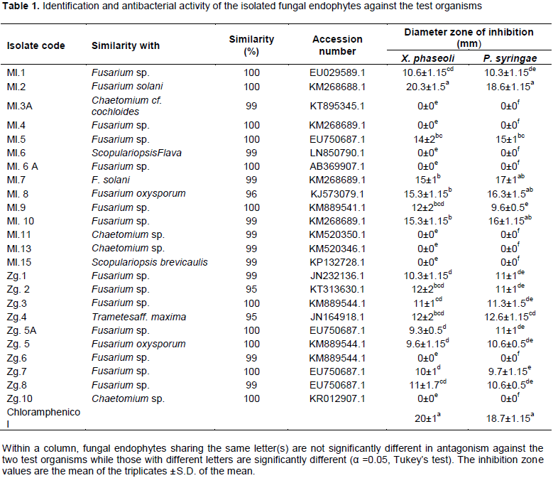
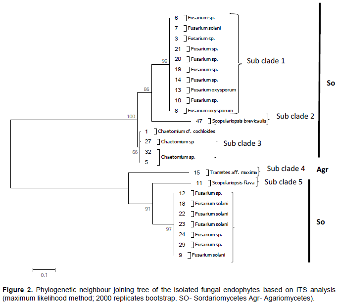
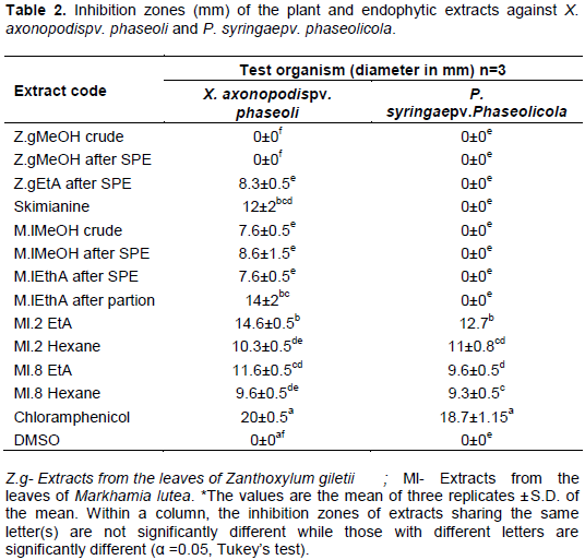
Antibacterial activity of extracts of Zanthoxylum gilletii, Markhamia lutea and their fungal endophytes were evaluated against bacterial pathogens of common bean: Xanthomonus axonopodis pv. phaseoli and Pseudomonus syringae pv. phaseolicola. The leaves of both plants were dried under shade, ground to fine powder and extracted using methanol. The methanol extracts were fractionated sequentially using ethylacetate and hexane to produce various fractions. Endophytic fungi were isolated from fresh leaves and identified by ITS-rDNA sequence analysis. Antibacterial screening of the fungal endophytes was done by dual culture assay. The most active endophytic fungi were fermented on rice media and extracted using methanol. Pure compounds were analyzed by a combination of mass spectrometry and spectroscopic techniques which included 1D and 2D NMR. Antibacterial activity of all the extracts was determined by disc agar diffusion assay against the test organisms. Twenty-four (24) fungal endophytes were isolated which included: Fusarium, Chaetomium, Scopulariopsis and Trametes. Endophytic Fusarium solani was the most active against X. axonopodis pv. phaseoli (20.3 mm inhibition zone) and P. syringae pv. phaseolicola (18.6 mm inhibition zone). The plant extracts were active against X. axonopodis pv. phaseoli with an inhibition zone ranging between 8-12 mm except the methanol extract from Z. gilletii which did not show any activity. The endophytic extracts were active against both test organisms with a zone of inhibition ranging from 9.3-14 mm. Phenolic compounds present in Fusarium species may have contributed to the antibacterial activity of this strain against the test organisms.

Key words: Common bean, medicinal plants, fungal endophytes, antibacterial activity, Xanthomonus axonopodis pv phaseoli, Pseudomonus syringae pv phaseolicola.


 INTRODUCTION

Common bean (Phaseolus vulgaris L.) is a major legume crop that is largely consumed among various communities in Kenya. It provides cheaper alternative source of protein and household food security to the  low-income earners in towns and the rural poor population (Gichangi et al., 2012). However, as noted over the years, its productivity is gradually declining (Katungi et al., 2010). This could be attributed, but not limited, to bacterial infections such as common bacterial blight caused by Xanthomonus axonopodis pv. phaseoli and halo blight caused by Pseudomonus syringae pv. phaseolicola. The effects of these bacteria can be easily spotted in the field which affects the leaves as well as the pods. This leads to a reduction in the productivity of common bean in Kenya. Currently, the methods used to control these pathogens in the field include the use of copper based foliar sprays, synthetic pesticides and antibiotics such as streptomycin. The indiscriminate and intensive use of these pesticides and antibiotics has caused many problems to the environment such as water, animals, soil and food contamination; elimination of non-target organisms; poisoning of farmers as well as selection of phytopathogens, weeds and pests (Stangarlin et al., 2011). There has also been incidences of the occurrence of pesticide residues in the farm produce (Sartori et al., 2004). Therefore, there is need to find alternative ways of controlling these pathogens using extracts from natural sources such as endophytic fungi and medicinal plants which are believed to be easily biodegradable and readily available.
 
The tropical ecosystem is a host to more than half the number of living species worldwide, and many bioactive metabolites are produced in this ecosystem. Therefore, most plant species in this ecosystem are known to possess medicinal properties (Suryanarayanan, 2011). These plants are inhabited intracellularly by either bacteria or fungi known as endophytes that do not cause any apparent disease symptom (Clay, 1990). The fungal endophytes produce secondary metabolites that have desirable antimicrobial properties such as antibacterial, antifungal, antiviral, antioxidant, somatic fat reducing, blood pressure regulating, anti-inflammatory among others. Zanthoxylum gilletii is an evergreen, aromatic deciduous shrub or tree that belongs to the family Rutaceae (Negi et al., 2011) while Markhamia lutea (Nile tulip) is an evergreen subtropical, flowering plant that belongs to the family Bignoniaceae (Orwa et al., 2009). Both plants possess medicinal properties and are commonly found and used in Kenya for various medicinal purposes. For instance, Z. gilletii is used traditionally for the treatment of urogenital infections, rheumatism and in the management of various parasitic infections (Gaya et al., 2013, Nyunja et al., 2009). M. lutea on the other hand has been used in the treatment of earache, skin infections, asthma, cough, gonorrhea as well as alleviation of AIDS symptoms among others (Lamorde et al., 2010). Most of the traditional uses of these plants are based on their importance on the alleviation of human pathogen. Therefore, this study aimed to determine the antibacterial   activity   of   these  plants  as  well  as  their endophytes against bean bacterial pathogens. This study is significant due to the reduction in the common bean productivity and the need for alternative sources from natural products to control these infections and thereby improving bean productivity in Kenya.


 MATERIALS AND METHODS
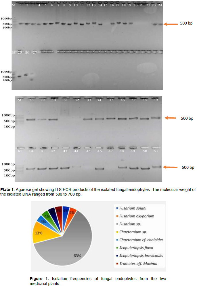
Collection of plant materials
 
Fresh leaves of Z.  gilletii and M. lutea were collected from Kakamega Tropical Rainforest which stretches from 0° 10ʹ to 0° 21ʹN and longitude 34° 44ʹ to 34°º 58ʹE and an altitude of 1524 m above sea level. The leaves were identified with the help of a taxonomist and were deposited at the Biological Sciences Department, Egerton University.
 
Isolation of the fungal endophytes
 
Endophytic fungi were isolated from the leaves of Z. gilletii and M. lutea within 8 h of collection using the procedure by Zinniel et al. (2002) with slight modifications. Briefly, the leaves were washed under running tap water and blotted dry using filter papers. Thereafter, they were sterilized for 2 min in 70% ethanol, 1% sodium hypochlorite for 3 min and rinsed three times in sterile distilled water. The leaves were then cut aseptically into sections approximately 1 by 4 mm and inoculated in Petri-dishes containing Sabourand Dextrose Agar (SDA) amended with streptomycin sulphate antibiotic (2 g/L). The plates were incubated at 25 ± 2°C for 1 to 4 weeks. Frequent monitoring was done to check for the growth of the endophytic fungi. The first visible hyphal tips were transferred to fresh SDA plates to prepare pure cultures. The cultures were then identified using molecular techniques.
 
Molecular identification of the isolated fungi
 
Pure cultures of the endophytes were grown in 30 ml of yeast Malt broth (pH 6.3) and incubated at 28°C on an orbital shaker for 3 to 4 days to allow the fungal mycelia to grow.
 
 
DNA extraction
 
The DNA extraction was performed using the BIO BASIC EZ-10 Genomic DNA kit following manufacturer’s instruction. Approximately, 6 to 10, 1.4 mm Precellys Ceramic Beads were added to a 1.5 ml screw cap reaction tube. Approximately, 60 mg of the fungal hyphae obtained from a 3 to 4 day old culture were added to the same tube. The sample was covered with 600 µl Plant Cell lysis buffer (PCB) and homogenized using a homogenizer (Precellys 24 lysis and homogenization, Peq lab, Bertin technologies). β-Mercaptoethanol (12 µl) was added to the sample, vortexed (IKA MS3 Digital) and incubated for 25 min at 65°C in a metal block (MTB 250). Chloroform (600 µl) was added and the mixture centrifuged (5430 R) at 10,000 rpm for 2 min. The supernatant was transferred to a clean Eppendorf tube and the rest discarded. Binding buffer  (BD  buffer) (200 µl)  was added and the mixture vortexed, followed by addition of 200 µl ethanol and again vortexed. The mixture was transferred into EZ-10 column placed in a 2 ml collection tube and centrifuged at 12,000 rpm for 1 min. The flow through was discarded and then 500 µl of PW solution was added. The mixture was centrifuged at 12, 000 rpm and the flow through discarded. Then, 500 µl of Wash solution was added and the mixture was again centrifuged at 12,000 rpm for 1 min and the flow through discarded again. The column was again centrifuged at 12,000 rpm for 2 min to remove any remaining wash solution. Finally, the column was transferred into an empty 1.5 ml Eppendorf tube and 70 µl of TE Buffer, pre-warmed to 60°C, added directly at the center of the EZ membrane to increase the elution efficiency. The sample was incubated for 2 min at room temperature and then centrifuged at 12,000 rpm for 2 min to elute the DNA. The DNA was stored at 4°C for further analysis.
 
 
Polymerase chain reaction (PCR) amplification
 
To a PCR tube, the following were added: 0.5 µl of forward primer ITS1F (CTTGGTCATTTAGAGGAAGTAA) and 0.5 µl of reverse primer ITS4 (TCCTCCGCTTATTGATATGC), 12.5 µL of the jump start ready mix that contained 20 mMTris-HCl pH 8.3, 100 mM KCl, 3 mM MgCl2, 0.002% gelatin, 0.4 mM dNTPs (dATP, dCTP, dGTP and dTTP), stabilizers, 0.1 unit/mL Taq DNA polymerase and JumpStart antibody. This was followed by 9.5 µl of distilled water and 2 µl of the template DNA to make a total volume of 25 µl of the mixture per sample. For a negative control, 2 µl of distilled water was used in the reaction mix instead of DNA template. The amplification was done in a thermocycler (Eppendorf® Mastercycler® nexus Thermal Cycler) under the following conditions; initial denaturation of 5 min at 94°C, followed by 34 cycles of denaturation for 30 s at 94°C, annealing at 52°C for 30 s and elongation for 1 min at 72°C. Then, a final elongation of 10 min at 72°C. The PCR products were pre-stained with midori green dyeand resolved in a 0.8% agarose gel. The visualization was done in a UV transilluminator (Nippon Genetics Europe GMbH) and photographs were taken. The amplified PCR products were purified using BIO-BASIC EZ-10 kit and stored at -4°C for further analysis.
 
DNA sequencing
 
The amplified DNA was sequenced by Illumina genome analyzer sequencing machine (applied Biosystems 3730 xl DNA analyzer). The forward and reverse primer sequences obtained from the sequencing were aligned by Genious R7 program to get the consensus sequences. The consensus sequences were deposited in NCBI GenBank and compared with those available in GenBank via BLAST searches. Phylogenetic analysis was conducted using the distance based neighbor joining methods in Molecular Evolutionary Genetics Analysis (MEGA) version 6.06 and the Neighbor joining (NJ) tree constructed using Tamura-Nei distance. All characters were equally weighted and unordered. Gaps and the missing data were treated as complete deletion. Support for the specific nodes on the NJ tree was estimated by bootstrapping 2000 replications. The substitution type was used for nucleotides and the pattern of lineage was homogeneous.
 
Antimicrobial activity of the isolated fungi
 
Test organisms
 
The test organisms used in this study were X. axonopodis pv phaseoli and P. syringae pv. phasiolicola which were provided by the Biological Sciences Department of Egerton University.
 
Dual culture assay
 
Inhibition of bacterial growth by the endophytic fungi was examined on Muller Hinton (MH) plates using dual culture assay as described by Srivastava and Anandrao (2015) with slight modification. Briefly, 100 µl of bacterial concentration of 5×10⁵ CFU/mL was swabbed evenly on the MH media on Petri-dishes using a sterile cotton swab and allowed to dry. A six-millimeter diameter of a 7-day old mycelia plug was placed in the MH media plate inoculated with the test bacteria. A standard chloramphenicol was used as a positive control. The plates were incubated at ± 32°C for 24 h and the zone of inhibition was measured in mm. The experiment was carried out in triplicates. The most active endophytic fungi were subjected to solid state fermentation for secondary metabolites extraction.
 
Fermentation and extraction of secondary metabolites
 
Fermentation of the endophyte was carried out using a procedure by Nascimento et al. (2012) with slight modification. The solid-state fermentation was carried out in 21, 500 ml Erlenmeyer flasks containing 90 g of rice in 90 ml of distilled water per flask which were twice autoclaved at 120°C for 40 min. Agar plugs of about 2 × 2 cm were cut from a 7-day old culture of the endophyte and then inoculated in the rice media. One flask without inoculum was kept as a control. The flasks were incubated for 21 days at 25°C under static conditions. The flasks were checked periodically for contamination.
 
Extraction of secondary metabolites from the endophytic fungi
 
After the incubation period, the fermentation was ended with the addition of 150 ml of methanol to each of the flasks and left to stand overnight. The cultures were cut into pieces with the aid of a spatula and the flask placed in an ultrasonic cleaner (SB-120 DTN) to allow complete extraction of the secondary metabolites. The mixture was filtered using a Whatman filter paper no. 1 followed by repeated extraction with methanol until exhaustion. The filtrate was evaporated under reduced pressure (BUCHI rotavapor R-205) to yield a methanol extract. The methanol extract was partitioned between hexane and ethylacetate to obtain the respective fractions. The fractions were subjected to antibacterial assay.
 
Extraction of secondary metabolites from the leaves of the medicinal plants
 
The collected leaves were dried under a shade for approximately 2 weeks. The leaves were ground into a fine powder and 700 g of each powder was soaked in 1.5 L of methanol overnight. The mixtures were then filtered using Whatman filter paper no. 1 and the filtrate evaporated under reduced pressure. The obtained fractions were partitioned using reverse phase solid phase extraction, followed by thin layer chromatography (TLC) and column chromatography. The fractions obtained were subjected to antibacterial assay.
 
Antibacterial assay
 
The antibacterial assay of both the endophytic and plant extracts was performed using agar disc diffusion method as described by Kajaria et al. (2012) with slight modification. The media used in this assay was Muller Hinton Agar (38 g/1000 ml of distilled water). A 24-h bacterial population of 1.5 x 10⁸ CFU/ml (1.0 x 10⁸ – 2.0 x 10⁸ CFU/ml) was spread on the plate containing media and left to dry. All  extracts  were  weighed  and  a  50 mg/ml  concentration  of the extracts made using DMSO. Blank sterile disc of Whatman filter paper No. 1 of 6 mm in diameter was impregnated with 10 µl of different extracts and plated against the test organisms. A standard chloramphenicol antibiotic was used as a positive control while the negative control was blank sterile disc soaked in DMSO. The plates were incubated at ±32°C overnight and zone of inhibition measured in millimeters.


 RESULTS

Isolation and identification of the fungal endophytes
 
A total of  24  fungal  endophytes  were  isolated  from  Z. gilletii and M. lutea, some of which are shown in Plate 1. Optimal PCR products of the isolates were obtained using primer pair ITS1F and ITS4 which varied in band sizes of 500-700 bp (Plate 1). All the 24 identified fungal endophytes belonged to the phylum Ascomycota except Trametes aff. maxima which belonged to the phylum Basidiomycota. The endophytes were divided into the following groups: 63% Fusarium species, 4% Fusarium solani, 4% Fusarium oxysporum, 4% Scopulariopsis flava, 4% Scopulariopsis brevacaulis, 13% Chaetomium cf. cochloides, 4% Chaetomium spp. and 4% Trametes maxima (Figure 1). The BLAST percentage similarity to sequences  in  NCBI  from  the  previously identified fungi ranged from 95 to 100% (Table 1).


The evolutionary relationships of the isolated fungi were determined by generation of a distance based neighbour   joining   phylogenetic   tree.   The   neighbour joining analysis placed the sequences into two groups: Sordariomycetes and Agariomycetes. The generated tree had two major clades that were divided into six sub clades of distinct species: sub-clade 1- Fusarium spp., sub-clade 2- Scopulariopsis sp., sub-clade 3- Chaetomium sp., sub-clade 4- Trametes sp., sub-clade 5- Scopulariopsis sp. and sub-clade 6- Fusarium spp. Approximately, 75% of the isolated endophytes belonged to the genus Fusarium (Figure 2).
 
 
Antimicrobial assay of the fungal endophytes
 
Dual culture assay was used to assess the antagonistic effects of the isolated fungal endophytes against X. axonopodis pv. phaseoli and P. syringae pv. Phaseolicola (Plate 2). As shown in Table 1, 15 out of 24 endophytes showed antagonistic activity against both  X.  axonopodis pv. phaseoli and P. syringe pv. Phaseolicol; 13 of which belong to the genus Fusarium. Endophytic fungus, F. solani (Ml.2) had the largest inhibition zone of 20.3±1.5 mm against X. axonopodis pv. phaseoli and 18.6±1.5 mm against P. syringae pv. phaseolicola
 
 
The one-way ANOVA-Leven’s test showed non-homogeneity of variance for the isolated endophytes with a p value of 0.001. The activity of fungal endophyte Ml.2 (F. solani) against the test organism had no significant difference in activity as compared to chloramphenicol standard.
 
 
Disc agar diffusion assay of the plant and endophytic extracts
 
Secondary metabolites were extracted from both the host plant and  the  isolated  fungal  endophytes. Extracts from both M. lutea and Z. gilletii showed some activity against X. axonopodis pv. phaseoli but this was not the case against P. syringae pv. phaseolicola (Table 2). The methanol crude extract of Z. gilletii showed no activity against both test organisms. This was also observed from the methanol extract after solid phase extraction (SPE). The ethyl acetate extract after the same procedure showed some activity against X. axonopodis pv.  phaseoli and no activity against P. syringae pv. phaseolicola.  The alkaloid skimmianine isolated from the Z. gilletii produced a zone of inhibition of 12±2 mm against X. axonopodis pv. phaseoli while it showed no activity against P. syringae pv. phaseolicola. All the extracts from M. lutea were active against X. axonopodis pv. phaseoli with the ethyl acetate extract after partitioning showing the highest activity   of  14±2 mm    inhibition    diameter   against   X. axonopodis pv. phaseoli. However, these extracts did not show any activity against P. syringae pv. phasolicola as similarly noticed in the extracts of Z. gilletii. 
 
 
The extraction from endophytic fungi F. solani Ml.2, yielded 0.4 g hexane extract and 1.24 g ethyl acetate extracts after partitioning while that of the second most active Ml.8 (F. oxysporum) yielded 1.69 g ethyl acetate extract and 0.6 g hexane extract. These extracts were then dissolved in DMSO to make a 50 mg/ml stock solution for the antimicrobial assay. The dual culture results of the endophytes were in line with the results from the extracts of the fungal endophytes with the ethyl acetate extracts of Ml.2 (F. solani) giving the highest zone of inhibition of 15 mm. This was followed by the hexane extract that produced a zone of inhibition of 10±2 mm (Table 2). The extracts from Ml.8 (F. oxysporum) showed a low activity as compared to Ml.2 given that they both belong to the genus Fusarium.
 
The one-way ANOVA-Levenes test revealed a non-homogeneity of variance by producing a p value of less than 0.005. Turkeys Honestly Significant Difference (HSD) test revealed that both the plant and endophytic extracts had significantly low activity as compared to the standard chloramphenicol with the most active being Ml.2 (F. solani) ethyl acetate extract as shown in Table 2.


 DISCUSSION

Isolation and identification of the fungal endophytes
 
Fresh leaves of the medicinal plants (Z. gilletii and M. lutea) were used in this study for the isolation of endophytes in SDA media as well as extraction of secondary metabolites. As compared to this work, several reports have indicated leaf tissues as a source of endophytic fungi (Suryanarayanan et al., 2009). The fungi may penetrate the plant tissues through the aerial interaction making the leaf the most favorable (Banhos et al., 2014).
 
Twenty-four (24) fungal isolates which are reported in this study were successfully amplified using PCR while the remaining (not reported) were not amplified. As noted by Paterson (2004), the production of PCR inhibitory metabolites such as humic acid and fluvic acid during growth of the fungi inhibits the amplification of the region of interest during PCR. Primer mismatch or bias may also impede PCR amplification (Ihrmark et al., 2012).
 
Fungal endophyte diversity in plants could be affected by environmental factors, host species as well as the host genotypes (Chen et al., 2010). Various research works show that endophytic fungi mostly consist of members of Ascomycota although some taxa of Basidiomycota, Zygomycota and Oomycote have also been reported (Guo et al., 2001). In this study, 95% of the isolated fungal endophytes were ascomycetes, while 5% were basidiomycetes, showing a combination of both  phyla as fungal endophytes. Of all the Ascomycetes obtained, 71% of the fungal endophytes belonged to the genus Fusarium. These results correlate with those obtained from a study by Bai et al. (2009), Chen et al. (2010) and Xing et al. (2011) which demonstrated that the dominant fungal endophyte strains isolated so far belong to the genus Fusarium. Although, Fusarium spp. are always considered as fungal pathogens on plants, they are often isolated as endophytes from various plants and they are also capable of producing various secondary metabolites with medicinal properties (Deng et al., 2009; Tayung et al., 2011). Bacon and Yates (2006) also notes that endophytic Fusarium species are capable of inducing plant host resistance to pathogens and increase the plants environmental fitness. This adaptation enables them to produce various secondary metabolites that have medicinal properties such as antimicrobial and anticancer (Shiono et al., 2007). It is worth noting that despite their biomedical importance, various Fusarium strains have not been identified to the species level and have not been phylogenetically characterized, hence making their phylogenetic identification quite difficult (Hidayat et al., 2016).
 
Chaetomium species is another group of fungi isolated in this study though they showed little activity against the test organisms. This group of fungi has been also isolated as endophytes from Ephedra fasciculate, Ginkgo biloba, Aegle marmelos among others (Bashyal et al., 2005; Qin et al., 2009). S. flava and S. brevacaulis are other species that were isolated in this study. These species have been isolated as fungal endophytes from lichens and marine sponge, Tethya aurantium respectively (Li et al., 2007; Wiese et al., 2011). Finally, Trametes aff. maxima, which is a white rot fungus, was also isolated as a fungal endophyte from Z. gilletii. This group of fungi have been isolated as endophytic fungi from Theobroma giler, T. cocoa, Podophyllum hexandrum and Taxus globose (Crozier et al., 2006; Puri et al., 2006; Rivera-Orduña et al., 2011). This study therefore revealed the presences of diverse species of endophytic fungi inhabiting these two medicinal plants.
 
Antibacterial activity of isolated fungal endophytes and extracts of the plants and the endophytes
 
Endophytic extracts used in the management of human and plant pathogens have gained a lot of interest (Ibrahim et al., 2017). Fungal endophytes that produce the same bioactive compounds as the host plant have been reported in the literature (Kusari et al., 2012). Different species of the isolated fungal endophytes displayed varied activity against the test organisms. As noted by Gong and Guo (2009) and Vaz et al. (2009), different Fusarium species exhibited different rates of activity. This trend was also seen in the activity of the isolated  Fusarium  species in this study. Species such as
Chaetomium and Scopulariopsis did not exhibit any significant activity against the test organisms which is in contrast to the results obtained by Momesso et al. (2008) and Rani et al. (2017). Trametes species showed activity against both test organisms. Species of this genus possess secondary metabolites that have broad spectrum antibacterial activity (Waithaka et al., 2017).
 
The two most active fungal endophytes (F. solani Ml.2, F. oxysporum Ml.8) were further examined using solid state fermentation. The two fractions (ethyl acetate and hexane) were both active against the two-test organisms and their inhibition zones were statistically different as compared to chloramphenicol standard. These results are in agreement  with those obtained by Devaraju and Satish (2011) and Specian et al. (2012) in which different extracts from fungal endophytes isolated from various plants were active against X. axonopodis pv. phaseoli.
 
The fractions obtained from the leaf extracts of the medicinal plants displayed varying levels of activity depending on the fractionation level. For instance, methanol extracts of Z. gilletii did not show any significant activity against both the test organisms while the ethyl acetate extract after SPE displayed activity against X. axonopodis pv. phaseoli. As explained by Tavares et al. (2014), the activity of an extract may depend on the percentage composition of the active secondary metabolite in the sample which may be the case in this instance. Alkaloids from the genus Zanthoxylum possess a broad spectrum antibacterial activity. The alkaloid Skimianine obtained from Z. gilletii displayed a significant activity against X. axonopodis pv. phaseoli. The extracts of M. lutea showed varying levels of activity for instance, ethyl acetate extract after partition which produced an inhibition zone of 14 mm against X. axonopodis pv. phaseoli while there was no activity against P. syringe pv. phaseolicola. All the medicinal plant extracts were not active against P. syringae pv. phaseolicola. The activity of the medicinal plant extracts against the test organism may depend on the secondary metabolite composition. As much as most of the extracts isolated in this study did not show any significant activity, some extracts from plants such as Ginkgo biloba have been shown to exhibit antibacterial activity against X. axonopodis pv. phaseoli (Sati and Joshi, 2011). Garlic extracts have also been shown to inhibit the growth of P. syringae pv. phaseolicola in vitro (Hassan Eman and El-Meneisy Afaf, 2014). This study therefore demonstrates that extracts of medicinal plants can be applied in the agricultural sector to manage common bean bacterial infections as well as other infections.


 CONCLUSION

This study demonstrated that the leaves of Z. gilletii and M. lutea are inhabited by different strains of endophytic fungi with promising benefits in controlling X.  axonopodis pv. phaseoli and P. syringae pv. phaseolicola. The results indicated that Fusarium species contains secondary metabolites that can be used as antibacterial agents against these two bacterial pathogens. The leaf extracts of both plants also contain secondary metabolites that can be used directly or incorporated in other available pesticides to control or manage these infections in common bean.


 CONFLICT OF INTERESTS

The authors have not declared any conflict of interests.


 ACKNOWLEDGEMENTS

The authors gratefully acknowledge the financial support provided by ERAfrica ASAFEM Project (ERAFRICA_RE-70). They thank the Helmholtz Zentrum FürInfektionforschung (HZI) in Braunschweig, Germany (especially Prof. Dr. Marc Stadler and Ms. Clara Chepkirui) for availing the laboratory space and guidance in conducting the molecular aspects of this study.


 REFERENCES

Bacon CW, Yates IE (2006). Endophytic root colonization by Fusarium species: histology, plant interactions, and toxicity. In. Microbial root endophytes. Springer, Berlin, Heidelberg pp. 133-152.
Crossref


Bai WD, Kai HL, Wen QC, Xiao WD, Xiu CX (2009). Fusarium solani, Tax-3, a new endophytic taxol-producing fungus from Taxus chinensis. World Journal of Microbiology and Biotechnology 25:139-143.
Crossref


Banhos EFD, Souza AQLD, Andrade JCD, Souza ADLD, Koolen HHF, Albuquerque PM (2014). Endophytic fungi from Myrcia guianensis at the Brazilian Amazon: distribution and bioactivity. Brazilian Journal of Microbiology 45(1):153-162.
Crossref


Bashyal BP, Wijeratne EK, Faeth SH, Gunatilaka AL (2005). Globosumones A− C, Cytotoxic Orsellinic Acid Esters from the Sonoran Desert Endophytic Fungus Chaetomium globosum. Journal of Natural Products 68(5):724-728.
Crossref


Chen XM, Dong HL, Hu KX, Sun ZR, Chen J, Guo SX (2010). Diversity and antimicrobial and plant-growth-promoting activities of endophytic fungi in Dendrobium loddigesii Rolfe. Journal of Plant Growth Regulation 29:328-337.
Crossref


Clay K (1990). Fungal endophytes of grasses. Annual Review of Ecology and Systematics 21(1):275-297.
Crossref


Crozier J, Thomas SE, Aime MC, Evans HC, Holmes KA (2006). Molecular characterization of fungal endophytic morphospecies isolated from stems and pods of Theobroma cacao. Plant Pathology 55(6):783-791.
Crossref


Deng BW, Liu KH, Chen WQ, Ding XW, Xie XC (2009). Fusariumá solani, Tax-3, a new endophytic taxol-producing fungus from Taxus áchinensis. World Journal of Microbiology and Biotechnology 25:139.
Crossref


Devaraju R, Satish S (2011). Endophytic Mycoflora of Mirabilis jalapa L. and Studies on Antimicrobial Activity of its Endophytic sp. Society of Applied Sciences 2(1):75-79.


Gichangi A, Maobe SN, Karanja D, Getabu A, Macharia CN, Ogecha JO, Nyang'au MK, Basweti E, Kitonga L (2012). Assessment of production and marketing of climbing beans by smallholder farmers in Nyanza region, Kenya. World Journal of Agricultural Sciences 8:293-302.


Gong L, Guo S (2009). Endophytic fungi from Dracaena cambodiana and Aquilaria sinensis and their antimicrobial activity. African Journal of Biotechnology 8(5):731-736.


Hassan Eman O. El-Meneisy Afaf ZA (2014). Biocontrol of halo blight of bean caused by Pseudomonas phaseolicola. International Journal of Virology 10:235-242.
Crossref


Hidayat I, Radiastuti N, Rahayu G, Achmadi S, Okane I (2016). Three Quinine and Cinchonidine producing Fusarium species from Indonesia. Current Research in Environmental and Applied Mycology 6:20-34.
Crossref


Ibrahim M, Kaushik N, Sowemimo A, Chhipa H, Koekemoer T, van de Venter M, Odukoya OA (2017). Antifungal and anti-proliferative activities of endophytic fungi isolated from the leaves of Markhamia tomentosa. Pharmaceutical Biology 55(1):590-595.
Crossref


Ihrmark K, Bödeker IT, Cruz-Martinez K, Friberg H, Kubartova A, Schenck J, Lindahl BD (2012). New primers to amplify the fungal ITS2 region–evaluation by 454-sequencing of artificial and natural communities. FEMS Microbiology Ecology 82(3):666-677.
Crossref


Kajaria D, Gangwar M, Kumar D, Sharma AK, Nath G, Tilak R, Tiwari SK (2012). Qualitative phytochemical characterization and antimicrobial evaluation of a polyherbal compound-Bharangyadi. Journal of Pharmacy Research 5:416-419.


Katungi E, Farrow A, Mutuoki T, Gebeyehu S, Karanja D, Alamayehu F, Sperling L, Beebe S, Rubyogo JC, Buruchara R (2010). Improving common bean productivity: An Analysis of socioeconomic factors in Ethiopia and Eastern Kenya. Baseline Report Tropical Legumes II. Centro Internacional de Agricultura Tropical-CIAT. Cali, Colombia.


Kusari S, Hertweck C, Spiteller M (2012). Chemical ecology of endophytic fungi: origins of secondary metabolites. Chemistry and Biology 19(7):792-798.
Crossref


Lamorde M, Tabuti JRS, Obua C, Kukunda-Byobona C, Lanyero H, Byakika-Kibwika P, Bbosa GS, Lubega A, Ogwal-Okeng J, Ryan M. (2010). Medicinal plants used by traditional medicine practitioners for the treatment of HIV/AIDS and related conditions in Uganda. Journal of Ethnopharmacology 130:43-53.
Crossref


Li W, Zhou J, Guo S, Guo L (2007). Endophytic fungi associated with lichens in Baihua mountain of Beijing, China. Fungal Diversity 25:69-80.


Momesso LDS, Kawano CY, Ribeiro PH, Nomizo A, Goldman GH, Pupo MT (2008). Chaetoglobosins produced by Chaetomiumglobosum, endophytic fungus found in association with Viguierarobusta Gardn (Asteraceae). Química Nova 31(7):1680-1685.
Crossref


Nascimento AMD, Conti R, Turatti IC, Cavalcanti BC, Costa-Lotufo LV, Pessoa C, Pupo MT (2012). Bioactive extracts and chemical constituents of two endophytic strains of Fusarium oxysporum. Revista Brasileira de Farmacognosia 22(6):1276-1281.
Crossref


Negi JS, Bisht VK, Bh AK, Singh P, Sundriyal RC (2011). Chemical constituents and biological activities of the genus Zanthoxylum: a review. African Journal of Pure and Applied Chemistry 5(12):412-416.


Nyunja ARO, Onyango JC, Erwin B (2009). The Kakamega forest medicinal plant resources and their utilization by the adjacent Luhya community. International Journal of Tropical Medicine 4:82-90.


Orwa C, Mutua A, Kindt R, Jamnadass R, Simons A (2009). Agroforestree database: a tree species reference and selection guide version 4.0. World Agroforestry Centre ICRAF, Nairobi, KE.


Paterson RR (2004). The isoepoxydon dehydrogenase gene of patulin biosynthesis in cultures and secondary metabolites as candidate PCR inhibitors. Mycological Research 108(12):1431-1437.
Crossref


Puri SC, Nazir A, Chawla R, Arora R, Riyaz-ul-Hasan S, Amna T, Ahmed B, Verma V, Singh S, Sagar R (2006). The endophytic fungus Trametes hirsuta as a novel alternative source of podophyllotoxin and related aryl tetralin lignans. Journal of Biotechnology 122:494-510.
Crossref


Qin JC, Zhang YM, Gao JM Bai MS, Yang SX, Laatsch H, Zhang AL (2009). Bioactive metabolites produced by Chaetomiumglobosum, an endophytic fungus isolated from Ginkgo biloba. Bioorganic & Medicinal Chemistry Letters 19(6):1572-1574.
Crossref


Rani R, Sharma D, Chaturvedi M, Yadav JP (2017). Antibacterial Activity of Twenty Different Endophytic Fungi Isolated from Calotropis procera and Time Kill Assay. Clinical Microbiology 6:3.
Crossref


Rivera-Ordu-a FN, Suarez-Sanchez RA, Flores-Bustamante ZR, Gracida-Rodriguez JN, Flores-Cotera LB (2011). Diversity of endophytic fungi of Taxus globosa (Mexican yew). Fungal Diversity 47:65-74.
Crossref


Sartori AF, Reis EM, Casa RT (2004). Quantificação da transmissão de Fusariummoniliforme de sementesparaplântulas de milho. Fitopatologia Brasileira 29(4):456-458.
Crossref


Sati SC, Joshi S (2011). Antibacterial activities of Ginkgo biloba L. leaf extracts. The Scientific World Journal 11:2237-2242.
Crossref


Shiono Y, Tsuchinari M, Shimanuki K, Miyajima T, Murayama T, Koseki T, Laatsch H, Funakoshi T, Takanami K, Suzuki K (2007). Fusaristatins A and B, two new cyclic lipopeptides from an endophytic Fusarium sp. Journal of Antibiotics 60:309.
Crossref


Specian V, Sarragiotto MH, Pamphile JA, Clemente E (2012). Chemical characterization of bioactive compounds from the endophytic fungus Diaporthe helianthi isolated from Luehea divaricata. Brazilian Journal of Microbiology 43(3):1174-1182.
Crossref


Srivastava A, Anandrao RK (2015). Antimicrobial potential of fungal endophytes isolated from leaves of Prosopis juliflora (sw.) Dc. An important weed. International Journal of Pharmacy and Pharmaceutical Sciences 7:128-136.


Stangarlin JR, Kuhn OJ, Assi L, Schwan-Estrada KRF (2011). Control of plant diseases using extracts from medicinal plants and fungi. Science against microbial pathogens: communicating current research and technological advances. Badajoz: Formatex 2:1033-1042.


Suryanarayanan TS (2011). Diversity of fungal endophytes in tropical trees. In. Endophytes of forest trees. Springer Netherlands pp. 67-80.
Crossref


Suryanarayanan TS, Thirunavukkarasu N, Govindarajulu MB, Sasse F, Jansen R, Murali TS (2009). Fungal endophytes and bioprospecting. Fungal Biology Reviews 23(1):9-19.
Crossref


Tavares LDC, Zanon G, Weber AD, Neto AT, Mostardeiro CP, Da Cruz IB, Morel AF (2014). Structure-activity relationship of benzophen anthridine alkaloids from Zanthoxylum rhoifolium having antimicrobial Activity. PloS one 9(5):e97000.
Crossref


Tayung K, Barik BP, Jha DK, Deka DC (2011). Identification and characterization of antimicrobial metabolite from an endophytic fungus, Fusarium solani isolated from bark of Himalayan yew. Mycosphere 2:203-213.


Vaz AB, Mota RC, Bomfim MRQ, Vieira ML, Zani CL, Rosa CA, Rosa LH (2009). Antimicrobial activity of endophytic fungi associated with Orchidaceae in Brazil. Canadian Journal of Microbiology 55(12):1381-1391.
Crossref


Waithaka PN, Gathuru EM, Githaiga BM, Onkoba KM (2017). Antimicrobial Activity of Mushroom (Agaricus bisporus) and Fungal (Trametes gibbosa) Extracts from Mushrooms and Fungi of Egerton Main Campus, Njoro Kenya. Journal of Biomedical Sciences, 6:3.
Crossref


Wiese J, Ohlendorf B, Blümel M, Schmaljohann R, Imhoff JF (2011). Phylogenetic identification of fungi isolated from the marine sponge Tethya aurantium and identification of their secondary metabolites. Marine Drugs 9(4):561-585.
Crossref


Xing YM, Chen J, Cui JL, Chen XM, Guo SX (2011). Antimicrobial Activity and Biodiversity of Endophytic Fungi in Dendrobium devonianum and Dendrobium thyrsiflorum from Vietman. Current Microbiology 62:1218-1224.
Crossref


Zinniel DK, Lambrecht P, Harris NB, Feng Z, Kuczmarski D, Higley P, Vidaver AK (2002). Isolation and characterization of endophytic colonizing bacteria from agronomic crops and prairie plants. Applied and Environmental Microbiology 68(5):2198-2208.
Crossref


          */?>