Full Length Research Paper

ABSTRACT
INTRODUCTION
Numerous members of the Helicobacter species are adapted to the harsh acidic environment of the stomach of humans and many animal species. The colonization of the human stomach by unidentified spiral organisms was reported as early as 1889 (Konturek, 2003), while Bizzozero reported the presence of similar organisms in canine gastric mucosa in 1893 (Danon and Lee, 2001). Interestingly, the role of these bacteria in causation of gastric ulcer disease in man was not established until 1984 (Marshall and Warren, 1984). The discovery stimulated the interest of researchers in this organism, thereby leading to the discovery of increasing numbers of members of this genus and the hosts they are adapted to. Helicobacter suis is a zoonotic organism and it has been reported to colonize up to 60% of the stomach of slaughtered pigs (Grasso et al., 1996; Park et al., 2004; Kopta et al., 2010). It is associated with gastric pathologies in these animal species (Haesebrouck et al., 2009; Choi et al., 2001; Roosendaal et al., 2000). It is also the most frequently detected non-Helicobacter pylori Helicobacter (NHPH) species in humans with gastric disease (De Groote et al., 2005; Van den Bulck et al., 2005) where it may cause gastritis, peptic ulcer disease and gastric mucosa-associated lymphoid tissue (MALT) lymphoma (Flahou et al., 2012; Morgner et al, 2000). The risk of developing MALT lymphoma in fact seems to be higher after infection with NHPH as compared to H. pylori infection in humans (Stolte et al., 2002). Predisposing factors to infection may include close contacts and consumption of uncooked or undercooked pork (De Cooman et al., 2014; Pasmans and Haesebrouck, 2014). There is dearth of information on H. suis infection in pigs in Nigeria. This study was therefore designed to provide information on the molecular evidence of infection and the frequency of detection of H. suis in pigs in Nigeria and to evaluate a possible association between its colonization and occurrence of gastric ulcers in pigs in Nigeria.
MATERIALS AND METHODS
Sampling
Stomach mucosal samples (approximately 2 cm2) were obtained from healthy pigs presented for slaughter at four abattoirs located in Lagos, Delta, Enugu and Plateau states of Nigeria between the months of November 2016 and March 2017. The pigs were of both sexes, between the ages of 12 and 18 months from intensively managed flocks and within the weight range of 50 to 90 kg. The samples were from the fundus of the stomachs of 160 pigs (40 samples from each abattoir). 50% of the stomachs purposively sampled were with ulcers in the fundus while the rest were from stomachs without gross lesions.
Statistical analysis
Data was explored using descriptive statistics and the association between gastric ulcers and H. suis infection was subjected to Chi-square test (p< 0.05).
DNA extraction and polymerase chain reaction (PCR)
DNA from 50 mg of tissue was isolated using the ZR Fungal/Bacterial DNA MiniPrepTM isolation kit (Zymo research corporation, USA) following the manufacturers instruction. For PCR assay, previously published primers targeting the 16S rRNA-coding gene of H. suis (De Groote et al., 2000; Proietti et al., 2010) were used (Table 1). The primers were manufactured by Integrated DNA Technologies, Belgium. Genomic DNA as positive controls was obtained from the Department of Pathology, Bacteriology and Avian Diseases, Faculty of Veterinary Medicine, University of Ghent, Belgium. Reactions were performed in a 25 µl consisting of 1 µl template DNA, 12.5 µl One Taq Quick-Load 2X Master Mix (20 mM Tris-HCl, 1.8 mM Mgcl2, 22 mM NH4Cl, 22 mM KCl, 0.2 mM dNTPs, 5% glycerol, 0.06% IGEPAL® CA-630, 0.05% Tween® 20, Xylene Cyanol FF, Tartrazine, 25 units/ml One Taq DNA Polymerase), 0.2 µM of each primer and 10.5 µl of nuclease free water in a G-Storm GS1 Thermal Cycler. Amplification of the 16S rRNA gene of H. suis was done after an initial 2 min denaturation at 95°C in 35 cycles of 94°C for 30 s, 52°C for 30 s, 68°C for 1 min with a final extension step at 68°C for 10 min and held at 4°C. The PCR products were analyzed by electrophoresis in 1.5% agarose gel containing 5 µl of GRGreen (Nucleic Acid Gel Stain, 10,000X in water) and examined by transillumination.
RESULTS AND DISCUSSION
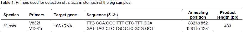
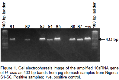
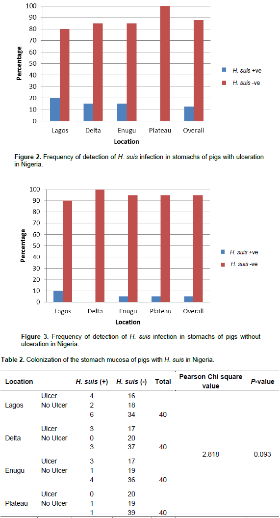
A 433 bp fragment of the 16S rRNA-coding gene of H. suis was amplified in 8.75% (14/160) of the samples from four states in Nigeria (Figure 1). This included 15% of the samples from Lagos State (6/40), 7.5% from Delta State (3/40), 10% from Enugu State (4/40) and 2.5% from Plateau State (1/40). H. suis was detected with overall occurrence of 12.5 and 5% in the stomachs with ulceration and without gross ulceration, respectively (Figures 2 and 3). There was no significant association between the occurrence of gastric ulcers and H. suis infection in the stomachs (Table 2). This study provides molecular evidence of infection and the frequency of detection of H. suis in pigs in Nigeria. The widespread detection of this organism in pigs calls for further search retrospectively and prospectively into its role in the causation of disease in pigs and man in Nigeria as it is an established pathogenic and zoonotic agent (Haesebrouck et al., 2009; Flahou et al., 2017). The current findings show that H. suis detection rates in pigs in Nigeria are relatively low (8.75%) as compared to other reports from Europe and Asia, where it often exceeds 60% in slaughter-age pigs (Park et al., 2004; Hellemans et al., 2007). This variation in colonization may be due to the influence of climatic conditions and management factors since the influence of environmental factors on H. pylori colonization in humans has been previously documented (Kusters et al., 2006). The ease of transfer to and adaptation of this organism in man makes it a threat to humans in close contacts with pigs as H. suis is the most prevalent NHPH affecting humans (Haesebrouck et al., 2009). This ease of transfer has been recently attributed to its primate origin (Flahou et al., 2017). H. suis is also known to cause indirect production losses in the swine industry (De Bruyne et al., 2016). In the assessment of the spread of the organism, H. suis was detected more frequently in stomachs with gastric ulcers in the fundus (12.5%) but there was no significant association between the occurrence of gastric ulcers and H. suis infection. This may infer that although the organism is associated with formation of gastric lesions in pigs, it may not be the sole factor responsible for the ulcerations observed in the fundus of the stomach of pigs in Nigeria. Other studies have conflicting reports on role of the organism in stomach pathologies in pigs (Monteiro, 2011; Queiroz et al., 1996; Roosendaal et al., 2000). Further studies on this organism in Nigeria should include the characterization of the circulating strains and their associated lesions in pigs. In addition, the frequency of occurrence of H. suis in gastric tissues of humans suffering from gastric disease should also be studied as there is previous documented evidence on direct human infection with field strains of H. suis from pigs (Joosten et al., 2013).
CONFLICT OF INTERESTS
The authors declare that there is no conflict of interest.
ACKNOWLEDGEMENTS
Support for this work was received through the Tertiary Education Trust Fund grant for Institutional Based Research 2017 (TETFUND IBR). The authors appreciate Prof. Freddy Haesebrouck and Dr Bram Flahou of the Department of Pathology, Bacteriology and Avian Diseases, Faculty of Veterinary Medicine, Ghent University, Merelbeke, Belgium for training support and provision of the H. suis genomic DNA used as positive control. The technical support of Mr John Odeyemi of the Biotechnology Laboratory, Department of Veterinary Medicine, University of Ibadan, is appreciated.
REFERENCES
|
Choi YK, Han JH, Joo HS (2001). Identification of novel Helicobacter species in pig stomachs by PCR and partial sequencing. J. Clin. Microbiol. 39:3311-3315. |
|
|
Danon SJ, Lee A (2001). Other Gastric Helicobacters and Spiral organisms. In. Helicobacter pylori: Physiology and Genetics. Mobley HLT, Mendz GL, Hazel SL(Eds.). ASM Press, Washington D.C.. ISBN-10: 1-55581-213-9. Available at: View -accessed 03/10/2016 |
|
|
De Bruyne E, Ducatelle R, Foss D, Sanchez M, Joosten M, Zhang G, Smet A, Pasmans F, Haesebrouck F, Flahou B (2016). Oral glutathione supplementation drastically reduces Helicobacter induced gastric pathologies. Sci. Rep. 6:20169. |
|
|
De Cooman L, Houf K, Smet A, Flahou B, Ducatelle R, De Bruyne E, Pasmans F, Haesebrouck F (2014). Presence of Helicobacter suis on pork carcasses. Int. J. Food Microbiol. 187:73-76. |
|
|
De Groote D, Ducatelle R, van Doorn LJ, Tilmant K, Verschuuren A, Haesebrouck F (2000). Detection of "Candidatus Helicobacter suis" in gastric samples of pigs by PCR: comparison with other invasive diagnostic techniques. J. Clin. Microbiol. 38:1131-1135. |
|
|
De Groote D, Van Doorn LJ, Van den Bulck K, Vandamme P, Vieth M, Stolte M (2005). Detection of non-pylori Helicobacter species in "Helicobacter heilmannii"- infected humans. Helicobacter, 10(5):398-406. |
|
|
Flahou B, Rossi M, Bakker J, Langermans J, Heuvelman E, Solnick J, Martin M, O'Rourke J, Ngoan L, Hoa NX, Nakamura M, Øverby A, Matsui H, Ota H, Matsumoto T, Foss DL, Kopta LA, Omotosho O, Franciosini M, Proietti PC, Guo A, Liu H, Borilova G, Bracarense AP, Lindén SK, De Bruyckere S, Zhang G, De Witte C, Smet A, Pasmans F, Ducatelle R, Corander J, Haesebrouck F (2017). Evidence for a primate origin of zoonotic Helicobacter suis colonizing domesticated pigs. ISME J. 145:1-10. |
|
|
Flahou B, Van Deun K, Pasmans F, Smet A, Volf J, Rychlik I, Ducatelle R, Haesebrouck F (2012). The local immune response of mice after Helicobacter suis infection: strain differences and distinction with Helicobacter pylori. Vet. Res. 43:75. |
|
|
Grasso GM, Ripabelli G, Sammarco ML, Ruberto A, Iannitto G (1996). Prevalence of Helicobacter-like organisms in porcine gastric mucosa: a study of swine slaughtered in Italy. Comp. Immunol. Microbiol. Infect. Dis. 19(3):213-217. |
|
|
Haesebrouck F, Pasmans F, Flahou B, Chiers K, Baele M, Meyns T, Decostere A, Ducatelle R (2009). Gastric helicobacters in domestic animalsand nonhuman primates and their significance for human health. Clin. Microbiol. Rev. 22:202-223. |
|
|
Hellemans A, Chiers K, Maes D, De Bock M, Decostere A, Haesebrouck F, Ducatelle R (2007). Prevalence of "Candidatus Helicobacter suis" in pigs of different ages. Vet. Rec. 161:189-92. |
|
|
Joosten M, Flahou B, Meyns T, Smet A, Arts J, De Cooman L, Pasmans F, Ducatelle R, Haesebrouck F (2013). Case Report: Helicobacter suis Infection in a Pig Veterinarian. John Wiley & Sons Ltd. Helicobacter, 18:392-396. |
|
|
Konturek JW (2003). Discovery by Jaworski of Helicobacter pylori and its pathogenetic role in peptic ulcer, gastritis and gastric cancer. J. Physiol. Pharmacol. 54(Suppl. 3):23-41. |
|
|
Kopta LA, Paquette, JA, Bowersock, TL, Choromanski LJ, Godbee TK, Galvin, JE, Foss DL (2010). Information of Helicobacter suis in pig-producing regions of North America. Conference of Research Workers in Animal Diseases, Chicago, Illinois, Dec 5-7. |
|
|
Kusters JG, van Vliet AHM, Kuipers EJ (2006). Pathogenesis of Helicobacter pylori infection. Clin. Microbiol. Rev. 19:449-490. |
|
|
Marshall BJ, Warren JR (1984). Unidentified curved bacilli in the stomach of patients with gastritis and peptic ulceration. Lancet, 1311-1315. |
|
|
Monteiro SAPS (2011). Prevalence and risk factors for gastric ulcers in swine. Porto: Universidade do Porto. [Online] View. Accessed on 10 August, 2015. |
|
|
Morgner A, Lehn N, Andersen LP, Thiede C, Bennedsen M, Trebesius K, Neubauer B, Stolte M, Bayerdörffer E (2000). Helicobacter heilmannii-associated primary gastric low-grade MALT lymphoma: complete remission after curing the infection. Gastroenterology, 118:821-828. |
|
|
Park JH, Seok SH, Cho SA, Baek MW, Lee HY, Kim DJ, Park JH (2004). The high prevalence of Helicobacter sp. in porcine pyloric mucosa and its histopathological and molecular characteristics. Vet. Microbiol. 104(3-4):219-25 |
|
|
Pasmans F, Haesebrouck F (2014). Presence of Helicobacter suis on pork carcasses. Int. J. Food Microbiol. 187:73-76 |
|
|
Proietti PC, Bietta A, Brachelente C, Lepri E, Davidson I and Franciosini MP (2010). Detection of Helicobacter spp. in gastric, fecal and saliva samples from swine affected by gastric ulceration. J. Vet. Sci. 11(Suppl. 3):221-225. |
|
|
Queiroz DMM, Rocha GA, Mendes E, Moura SB, Rocha De Oliveira AM, Miranda D (1996). Association between Helicobacter and gastric ulcer disease of the pars oesophagea in swine. Gastroenterology, 111:19-27. |
|
|
Roosendaal R, Vos JH, Roumen T, van Vugt R, Cattoli G, Bart A, Klaasen HL, Kuipers EJ, Vandenbroucke-Grauls CM, Kusters JG (2000). Slaughter pigs are commonly infected byclosely related but distinct gastric ulcerative lesion-inducing gastrospirilla. J. Clin. Microbiol. 38:2661-2664. |
|
|
Stolte M, Bayerdörffer E, Morgner A, Alpen B, Wündisch T, Thiede C, Neubauer A (2002). Helicobacter and gastric MALT lymphoma. Gut. 50(Suppl 3):19-24. |
|
|
Van den Bulck K, Decostere A, Baele M, Driessen A, Debongnie JC, Burette A, Stolte M, Ducatelle R, Haesebrouck F (2005). Identification of non-Helicobacter pylori spiral organisms in gastric samples from humans, dogs and cats. J. Clin. Microbiol. 43(5):2256-2260. |
|
Copyright © 2024 Author(s) retain the copyright of this article.
This article is published under the terms of the Creative Commons Attribution License 4.0


