Full Length Research Paper

ABSTRACT
This study histologically and histochemically assess the effect of ethanol fruit extract of Phoenix dactylifera L. (EFPD) on the cerebral cortex of lead acetate exposed Wistar rats. Twenty rats were grouped into five groups (A to E, n=4). Group A (control) was administered distilled water (2 ml/kg), while groups B to E were treatment groups. Cerebral damage was induced in rats by the administration of lead acetate (120 mg/kg). Groups B, C, D and E were administered lead acetate (120 mg/kg) for a period of 3 weeks, after which groups C and D were administered EFPD (500 and 1000 mg/kg, respectively) and group E was administered dimercaptosuccinic acid (10 mg/kg) for 2 weeks. All administrations were via oral route, once daily. Microscopic examination of cerebral sections of lead acetate-treated rats revealed histo-architectural alteration; cortical degenerative changes, such as, necrosis, satellitosis, vacuolation and neuronal cytoplasmic shrinkage. However, administration of EFPD remarkably ameliorated lead acetate-induced cortical cerebral degenerative changes in the rats, in a dose dependently manner, as compared to the reference drug dimercaptosuccinic acid. Results suggest that EFPD is a potential therapeutic agent against lead acetate-induced cortical cerebral alterations in Wistar rats.
Key words: Cerebrum, lead acetate, Phoenix dactylifera L, Wistar rats.
INTRODUCTION
Human and animal populations interact with their environment via food, air and water on a daily basis, as such exposes them to toxic substances, such as chemicals and heavy metals, capable of causing harm or even death (Wade et al., 2002; Burger et al., 2013). Heavy metals are natural constituents of the earth crust, their biochemical balance are easily altered by human needs for improving quality of live and well-being (Das et al., 2014). Once present in the environmenteven in trace amount can pose a serious problem for all organism and prolong exposure creating a deleterious health effect in humans, since they cannot be degraded or destroyed (Chen and Chen, 2001; Sedbrook, 2016). Lead is one of the common toxic heavy metals due to the ease in mining and refining. It is used in building construction, making of water pipes, lead-acid batteries, bullets and shot, weights, as part of solders, pewter’s fusible alloys, as well as radiation shield (Duah et al., 2012). The wide use of lead had turned lead poisoning into an ever present environmental and health challenge, hence the increased blood-lead level (Ahmed et al., 2013). Lead exposure affect both central and peripheral nervous system resulting to nerve cell degeneration and demyelination (Sanders et al., 2009; Abeer, 2012; Assi et al., 2016).
The cerebrum is rostral most part of the brain responsible for higher brain function, such as motor movements, perception of stimuli, emotions, problem solving and recognition (Singh, 2002; Owolabi et al., 2014). The cerebrum is vulnerable to damage from a variety of sources such as developmental defects, degenerative diseases, infectious processes, trauma and tumors (Klementiev et al., 2007). Heavy metals exposure which lead happen to be one, have been reported to be one of the leading cause of cerebral injuries (Korogi et al., 2011; Fonfria et al., 2005; Wagner et al., 2010; Owoeye and Farombi, 2015).
Pharmacotherapy and psychoactive drugs in the last two decades have gain recognitions, due to its efficacy in the management of neurological related disorders. However, several studies have revealed that such relieve are temporal with manifestation of various side effects (Handa, 1995; Mireille et al., 2017). Traditional medical practice has gained interest in the world over due to the wide spread usage of medicinal plants and its consumption, especially in developing countries (Ashafa and Olunu, 2011; Sujith et al., 2012).
Phoenix dactylifera L. (date palm) and its various parts are widely used in folk medicine for the treatment of various ailment and disorders, such as memory disturbance, fever, inflammation, paralysis, and even nervous disorders (Nadkarni, 1976; Elgindi et al., 2015; Alhaider et al., 2017). Several researchers have documented on the rich nutritional value, high dietary fibre and essential mineral of date palm, such as phosphorus, iron, potassium and a significant amount of calcium and vitamins (Mohamed and Al-Okbi, 2004; Usama et al., 2009; Yusuf et al., 2017). Several studies on extracts of date palm have indicated the presence of antioxidant properties (Mansouri et al., 2005; Al-Qarawi et al., 2008; Agbon et al., 2016); these antioxidant activities are attributed to a wide range of phenolic and flavonoid compounds and some Vitamin in date palm (Vayalil, 2012; Benmeddourt et al., 2013).
The aim of this study was to histologically and histochemically assess the therapeutic effect of ethanol fruit extract of P. dactylifera (EFPD) against lead acetate-induced cerebral alterations in Wistar rats.
MATERIALS AND METHODS
Plant collection and identification
Dried P. dactylifera (date palm) fruits were obtained at a local market (Samaru) in Zaria, Nigeria and was authenticated and given a Voucher Specimen Number of 7130, at the Herbarium Unit of the Department of Botany, Faculty of Life Sciences, Ahmadu Bello University, Zaria, Kaduna State, Nigeria.
Extract preparation and phytochemistry
Extraction of P. dactylifera fruit and phytochemical screening were conducted in the Department of Pharmacognosy and Drug Development, Faculty of Pharmaceutical Sciences, Ahmadu Bello University, Zaria. The method of maceration as reported by Agbon et al. (2013) for the preparation of ethanol fruit extract of P. dactylifera was adopted. The method of Trease and Evans (2002) as reported by Oni et al. (2015) was adopted for photochemical screening.
Experimental animals
Twenty Wistar rats (male and female; 100 to 180 g) were obtained from Animal House of the Department of Human Anatomy, Faculty Basic Medical Sciences, College of Health Sciences, Ahmadu Bello University, Zaria and housed in new wired cages in the same animal house were rats acclimatized for two weeks prior to the commencement of the experiment. The rats were separated into five groups; one control and four treatment groups. The rats were housed under standard laboratory condition, light and dark cycles of 12 h and were provided with standard rodent pellet diet and water ad libitum. The treatment groups were administered, in addition to feed and water, lead acetate/EFPD/DMSA for a period of five weeks. The rats were weighed before and after the experiment and weight changes were computed and analysed.
Drug
Lead acetate (Analytical) manufactured by British Drug Houses (BDH) Laboratory Chemicals Division, Poole, England, was obtained and used as neurotoxin for the experiment.
Dimercaptosuccinic acid (DMSA, Analytical) manufactured by Best of Chemical (BOC) Sciences, New York, USA was obtained and used for the experiment as standard chelating drug.
Experimental procedure
Twenty Wistar rats were grouped into five groups (A to E) of four rats each. Group A (control) was administered distilled water (2 ml/kg), while groups B to E were treatment groups. Cerebral damage was induced in rats by the administration of lead acetate (120 mg/kg; 20% LD50) (Sujatha et al., 2011) as reported by Yusuf et al. (2017). Groups B, C, D and E were administered lead acetate (120 mg/kg) for a period of 3 weeks, after which groups C and D were administered ethanol fruit extract of P. dactylifera (500 and 1,000 mg/kg, respectively, that is, 10 and 20% of LD50 oral in rats (Agbon et al., 2017) and group E was administered DMSA (10 mg/kg) (Chen et al., 1999) for an additional period of 2 weeks. All administrations were via oral route, once daily.
Histological and histochemical studies
At the end of the experiment, rats were euthanized and brain organs were harvested. Harvested brain organs were fixed in Bouin’s fluid and tissues processed using routine histological techniques, stained with Haematoxylin and Eosin (H&E) and histochemical stains (Cresly Fast Violet [CFV] for Nissl substance and Bielschowsky for neurites/nerve fibres) for light microscopic examination.
Data analysis
Results obtained were analysed using the statistical software, Statistical Package for Social Sciences (IBM SPSS version 20.0) and results were expressed as mean ± standard error of mean (SEM). Presence of significant difference among mean of the groups was determined using one-way analysis of variance (ANOVA) with Tukey post hoc test. Paired sample t-test was employed for the comparison of means as appropriate. Values were considered significant when p Ë‚ 0.05.
RESULTS
Phytochemical analysis
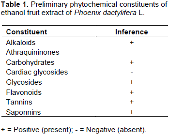
Phytochemical analysis of ethanol fruit extract of P. dactylifera (EFPD) produced positive reaction for secondary metabolite and negative for some shown in Table 1.
Physical observation
During the period of administration, physical activities of the rats were observed. Rats in the control group were observed to exhibit normal physical activities, such as movement and playfulness, whereas rats in the treatment groups exhibited aggressiveness and decreased activity, especially in lead acetate-treated group.
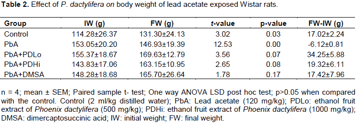
The weight of the rats in all groups were observed to have increased (p>0.05), except in the lead acetate-treated group (p<0.05) when initial and final weights were compared. However, there was no significant difference (p>0.05) in weight change (difference in initial and final weights) when treated groups were compared with the control (Table 2).
Histological and histochemical examination
Histological examination of sections of cerebral cortex of rats stained with routine (H&E) histological and histochemical (CFV and Bielschowsky) stains revealed the following.
The cerebral sections of rats in the control group revealed normal histoarchitecture of the cerebral cortex with distinctive appearance of cortical neurones arranged in six layers (I to VI). Morphology of the ganglionic layer (Layer V) revealed dense distribution of large pyramidal cells; ganglion or Betz cells and glial cells. Histochemical (CFV) staining for Nissl substance revealed normal appearance of distinct intensely stained pyramidal cells and Bielschowsky revealed normal neuronal fibres (Plate 1A).
The cerebral sections of lead acetate-treated rats revealed histo-architectural degeneration of neurons such as satellitosis, perineuronal vacuolations, necrosis and neuronal cytoplasmic shrinkage. CFV staining revealed chromatolysis, cytoplasmic shrinkage and indistinct staining intensity and Bielschowsky stain revealed pyramidal neuronal degeneration as loss of neuronal fibres when compared with the control (Plates 1B, 2B and 3B).
Examination of the cerebral sections of ethanol fruit extract of P. dactylifera (500 and 1,000 mg/kg)-treated rats, revealed mild cerebral cortex histo-architectural distortions such as perineuronal/cytoplasmic vacuolation and pkynosis; CFV staining showed reduced staining intensity and Bielschowsky staining revealed loss of neuronal fibres when compared with the severe distortions of the lead acetate-treated group (Plates 1C, 1D, 2C and 2D).
The histological features of the DMSA-treated group revealed relatively similar histo-architecture, with mild distortion, when compared with control (Plates 1E, 2E and 3E).


DISCUSSION
In this study, phytochemical analysis of ethanol fruit extract of P. dactylifera revealed the presence of secondary metabolites such as, flavonoids, saponins, tannins and alkaloids which have been reported to have neuroprotective activities (Chan et al., 2012; Wan Ismail and MohdRadzi, 2013; Hwang et al., 2015). This finding is in agreement with the reported phytochemical constituents in fruit extract of P. dactylifera L. (Faqir et al., 2012; Abiola et al., 2017).
Reduced physical activity observed among the lead acetate-treated rats, reflects treatment-related toxicity. This agrees with the findings on drug-related toxicity and physical activity as reported by Agbon et al. (2014) and Yusuf et al. (2017). Body weight changes are strong pointers of general health status and drug related toxicity in animals (Mukinda, 2007; Salawu et al., 2009).
Significant weight decrease observed in lead acetate-treated rats is indicative of heavy metal related toxicity. Heavy metals intoxication, have been implicated for poor appetite and malabsorption of nutrients in animal models (Jadhav et al., 2007; Wadaan, 2009). Finding is in accordance with the report of Yusuf et al. (2017) which observed remarkable decrease in weight in lead-exposed rats. Body weight gain observed in EFPD-treated rats could be attributed to the plant’s wide range of essential nutrients with high caloric value, particularly carbohydrates and lipids (Shaba et al., 2015; Punia, 2016; Megbo et al., 2017). This finding is in agreement with the report of Wahab et al. (2010) and Haouas et al. (2014).
Neuronal degeneration have been identified as one of the major causes of neuronal cell apoptosis (cell death), which could be as a result of disease (pathological) or natural (aging) condition (Mazanetz and Fischer, 2007; Kumar and Khanum, 2012) exerting extrinsic insults or traumatic stresses on the cells (Kumar et al., 2009; Sivanandam and thakur, 2012). Characteristic neuronal histo- and cyto-architectual distortions, such as neuronal shrinkage, perineuronal vacuolation, satellitosis, and indistinct staining intensity, loss of pyramidal neuron processes, necrosis and pkynosis observed in this study are indicators of neurodegenerative changes. Satellitosis observed in this study is indicative of treatment related toxicity, a condition marked by an accumulation of neuroglia cells around damaged or necrosed neurones of the central nervous system, often as a prelude to neuronophagia (Manickam et al., 2009). Findings are in consistence with the reports on neurodegenerative changes following heavy metals (lead, mercury, cadmium) exposure. These metals have capacity to induce nervous tissue insult (Amal and Mona, 2009; Fakunle et al., 2013; Wani et al., 2015; Butt et al., 2018) and disrupt the release mechanism of calcium-dependent neurotransmitter and DNA repairs (Hartwig, 1994; Sharma et al., 2014).
In light microscopy, rough endoplasmic reticulum and free ribosomes appear as basophilic granular areas (Nissl bodies) with Cresy Fast Violet staining. Neuronal degeneration has been related to reduction in Nissl substances (Akassoglou et al., 2004) involving degradation of β-tubulin, an important component of the neuronal cytoskeleton, and these effects are evoked by N-methyl D-aspartate receptor (NMDAR) function (Xu et al., 2012). Findings in these studies are in accordance with the reports of Ajibade et al. (2012) and Agbon et al. (2014) with loss of Nissl substance in cerebellar neurones and shrinkage of the nucleus following chemically induced toxicity. The brain among other tissues and organ with high lipid content is the most sensitive target of heavy metal intoxication. Lead neurotoxic effect results from its ability to cross the blood brain barrier readily and influence alteration of certain membrane bound enzymes responsible for protecting the biological systems against reactive oxygen species (ROS) and oxidative stress (Xu et al., 2008; Yun et al., 2011; Fakunle et al., 2013; Ibegbu et al., 2013). Naqi (2015) and Owolabi et al. (2017) reported cortical cerebellar histo-architectural distortions on exposure to lead in rats. Bielschowsky staining technique demonstrates neuronal processes of the central nervous system. Loss of neuronal processes observed in the lead-treated rats is indicative of treatment related toxicity. Heavy metals have been reported to disrupt micro-skeletal like structures in the brain cells (Leong et al., 2001).
Neuronal injury may result in reversible or irreversible cell damage or cell death (Kumar et al., 2006). Mild cortical cerebral histo-architectural distortion in rats treated with DMSA and EFPD (500 and 1000 mg/kg) after exposure to lead acetate, was observed when compared with the severe histo-architectural distortions observed in the lead acetate-treated group.
In this study, histo-architectural features of DMSA (succimer)-treated group were comparable with the control. Thus, indicates the therapeutic activity of succimer. Succimer is an established metal ion chelating agent recommended for the treatment of heavy metal toxicity, such as lead (Kalia and Flora, 2005; Lowry, 2010).
Natural agents with antioxidant properties are beneficial in attenuating drug-induced oxidative stress in biological systems (Musa et al., 2012; Bauchi et al 2016). P. dactylifera antioxidant activities are attributed to a wide range of phenolic phytochemical constituents (Vayalil, 2012; Benmeddourt et al., 2013). Flavonoid, a polyphenolic compound, found in P. dactylifera fruits have been reported to have strong ROS scavenging and metal ions chelating activities; an antioxidant that shields neurones from lethal damage and has ability to suppress neuroinflammation (Pujari, 2011; Rice, 2001; Komaki et al., 2015). Findings are in line with the reported ameliorative potentials of P. dactylifera fruit extract; Agbon et al. (2017) reported ameliorative activity of ethanol fruit extract against inorganic mercury induced cerebral and cerebellar alterations in Wistar rats and Yusuf et al. (2017) reported neuroprotective activity on lead acetate-induced toxic effects in cerebellum. Thus, indicating the therapeutic potentials of EFPD on heavy metals induced cortical cerebral alterations.
CONCLUSION
In light of the observed results of the present study, it could be concluded that ethanol fruit extract of P. dactylifera possess therapeutic potentials against lead acetate-induced cortical cerebral alterations in Wistar rats. The therapeutic property of the extract, comparable to the standard drug, DMSA, could be attributed to the antioxidant properties of its constituent phytochemicals, such as flavonoid.
CONFLICT OF INTERESTS
The authors have not declared any conflict of interests.
ACKNOWLEDGMENTS
The authors thank the Ahmadu Bello University, Zaria, Kaduna State, Nigeria for providing an enabling environment to conduct this study and appreciate the following research assistants: Ivang, Andrew, Usman, M. Ibe, and Makena Wusa for their technical support and the Department of Human Anatomy Department, Faculty of Basic Medical sciences, College of Health Sciences, Ahmadu Bello University, Zaria for providing the facilities to conduct this study.
REFERENCES
|
Abeer M (2012). Grape seed extract (Vitisvinifera) alleviate neurotoxicity and hepatotoxicity induced by lead acetate in male albino rats. Journal of Behavioral and Brain Science 2(02):176-184. |
|
|
Abiola T, Dibie DC, Akinwale OJ, Shomuyiwa OA (2018). Assessment of the antidiabetic potential of the ethanolic extract of date palm (Phoenix Dactylifera) seed in alloxan-induced diabetic rats. Journal of Diabetes and Metabolism 9(784):1-9. |
|
|
Agbon AN, Abubakar MG, Enemeli FU, Mahdi O, Bobb KA, Sule H, Yahaya MH and Okah CC (2017). Assessment of Ethanol Fruit extract of Phoenix dactylifera L. (Date Palm) on mecury chloride - induced cerebral and cerebellar alterations in wistar rats damage in Wistar rats. Journal of Anatomy 8(1):188-201. |
|
|
Agbon AN, Ingbian SD, Dahiru AU (2014). Preliminary histological and histochemical studies on the neuroprotective effect of aqueous fruit extract of Phoenix dactyliferaL. (Date Palm) on atesunate-induced cerebellar damage in wistar rats. Sub-Saharan African Journal of Medicine 1(4):204. |
|
|
Agbon AN, Kwaneshie HO, Hamman WO (2013). Antidiarrheal activity of aqueous fruit extract of Phoenix dactylifera (date palm) in Wistar rats. British Journal of Pharmacology and Toxicology 4(3):121-127. |
|
|
Agbon AN, Kwaneshie HO, Hamman WO, Ibegbu AO, Akpulu PS, Ogirima NA, Festus F (2016). Histological and histochemical studies on the neuroprotective effect of aqueous fruit extract of Phoenix dactylifera (date palm) on mercury-induced cerebellar damage in Wistar rats. Journal of Anatomical Sciences 7(2):44-54. |
|
|
Ajibade AJ, Fakunle PB, Shallie PD (2012). Some histological observations and microstructural changes in the nissl substances in the cerebellar cortex of adult wistar rats following artesunate administration. Current Research in Neuroscience 2:1-10. |
|
|
Alhaider IA, Mohamed ME, Ahmed KKM, Kumar AHS (2017). Date palm (Phoenix dactylifera) fruits as a potential cardioprotective agent: The role of circulating progenitor cells. Frontiers in Pharmacology 8:592:1-11. |
|
|
Al-Qarawi AA, Abdel-Rahman H, Mousa HM, Ali BH, El-Mougy SA (2008). Nephroprotective action of Phoenix- dactylifera, Inin gentamicin-induced nephrotoxicity. Pharmaceutical Biology 46(4):227-230. |
|
|
Amal EA, Mona HM (2009). Protective effect of some antioxidants on the brain of adult male albino rats, Rattus rattus, exposed to heavy metals. Bioscience Research 6(1):12-19. |
|
|
Ashafa AO, Olunu O (2011). Toxicological evaluation of ethanolic root extract of Morinda lucida (L.) Benth. (Rubiaceae) in male Wistar rats. Journal of Natural Pharmaceuticals 2(2):108-108. |
|
|
Assi MA, Hezmee MNM, Haron AW, Sabri MY, Rajion MA (2016) The detrimental effects of lead on human and animal health. Veterinary World 9(6):660-671. |
|
|
Bauchi ZM, Kizito D, Alhassan AW, Akpulu SP, Timbuak JA (2016). Effect of aqueous seed extract of Nigella sativa on lead-induced cerebral cortex toxicity in lLong Evans rats. Bayero Journal of Pure and Applied Sciences, 9(1):48-52. |
|
|
Benmeddour Z, Mehinagic E, Le -Meurlay D, Louaileche H (2013). Phenolic composition and antioxidant capacities of ten Algerian date (Phoenix dactylifera L.) cultivars: a comparative study. Journal of Functional Foods 5(1):346-354. |
|
|
Burger P, Hardy F, Bohmer AE, Aoki D, Wolf T, Schweiss P, Heid R, Adelmann P, Yao YX, Kotliar G, Schmalian J, Meingast C (2013). Evidence of Strong Correlation and Coherence-Incoherence Crossover in the Iron Pnictide Superconductor. Physical Review Letters 111(2):027002. |
|
|
Butt UJ, Ali-Shah SA, Ahmed T, Zahid S (2018). Protective effects of Nigella sativa L. seed extract on lead induced neurotoxicity during development and early life in mouse models. Toxicological Research 7:32-40. |
|
|
Chen S, Golemboski KA, Sanders FS, Dietert RR (1999). Persistent effect of in utero meso-2,3, 3-dimercaptosuccinic acid (DMSA) on immune function and lead-induced immunotoxicity. Toxicology. 132:67-79. |
|
|
Chen YC, Chen MH (2001). Heavy metal concentrations in nine species of fishes caught in coastal waters off Ann-Ping, SW Taiwan. Journal of Food and Drug Analysis 9(2):219. |
|
|
Duah S (2012). Determining the Level of Heavy Metal in Orange (Citrus sinensis) within AngloGold Ashanti, Obuasi Mine. |
|
|
Elgindi MR, Singab AN, El-Taher EMM, Kassem MES (2015). A Comprehensive review of phoenix (Arecaceae). RJPBCS 6(3):966-974. |
|
|
Faqir MA, Sardar IB, Ahmad HE, Muhammad IK, Muhammad N (2012). Phytochemical characteristics of date palm (Phoenix dactylifera) fruit extracts. Pakistan Journal of Food Sciences 223:117-127. |
|
|
Fonfria E, Vilaró MT, Babot Z, Rodríguezâ€Farré E, Sunol C (2005). Mercury compounds disrupt neuronal glutamate transport in cultured mouse cerebellar granule cells. Journal of Neuroscience Research 79(4):545-553. |
|
|
Handa SS (1995). Plants and plant products for mental health. Decade of the brain. Rockville, MD: US. Department of Health and Human Services pp.163-A171. |
|
|
Haouas Z, Sallem A, Zidi I, Hichri H, Mzali I, Mehdi M (2014). Hepatotoxic Effects of lead acetate in rats: histopathological and cytotoxic studies. Journal of Cytology and Histology 5(5):1000256. |
|
|
Hartwig A (1994). Role of DNA repair inhibition in lead-and cadmium-induced genotoxicity: a review. Environmental Health Perspectives, 102(Suppl 3):45. |
|
|
Hwang SL, Shih PH, Yen GC (2015). Citrus Flavonoids and Effects in Dementia and Age-Related Cognitive Decline. In Diet and Nutrition in Dementia and Cognitive Decline pp. 869-878. |
|
|
Ibegbu AO, Animok, AA, Ayuba M, Brosu D, Adamu SA, Akpulu P, Hamman WO, Umana UE, Musa SA (2013). Effect of Ascorbic Acid on Mercuric Chloride Induced Changes on the Cerebral Cortex of Wistar Rats. African Journal of Cellular Pathology 1:23-29. |
|
|
Jadhav SH, Sarkar SN, Patil RD, Tripathi HC (2007). Effects of subchronic exposure via drinking water to a mixture of eight water-contaminating metals: a biochemical and histopathological study in male rats. Archives of Environmental Contamination and Toxicology 53(4):667-677. |
|
|
Kalia K, Flora SJ (2005). Strategies for safe and effective therapeutic measures for chronic arsenic and lead poisoning. Journal of Occupational Health 47(1):1-21. |
|
|
Klementiev B, Novikova T, Novitskaya V, Walmod PS, Dmytriyeva O, Pakkenberg B, Bock E (2007). A neural cell adhesion molecule–derived peptide reduces neuropathological signs and cognitive impairment induced by Aβ25-35. Neuroscience 145(1):209-224. |
|
|
Komaki A, Hoseini F, Shahidi S, Baharlouei N (2015). Study of the effect of extract of Thymus vulgaris on anxiety in male rats. Journal of Traditional and Complementary Medicine pp.1-5. |
|
|
Korogi Y, Takahashi M, Shinzato J, Okajima T (1994). MR findings in seven patients with organic mercury poisoning (Minamata disease). American Journal of Neuroradiology 15(8):1575-1578. |
|
|
Kumar GP, Khanum F (2012). Neuroprotective potential of phytochemicals. Pharmacognosy Reviews, 6(12):81. |
|
|
Kumar V, Abbas AK, Fausta NR (2006). Cotran. Pathological Basis of Diseases. 7th edition: Elsevier Singapore P 289. |
|
|
Kumar V, Abbas AK, Fausto N, Aster JC (2009). Robbins and cotran pathologic basis of disease, Professional Edition: Expert Consult-Online. St. Louis, MO: Elsevier Health Sciences. |
|
|
Liu JT, Chen BY, Zhang JQ, Kuang F, Chen LW (2015). Lead exposure induced microgliosis and astrogliosis in hippocampus of young mice potentially by triggering TLR4–MyD88–NFκB signaling cascades. Toxicology Letters 239(2):97-107. |
|
|
Lowry JA (2010). Oral chelation therapy for patients with lead poisoning. American Academy of Pediatrics 116:1036-1046. |
|
|
Manickam B, Sajitha IS, Lakshmi R, Nair ND (2009). Prevalence, gross and histopathological study of brain disorders in Cattle-Kerala State, India. International Journal of Tropical Medicine 4(1):9-20. |
|
|
Mansouri A, Embarek G, Kokkalou E, Kefalas P (2005). Phenolic profile and antioxidant activity of the Algerian ripe date palm fruit (Phoenix dactylifera). Food Chemistry 89(3):411-420. |
|
|
Mazanetz MP, Fischer, PM (2007). Untangling tau hyperphosphorylation in drug design for neurodegenerative diseases. Nature Reviews Drug Discovery 6(6):464. |
|
|
Megbo BC, Samuel AM, Dio DW (2017). Phoenix Dactylifera fruit: A Nutraceutical Agent In the treatment of Diarrhea. Innovat International Journal of Medical and Pharmaceutical Sciences 2:3. |
|
|
Mireille KP, Desire DDP, Pierre K (2017). Male Sexual Disorders in Patients with Parkinson Disease: Treatment with Natural Remedies. Advances in Tissue Engineering and Regenerative Medicine 3(2):00061. |
|
|
Mohamed DA, Al-Okbi SY (2004). In vivo evaluation of antioxidant and anti-inflammatory activity of different extracts of date fruits in adjuvant arthritis. Polish Journal of Food and Nutrition Sciences 13:397-402. |
|
|
Mukinda JT, Syce JA (2007). Acute and chronic toxicity of the aqueous extract of Artemisia afra in rodents. Journal of Ethnopharmacology 112(1):138-144. |
|
|
Musa SA, Omoniye IM, Hamman WO, Ibegbu AO, Umana UE (2012). Preventive activity of ascorbic acid on lead acetate induced cerebellar damaged in adult wistar rats. Medicine and health JournalsMed and Health Journal 13:99-104. |
|
|
Nadkarni IM (1976). Popular Prakashan Pvt Ltd. Tardeo, Mumbai. 400:034. |
|
|
Naqi SZ (2015). A comparative study of the histological changes in cerebral cortex, hippocampus, cerebellum, pons and medulla of the albino rat due to lead toxicity. International Journal of Anatomy and Research 3(2):1173-1178. |
|
|
Oni SO, Adeosun AM, Ladokun OA, Ighodaro OM, Oyedele MO (2015). Nutritional and phytochemical profile of niger cultivated date palm (Phoenix dactilyfera L). Journal of Food and Nutrition Sciences 3:114e118. |
|
|
Owoeye O, Farombi EO (2015). Tomato pomace protects against mercuric chloride-induced neurodegeneration and motor abnormality in adult rat. International Journal of Biological and Chemical Sciences 9(3):1142-1153. |
|
|
Owolabi J, William F, Olanrewaju J, Etibor T, Fabiyi O (2014). Histomorphological Evidences of Moringa oleifera's Ameliorative Effects against Lead Toxicity in Cerebral Cortex. World Journal of Life Sciences and Medical Research 3(2):53. |
|
|
Owolabi JO, Ogunnaike PO, Adeyeye JA (2017). Lead poisoning causes histoarchitectural disruptions in blood marrow, brain regions and muscles; Histological Observations of Lead Poisoning Effects on Vital Body Tissues of Murine Models: Part I. Pharmaceutical Chemistry Journal 4(5):164-173. |
|
|
Pujari RR, Vyawahare NS, Kagathara VG (2011). Evaluation of antioxidant and neuroprotective effect of date palm (Phoenix dactylifera L.) against bilateral common carotid artery occlusion in rats. Indian Journal of Experimental Biology, 49(8):627-633. |
|
|
Punia D (2016). Nutritional composition of fruit of four date palm (Phoenix dactylifera L.) cultivars grown in Haryana, India. Asian Journal of Dairy and Food Research 35(4). |
|
|
Rice Evans CA (2001). Flavonoids antioxidants. Current Medicinal Chemistry 8:797. |
|
|
Salawu OA, Chindo BA, Tijani AY, Obidike IK, Salawu TA, Akingbasote AJ (2009). Acute and sub-acute toxicological evaluation of the methanol stem bark extract of Crossopteryx febrifuga in rats. African Journal of Pharmacy and Pharmacology 3:621-626. |
|
|
Sanders T, Liu Y, Buchner V, Tchounwou PB (2009). Neurotoxic effects and biomarkers of lead exposure: A review. Reviews on Environmental Health 24(1):15-46. |
|
|
Sedbrook D (2016). Everything you need to know about lead. Natural Resources Defense Council (NRDC), USA. View. |
|
|
Shaba EY, Ndamitso MM, Mathew JT, Etsunyakpa MB, Tsado AN, Muhammad SS (2015). Nutritional and anti-nutritional composition of date palm (Phoenix dactylifera L.) fruits sold in major markets of Minna Niger State, Nigeria. African Journal of Pure and Applied Chemistry 9(8):167-174. |
|
|
Sharma B, Singh S, Siddiqi NJ (2014). Biomedical Implications of Heavy Metals Induced Imbalances in Redox Systems. BioMed Research Internationa 640(75)4-26. |
|
|
Singh S, Bohn D, Carlotti AP, Cusimano M, Rutka JT, Halperin ML (2002). Cerebral salt wasting: truths, fallacies, theories, and challenges. Critical care Medicine 30(11):2575-2579. |
|
|
Sivanandam TM, Thakur MK (2012). Traumatic brain injury: a risk factor for Alzheimer's disease. Neuroscience and Biobehavioral Reviews 36(5):1376-1381. |
|
|
Sujatha S, Joseph B (2011). Effect of few marine sponges and its biological activity against Aedes aegypti Linn. Musca domestica (Linnaeus, 1758) (Diptera: Culicidae). Journal of Fish.eries and Aaquatic. Science, 6(2): 170-177. |
|
|
Sujith K, Darwin CR, Suba V (2012). Memory-enhancing activity of Anacyclus pyrethrum in albino Wistar rats. Asian Pacific Journal of Tropical Disease 2(4):307-311. |
|
|
Trease GE, Evans WC (2002). Phytochemicals In: Pharmocognosy, 15th Edn., WB Sanders Publishers London. |
|
|
Usama B, El-Gazzar AH, El-Far Hussein AA (2009). The Ameliorative effect of Phoenix dactylifera extract on CCl4 hepatotoxicity in New Zealand rabbits. Journal of Applied Sciences Research 5(9):1082-1087. |
|
|
Vayalil PK (2012). Date fruits (Phoenix dactylifera Linn): An emerging medicinal food. Critical reviews in food science and nutrition 52(3):249-271. |
|
|
Wade MG, Foster WG, Younglai EV, McMahon A, Leingartner K, Yagminas AL, Hughes CL (2002). Effects of subchronic exposure to a complex mixture of persistent contaminants in male rats: Systemic, immune, and reproductive effects. Toxicological Sciences 67(1):131-143. |
|
|
Wagner C, Vargas AP, Roos DH, Morel AF, Farina M, Nogueira CW, Rocha JB (2010). Comparative study of quercetin and its two glycoside derivatives quercitrin and rutin against methylmercury (MeHg)-induced ROS production in rat brain slices. Archives of Toxicology 84(2):89-97. |
|
|
Wahab AA, Mabrouk MAA, Joro JM, Oluwatobi SE, Bauchi ZM, John AA (2010). Ethanolic extract of Phoenix dactylifera L. prevents lead induced hematotoxicity in rats. Continental J. Biomedical Sciences 4:10-15. |
|
|
Wan Ismail WI, Mohd Radzi MNF (2013). Evaluation on the Benefits of Date Palm (Phoenix dactylifera) to the Brain. Alternative and Integrative Medicine, 2(4):2-3. |
|
|
Wani A, Ara A, Usmani JA (2015). Lead toxicity: A review. Interdisciplinary Toxicology 8(2):55-64. |
|
|
Xu F, Farkas S, Kortbeek S, Zhang FX, Chen L, Zamponi GW, Syed NI (2012). Mercury-induced toxicity of rat cortical neurons is mediated through N-methyl-D-Aspartate receptors. Molecular Brain 5(1):30. |
|
|
Xu J, Lian LJ, Wu C, Wang XF, Fu WY, Xu LH (2008). Lead induces oxidative stress, DNA damage and alteration of p53, Bax and Bcl-2 expressions in mice. Food and chemical toxicology, 46(5):1488-1494. |
|
|
Yun H, Kim I Kwon SH, Kang JS, Om AS (2011). Protective effect of Chlorella vulgaris against lead-induced oxidative stress in rat brains. Journal of Health Science 57(3):245-254. |
|
|
Yusuf AO, Buraimoh AA, Agbon AN, Raji KB, Akpulu PS (2017). Preliminary Histological Studies on the Effect of Aqueous Fruit Extract of phoenix dactylifera L. (Date Palm) on Lead Acetate-Induced Cerebellar Damages in Wistar Rats. African Journal of Cellular Pathology 8(1):1-8. |
|
Copyright © 2024 Author(s) retain the copyright of this article.
This article is published under the terms of the Creative Commons Attribution License 4.0


