African Journal of
Environmental Science and Technology

  • Abbreviation: Afr. J. Environ. Sci. Technol.
  • Language: English
  • ISSN: 1996-0786
  • DOI: 10.5897/AJEST
  • Start Year: 2007
  • Published Articles: 1126

Full Length Research Paper

Characterization of cyanobacteria microcystins (cyanotoxins) blooming in the Dams of Northern Morocco

Mustapha Ouhsassi
  • Mustapha Ouhsassi
  • Laboratory of Biology and Health, Biotechnology and Applied Microbiology, Faculty of Sciences, Abdelmalek Essaadi University, Tétouan, Morocco.
  • Google Scholar
El Ouardy Khay
  • El Ouardy Khay
  • Laboratory of Biology and Health, Biotechnology and Applied Microbiology, Faculty of Sciences, Abdelmalek Essaadi University, Tétouan, Morocco.
  • Google Scholar
Anass El Laghdach
  • Anass El Laghdach
  • Laboratory of Water, Environmental Studies and Analysis, Department of Chemistry, Faculty of Sciences, Abdelmalek Essaâdi University, Tétouan, Morocco.
  • Google Scholar
Farid Ben Abdelouahab
  • Farid Ben Abdelouahab
  • Laboratory Materials and Radiation, Department of Physique, Faculty of Science, Tétouan, Faculty of Science, Abdelmalek Essaâdi University, BP 2121, Tétouan 93002, Morocco.
  • Google Scholar
Abdeltif El Ouahrani
  • Abdeltif El Ouahrani
  • Laboratory of Biology and Health, Biotechnology and Applied Microbiology, Faculty of Sciences, Abdelmalek Essaadi University, Tétouan, Morocco.
  • Google Scholar
Mohamed Idaomar
  • Mohamed Idaomar
  • Laboratory of Biology and Health, Biotechnology and Applied Microbiology, Faculty of Sciences, Abdelmalek Essaadi University, Tétouan, Morocco.
  • Google Scholar
Jamal Abrini
  • Jamal Abrini
  • Laboratory of Biology and Health, Biotechnology and Applied Microbiology, Faculty of Sciences, Abdelmalek Essaadi University, Tétouan, Morocco.
  • Google Scholar


  •  Received: 10 December 2020
  •  Accepted: 17 February 2021
  •  Published: 31 March 2021

 ABSTRACT

Cyanobacteria thrive in eutrophic freshwaters and impose a serious problem for the management of water bodies. Some Cyanobacteria species impose even a risk for public health due to the production of intracellular toxins. This study is a qualitative approach to determine the degree of toxicity and the toxicological aspect of cyanotoxins in order to setup a monitoring program for cyanobacteria blooms and the management of cyanotoxins thriving in three water bodies in Northern Morocco. Water samples were collected from three major water reservoirs/dams near the city of Tétouan (SMIR, BELMEHDI and NAKHLA). These water samples were screened for possible Cyanobacteria using specific culture media (BG13 & Z8). Three cyanobacteria species (Microcystis aeruginosa, Pseudanabaena galeata and Oscillatoria tenuis) were isolated, purified and lyophilized. Using gas chromatography coupled with mass spectrometry, nine types of microcystins were characterized namely: (MC-LR); (MC-YR); (MC-LA); (MC-FR); (MC-RF), [Mser7]MC-LR; [Dha7]MC-LR; MC-YAba; and [Mser7]MC-YR. Our results strongly recommend and urge different stakeholders to consider the various health risks potentially generated by these toxins during water use and management. In addition, this study is a contribution to raise awareness of the toxicological aspect of the cyanobacteria inhabiting the water bodies of Northern Morocco.
 
Key words: Blue algae, gas chromatography coupled to mass spectrum, bio-toxins, water dam.


 INTRODUCTION

The problem of water quality degradation in dams and reservoirs is due essentially to different sources of pollution that cause nutritive elements (nitrogen and phosphorus) enrichment causing the anarchic development of algae, which indicates an advanced state of water quality degradation. Furthermore, soil erosion brings additional elements that may accelerate the alteration of water quality (Issaka and Ashraf, 2017; Rose et al., 2010). The ecosystem imbalance caused by such phenomenon promotes the development of algae, in particular blue algae (cyanobacteria) which are responsible for the organoleptic and esthetic alteration of water, as well as the production of cyanotoxins within these waters (self-purification phenomenon).
 
Previous works on lakes and reservoirs located in warm climate zones, such as dams in the Mediterranean, show that the latter are distinguished by particular hydrological, physico-chemical and biological characteristics (Loudiki, 1990; Loudiki et al., 1994; Cherifi and Loudiki, 2002). Among the determining factors, the unpredictability of the climate (flash floods, droughts and very variable low water levels) and the irregularity of rainfall and erosion materials play a predominant role.
 
Morocco is a Mediterranean country characterized by a semi-arid climate  (Perrin et al., 2014, Ouhamdouch et al. 2019), with clear spatiotemporal disparities in rainfall towards the southern region of the country. Moreover, the country is likely to experience 20% on average net reduction in rainfall by the end of this century (IPCC, 2007); this will boost cyanobacteria in inland water bodies such as dams (Gophen, 2021). 
 
In Morocco, the supply of drinking water is mainly ensured by rainfall collected in water reservoirs or dams. This strategic approach was adopted since the 1940s, in order to mobilize water resources through the construction of several large dams to provide drinking water and other services (El Ghachtoul et al., 2005). Nevertheless, these dams recognize in the summer period phenomena of eutrophication due to nutrient enrichment mainly nitrogen and phosphorus (El Ghachtoul et al., 2005).
 
In fact, recent climate change and anthropogenic impact on water environment by either intense withdrawal and diversion or chemical pollution and nutrient enrichment promoted a worldwide proliferation of cyanobacteria blooms often harmful to ecological and human health (Paerl, 2016). Certainly, the massive proliferation of cyanobacteria in dam waters is increasingly frequent phenomenon worldwide (Huisman et al., 2018), accompanied by the release of toxic substances in the form of secondary metabolites (cyanotoxins). These cyanotoxins cause harmful ecological, health and socio-economic effects leading to a degradation of water quality and a reduction in the productivity of the aquatic environment (Wiegand and Pflugmacher, 2005; Jacoby and Kann, 2007; Plaas and Paerl, 2016). 
 
Most cyanotoxins are called Microcystins. According to some studies the first Microcystin was isolated from Microcystis aeruginosa (Carmichael, 1992a; Namikoshi et al., 1992, 1995). Dittmann and Börner (2005) and Hotto et al. (2007) indicated that Microcystins are toxins produced by the most common cyanobacteria and are present in most of the world's water reservoirs. These cyanotoxins are structurally cyclic heptapeptide amino acids (Van Apeldoorn et al., 2007; Shimizu, 2014). According to the structure of Microcystins established by Chorus et al. (1999) the toxicity role of the acid group (3-amino-9-methoxy-2,6,8-trimethyl-10-phenyldeca-4,6-dienoic acid) recognized under the name Adda is primordial. In addition, several studies reveal the carcinogenic potential of Microcystins under long-term exposure (Yu, 1995; Codd, 2000; Carmichael et al., 2001; ADH, 2006).
 
Different Microcystins have been isolated from several species of cyanobacteria and several of them can be produced by a single bloom (Zastepa et al., 2017). Studies to date have focused solely on assessing the level of cyanotoxins in blooms rather than characterizing the water-soluble fractions of the toxins (Ai et al., 2020). In this context, this study is a qualitative approach to determine the degree of toxicity and the toxicological aspect of cyanotoxins in the studied reservoirs in order to setup a monitoring program for cyanobacteria blooms and the management of cyanotoxins produced in water bodies in Northern Morocco. The specific objectives are to extract and characterize the intracellular Microcystins using gas chromatography coupled with mass spectrometry analysis.


 MATERIALS AND METHODS
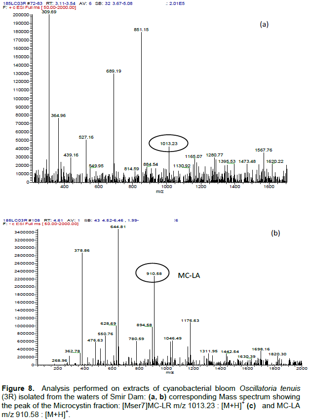
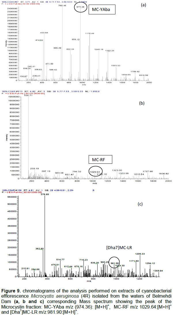
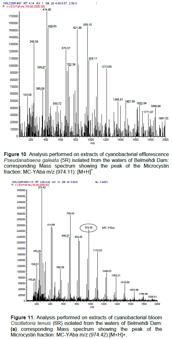
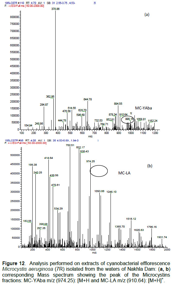
Study site
 
Water samples were collected from three major water bodies supplying drinking water to the city of Tetouan (northern Morocco). The three dams are Smir (35°40'46.9 "N 5°23'33.8 "W) (Figure 1a), Belmehdi (35°42'33.2 "N 5°30'28.0 "W) (Figure 1b) and Nakhla (35°42'01.9 "N 5°30'11.7 "W) (Figure 1c). Their characteristics are noted in Table 1. These reservoirs are experiencing the phenomenon of eutrophication due to the accumulation of two highest nutrients (nitrogen and phosphorus), which lead to the proliferation of cyanobacteria blooms, a sign of water quality degradation. 
 
Sampling method
 
Sampling was performed in the morning (around 10:00 am) on a monthly basis, from June to December 2017. Three sampling points were selected separately following the long transect of the reservoir lake. We collected approximately 50 liters of surface water from each water reservoirs. The water samples were carried in sterilized special plastic laboratory bottles of 5 L. To allow a good conservation, these bottles were transported in a cooler box of 4°C. These raw water samples were used for isolation and purification of cyanobacteria, which requires a preliminary culture in synthetic media (Saoudi, 2008).
 
Culture media and Isolation conditions
 
Two culture media BG13 (Ferris and Hirsch, 1991) and Z8 (Kotai, 1972;  Rippka  et  al., 1979) were prepared in liquid and solid forms, autoclaved for 20 min at 120°C and a pressure of 1.1 kg/cm². Afterwards, we added 100 mg of cycloheximide per litter to both liquid and solid culture media using a sterile syringe with a 0.2 µm porosity acrodisc, to remove most eukaryotes and obtain a monoalgal culture.
 
 
Water samples were filtered through Millipore membranes with a porosity of 45 µm by means of a pressurized vacuum pump (Figure 2). These membranes were transferred into two liquid media BG13 (Ferris and Hirsch 1991) and Z8 (Kotai, 1972; Rippka et al., 1979), with pH adjusted to 8, and incubated for three weeks under ambient temperature (20 - 25°C), fluorescent lamps of 2000 lumens intensity and a photoperiod of 12 h. Afterwards, these membranes were transferred into Petri dishes containing solid media (BG13 and Z8) for purification  phase.  After  three  weeks,  a  tiny  fragment  of  the growing strain was transferred into new solid media, and this step is repeated until purified strains were observed. These purified strains were then transferred to sterile bottles containing 100 ml of liquid culture media (BG13 and Z8). These bottles were sealed with a stopper of 0.2 µm porosity filter to allow airflow. By repeating these manipulations 3 to 4 times, we obtained an axenic strain after 12 weeks. The purified strains would then be subject to a preliminary morphological identification. Once verified, we transferred the axenic strains to a vial containing 2L of sterile culture medium (BG13 and Z8) under illumination and continuous aeration to obtain a large mass of each strain (massification phase). Two weeks later, the biomass of each cyanobacterial strains collected during the massification phase was centrifuged (4000 g, 20 min), then lyophilized  and  stored  at  -20°C f or  cyanotoxins  extraction.
 
 
All manipulations (transplantation, purification, and massification) were carried out under a laminar flow hood at 25°C. The materials necessary for these manipulations (Pasteur pipettes, platinum wire, and others) were sterilized. The species were observed, measured and morphologically identified using a light microscope according to criteria-based taxonomy using several specialized cyanobacterial florae and a multitude of works dealing specifically with these organisms (Bourrelly, 1985; Lund and Lund, 1995, Komárek and Anagnostidis, 2005; Komárek, 2016). This identification focused on the definition of many morphological criteria according to universally accepted identification keys:
 
(i) The color and structure of cyanobacteria (unicellular or colonial), 
(ii) The shape of the colony or trichome,
(iii) The size of the cells 
(iv) The presence or absence of gelatinous sheath (color, appearance and size), akinetes, heterocysts and gas vacuoles (pseudovacuoles). 
 
Furthermore, the morphological identification was confirmed by analyzing the conservative fatty acid composition profile according to previous studies (Passaquet et al., 1989; Hayakawa et al., 2002; Thajuddin and Subramanian, 2005; Sharathchandra and Rajashekhar, 2011; Mahapatra and Ramachandra, 2013;  Ouhsassi et al., 2017). The composition of fatty acid is used as a phylogenetic marker for cyanobacteria (Kenyon and Stanier, 1970; Murata et al., 1992; Sarsekeyeva et al., 2014; Los and Mironov, 2015). 
 
Extraction and pre-purification of Microcystins 
 
To extract and purify Microcystins, we followed the method described by Lawton et al. (1994). For each study site, the lyophilizate (250 mg) was recovered in the stationary phase from each pure culture. For each lyophilizate, the extraction is done three times with 70% methanol (Figure 3). After each extraction, the suspensions were centrifuged (4000 × g, 10 min, +4°C). The total extract was diluted with ultrapure water (milli-Q, Millipore) to obtain an extract with 20% methanol. For pre-purification of microcystins, the final extract was passed through an ODS silica gel column (Sep-Pak Vac C18, Waters Corporation, Milford, MA, USA). The last recovered fraction containing the microcystins is completely evaporated at 40°C, dissolved in 1 mL methanol /ultrapure water (50:50, v/v), and filtered through a 0.2 µm filter (Acrodisc, Nylon, Gelman Sciences Inc.) before being analyzed by gas chromatography coupled with a mass spectrometer (GC-MS).


 RESULTS

The morphological identification, confirmed by conservative fatty acid method, allows identifying three major cyanobacteria species with the same characteristics at the level of the studied water bodies (SMIR, BELMEHDI and NAKHLA): 
 
Microcystis aeruginosa, belonging to Microcystaceae, isolated from Smir (1R), Belmehdi (4R) and Nakhla (7R), is a unicellular cyanobacterium with spherical colonies; it is grouped as an envelope and floated using gaseous vacuoles (Figure 4a).
 
Pseudanabaena galeata, belonging to Pseudanabaenaceae, isolated from SMIR (2R), BELMEHDI (5R) and NAKHLA (8R), is a filamentous cyanobacterium with solitary, mobile and without sheathing trichomes. The cells are distant from each other and joined by a gelatinous bridge. There are no kinetes or heterocysts (Figure 4b).
 
Oscillatoria tenuis, belonging to Oscillatoriaceae, isolated from SMIR (3R), BELMEHDI (6R) and NAKHLA (9R), is a filamentous cyanobacteria with a free, solitary and sheathless trichome (Figure 4c). The movement and helical displacement of the apex are characteristic of this genus. The analysis of GC-MS chromatograms revealed the presence of a wide variety of Microcystins in all studied cyanobacteria strains. The data (Tables 2 to 4) regroup Microcystins elaborated by M. aeruginosa, P. galeata and O. tenuis isolated from the three studied dams: SMIR, BELMEHDI and NAKHLA. 
 
The chromatograms show the retention times recorded by different microcystins elaborated by M. aeruginosa (Figure 5), and they are: 
 
(i) MC-LR and a demethylated form MC-YAab in the case of SMIR reservoir (Figures 6a and 6b); 
(ii) MC-RF and two demethylated forms MC-YAab and [Dha7]MC-LR in the case of BELMEHDI reservoir (Figures 9b to d); 
(iii) MC-LA and a demethylated form MC-YAab in the case of NAKHLA reservoir (Figure 12b and c). 
 
In the case of P. galeata, the retention times are shown respectively for the three studied dams (Figure 5). The elaborated Microcystins by this species are:
 
(i) MC-YR and a demethylated form [Mser7]MC-YR (Figure 7a and b) in the case of SMIR reservoir; 
(ii) MC-YR and a demethylated form MC-YAab (Figure 10a) in the case of BELMEHDI dam; 
(iii) MC-LA and a demethylated form MC-YAba (Figure 13a and 13b) in the case of NAKHLA reservoir. 
 
In the case of O. tenuis, the retention times recorded by different Microcystins are shown in Figure 5. The elaborated Microcystins are: 
 
(i)  MC-LA   and    a    demethylated   form  [Mser7]MC-LR  (Figure 8a and 8b) case of SMIR dam; 
(ii) a single demethylated form MC-YAba (Figure 11a) case of BELMEHDI dam; 
(iii) two demethylated forms MC-YAba and ([Mser7]MC-LR (Figure 14a and 14b) case of Nakhla dam.


 DISCUSSION

Characterization of cyanotoxins has been performed (Poon et al., 1993; Diehnelt et al., 2005, 2006; Miles et al., 2013) using GC-MS or LC-MS or other mass spectrometry methods, which use precise mass measurements showing the connectivity of amino acids in different Microcystins. By standardizing the base peak intensity to 100%, the appearance of a mass spectrum becomes independent of the absolute amount of sample. Thus, mass spectra can be compared even when they have been generated from different sample quantities and/or different instruments. A list of m/z values (m = mass; z = atomic number) and intensity is useful for a more detailed analysis of a spectrum. The signal resulting from molecular dissociation has an ion mass (m/z) and a spectrum that normally reflects the corresponding molecular ion, usually called a molecular ion peak. The accompanying signals represent ion fragments. For example, dissociation of MC-LR exhibits a molecular ion peak break ([M.H]+, 995.5546), dissociation of MC-RR exhibits a molecular ion peak break ([M.H]+, 1038.5), and dissociation of MC-YR exhibits a molecular ion peak break ([M.H]+, 1045.5358).
 
Some authors have shown that cyanobacteria blooms in Morocco appear in summer and reach their maximum proliferation in October with a significant annual variability in biomass. Sbiyyaa (1997) and Oudra et al. (1998) indicated that the main species responsible for blooms in Moroccan dams is attributed to three main species: M. aeruginosa, M. aeruginosa flos-aquae, and P. muscicola. (Malki, 1994) are reported from the Al Massira reservoir. M. aeruginosa proliferates regularly and dominates the phytoplankton each year between November and December with a toxic bloom of cyanobacteria. Oudra et al. (2002a) reported that several species of Microcystaceae often bloom in numerous dams, and are therefore the most studied and geographically the most distributed.
 
The toxicity of a single bloom is a function of time and space. In this study, the monitoring of cyanobacterial
 
strains toxicity revealed that the studied species generate a wide variety of cyanotoxins. Toxic blooms of the genus Microcystis were demonstrated for the first time in the Oued el Mellah dam in 1997 (Loudiki  et al., 2002). As well as in other Moroccan aquatic systems, particularly in dams intended to provide drinking water to urban populations. Species of this genus are isolated from other Moroccan water reservoirs, including Imfout, Takerkoust, and Almassira (Oudra et al., 2001a, 2001b; Loudiki et al., 2002; Sabour,   2002). This genus is known to form cyanobacterial blooms in stagnant waters (Douma et al., 2010). In fact, in the studied water bodies, Microcystis spp produces more cyanotoxins compared to other species. Thus, Microsystis aeruginsa produces MC-LR and MC-YAba in SMIR dam; MC-FR, MC-RF, and [Dha7], MC-LR in BELMEHDI dam; and MC-YAba, MC- LA, and MC-YAba in NAKHLA dam. These varieties of cyanotoxins are recognized by their acute toxicity. Studies have revealed that the toxicity of Microcystis is due to the disposition of the gene coding for microcystin (Carmichael, 1995; Rouhiainen et al., 1995; Dittmann et al., 1997). P. galaeta filamentous species is also incriminated in dam waters and produces cyanotoxins also recognized by their acute toxicity either in SMIR (MC-YR, [Mser7] MC-YR), BELMEHDI (MC-YAba) and NAKHLA (MC-LA and MC-YAba). Studies in this context affirm that several species of Pseudanabaenaceae have been inventoried in Moroccan water bodies, including Pseudanabaena mucicola. This family is dominant in lakes and shallow ponds, and this dominance can sometimes  persist  throughout  the  year  in  turbid  water  bodies and when the winter is not very cold (Sas, 1989; Oudra et al., 2002a; Loudiki et al., 2002).
 
O. tenuis is also inventoried in Moroccan water bodies (Oudra et al., 2002a). This species develops variant cyanotoxins also recognized by its acute toxicity. In our study, it is found in SMIR ([Mser7]MC-LR,  MC-LA), in BELMEHDI (MC-YAba), and in NAKHLA ([Mser7]MC-LR and MC-YAba). This species is less frequent than the other species but it also has the capacity to generate large varieties of cyanotoxins.
 
Microcystis, Pseudanabaena and Oscillatoria species are the most common and potentially toxic cyanobacteria found in Moroccan freshwater and are the main producers of cyanotoxins. This is also evident in Euro-Mediterranean countries (Filatova et al., 2020), such as in France (Lac de Grand-Lieu), Portugal (Vasconcelos et al., 1996; Moreira et al., 2020), Spain (Quesada et al., 2004), Greece (Christophoridis et al., 2018), and Italy (Bruno et al., 1992), African countries such as Algeria (Saoudi et al., 2017), Ethiopia (Major et al., 2018), and Nigeria (Kadiri et al., 2020).
 
The presence of microcystins in freshwater species of the genus Oscillatoria is not only an ecological problem, but also presents a health risk when water is used for drinking purposes (Lindholm et al., 1989). On the other hand, some strains, known for their toxicological potential, are considered to be of biotechnological interest, such as Anabaena variabilis. This species is used for its hydrogen production (Yoon et al., 2006) and can remove phenols and its derivatives (Hirooka et al., 2003) as well as heavy metals (Nagase et al., 2005) from the environment and industrial wastewater (Yoon et al., 2006).


 CONCLUSION

This study contributes to the knowledge of the systematics and biogeography of toxic cyanobacteria and their toxins quality in the water bodies of Northern Morocco. It is a qualitative analysis of cyanotoxins produced by cyanobacteria species thriving in three water reservoirs near the City of Tetouan, namely SMIR, BELMEHDI and NAKHLA. The results show that the water bodies of Northern Morocco are exposed to cyanobacterial proliferation exposing these water bodies to numerous variants of Microcystins (MC-YR, [Mser7]MC-YR, MC-YAba, MC-LA, [Mser7]MC-LR, MC-FR, MC-RF, [Dha7]MC-LR) produced by the three major species (Microsystis aeruginosa, Pseudanabaena galeata, Oscillatoria tenis). These cyanotoxins are recognized by their acute toxicity and reflect a permanent threat to the health of the consumer (drinking water) mainly in late summer time. Thus, the need for action plan to monitor the levels and seasonal variations of these toxins and the biochemical factors involved directly or indirectly in the proliferation of cyanobacterial blooms.


 CONFLICT OF INTERESTS

The authors have not declared any conflict of interests.


 REFERENCES

ADH (2006). Les grands barrages au Maroc. Publication de l'administration de l'hydraulique, Rabat, Maroc 123 p.


Ai Y, Lee S, Lee J (2020). Drinking water treatment residuals from cyanobacteria bloom-affected areas: Investigation of potential impact on agricultural land application. Science of the Total Environment 706:1-11.
Crossref


Bourrelly P (1985). Les algues d'eau douce: Initiation à la systématique. Tome III: Les algues bleues et rouges. Les Eugléniens, Péridiniens et Cryptomonadines. Société Nouvelle des Editions Bouée.


Bruno MP, Gucci PMB, Pierdominici E, Sestili P, Ioppolo A, Sechi N, Volterra L (1992). Microcystin-like toxins in different freshwater species of Oscillatoria. Toxicon 30(10):1307-1311.
Crossref


Carmichael WW (1992a). Cyanobacteria secondary metabolites the cyanotoxins. Journal of Applied Bacteriology 72(6):445-459.
Crossref


Carmichael WW (1995). Toxic Microcystis and the environment. Toxic microcystis pp.1-11.


Carmichael WW, Azevedo SM, An JS, Jochimsen EM, Lau S, Eaglesham GK (2001). Human fatalities from cyanobacteria: chemical and biological evidence for cyanotoxins. Environmental Health Perspectives 109(7):663-668.
Crossref


Cherifi O, Loudiki M (2002).Variations de la structure du lac-réservoir oligotrophe Bin El Ouidane (Maroc). Revue des Sciences de l'Eau 15(1):193-208.
Crossref


Chorus I, Bartram J, Editors (1999). Toxic Cyanobacteria in Water-A Guide to Their Public Health Consequences, Monitoring, and Management, E & FN Spon, published on behalf of the World Health Organization, New York.
Crossref


Christophoridis C, Zervou SK, Manolidi K, Katsiapi M, Moustaka-Gouni M, Kaloudis T, Hiskia A (2018). Occurrence and diversity of cyanotoxins in Greek lakes. Scientific Reports 8(1):1-22.
Crossref


Codd GA (2000). Cyanobacterial toxins, the perception of water quality, and the prioritisation of eutrophication control. Ecological Engineering 16(1):51-60.
Crossref


Diehnelt CW, Dugan NR, Peterman SM, Budde WL (2006). Identification of Microcystin Toxins from a Strain of Microcystis aeruginosa by Liquid Chromatography Introduction into a Hybrid Linear Ion Trap-Fourier Transform Ion Cyclotron Resonance Mass Spectrometer. Analytical Chemistry 78(2): 501-512.
Crossref


Diehnelt CW, Peterman SM, Budde WL (2005). Liquid chromatography-tandem mass spectrometry and accurate m/z measurements of cyclic peptide cyanobacteria toxins. Trends in Analytical Chemistry 24(7):622-634.
Crossref


Dittmann E, Börner T (2005). Genetic contributions to the risk assessment of microcystin in the environment. Toxicology and Applied Pharmacology 203(3):192-200.
Crossref


Dittmann E, Neilan BA, Erhard M, Von Döhren H, Börner T (1997). Insertional mutagenesis of a peptide synthetase gene that is responsible for hepatotoxin production in the cyanobacterium Microcystis aeruginosa PCC 7806. Molecular microbiology 26(4):779-787.
Crossref


Douma M, Ouahid Y, del Campo FF, Loudiki M, Mouhri Kh, Oudra B (2010). Identification and quantification of cyanobacterial toxins (microcystins) in two Moroccan drinking-water reservoirs (Mansour Eddahbi, Almassira). Environmental Monitoring and Assessment 160(1-4):439-450.
Crossref


El Ghachtoul Y, Alaoui Mhamidi M, Gabi H (2005). Eutrophisation des eaux des retenues des barrages Smir et Sehla (maroc): causes, conséquences et consignes de gestion. Revue des Sciences de l'Eau 18:75-89.
Crossref


Ferris JM, Hirsch CF (1991). Method for isolation and purification of Cyanobacteria. Applied and Environmental Microbiology 57(5):1448-1452.
Crossref


Filatova D, Picardo M, Núñez O, Farré M (2020). Analysis, levels and seasonal variation of cyanotoxins in freshwater ecosystems. Trends in Environmental Analytical Chemistry 26:e00091.
Crossref


Gophen M (2021). Climate Change-Enhanced Cyanobacteria Domination in Lake Kinneret: A Retrospective Overview. Water 13(2):163.
Crossref


Hayakawa K, Tsujimura S, Napolitano GE, Nakano SI, Kumagai M, Nakajima T, Jiao C (2002). Fatty acid composition as an indicator of physiological condition of the cyanobacterium miicrocystis aeruginosa. Limnology 3(1):29-35.
Crossref


Hirooka T, Akiyama Y, Tsuji N, Nakamura T, Nagase H, Hirata K, Miyamoto K (2003). Removal of hazardous phenols by microalgae under photoautotrophic conditions. Journal of Bioscience and Bioengineering 95(2):200-203.
Crossref


Hotto AM, Satchwell MF, Boyer GL (2007). Molecular characterization of potential microcystin-producing cyanobacteria in Lake Ontario embayments and nearshore waters. Applied and Environmental Microbiology 73(14):4570-8.
Crossref


Huisman J, Codd GA, Paerl HW, Ibelings BW, Verspagen JM, Visser PM (2018). Cyanobacterial blooms. Nature Reviews Microbiology 16(8):471-83.
Crossref


IPCC (2007). Climate change 2007: the physical sciences basis. Contribution of the Working Group I to the Fourth Assessment report of The IPCC [Solomon S, Qin D, Manning M, Chen Z, Marquis M, Averty KB, Tignor M, Miller HL (eds). Cambridge University Press, Cambridge, United Kingdom and New York, USA.


Issaka S, Ashraf MA, (2017). Impact of soil erosion and degradation on water quality: a review. Geology, Ecology and Landscapes 1(1):1-11.
Crossref


Jacoby JM, Kann J (2007). The occurrence and response to toxic cyanobacteria in the Pacific Northwest, North America. Lake and Reservoir Management 23(2):123-143.
Crossref


Kadiri MO, Isagba S, Ogbebor JU, Omoruyi OA, Unusiotame-Owolagba TE, Lorenzi AS, Chia MA (2020). The presence of microcystins in the coastal waters of Nigeria, from the Bights of Bonny and Benin, Gulf of Guinea. Environmental Science and Pollution Research 27(28):35284-35293.
Crossref


Kenyon CN, Stanier RY (1970). Possible evolutionary significance of Polyunsaturated fatty acids in blue-green algae. Nature 227(5263):1164-1166.
Crossref


Komárek J (2016). Review of the cyanobacterial genera implying planktic species after recent taxonomic revisions according to polyphasic methods: state as of 2014. Hydrobiologia 764 (1):259-270.
Crossref


Komárek J, Anagnostidis K (2005). Cyanoprokaryota, freshwater flora of Central Europe, Gustav Fischer, Jena Stuttgart Lübeck Ulm 548 p.


Kotai J (1972). Instructions for preparation of modified nutrient solution Z8 for algae. Norwegian Institute for Water Research, Oslo 11(69):5.


Lawton LA, Edwards C, Codd, GA (1994). Extraction and high-performance liquid chromatographic method for the determination of microcystins in raw and treated waters. Analyst 119(7):1525-1530.
Crossref


Lindholm T, Eriksson JE, Meriluoto JAO (1989). Toxic cyanobacteria and water quality problems: Examples from a eutrophic lake on Aland, South west Finland. Water Research 23(4):481-486.
Crossref


Los DA, Mironov KS (2015). Modes of fatty acid desaturation in cyanobacteria: an update. Life 5(1):554-567.
Crossref


Loudiki M (1990). Etude limnologique d'un hydrosystème récemment aménagé dans la région de Marrakech (Maroc): apports du bassin versant, hydrochimie, algues et macrophytes (Doctoral dissertation, Aix-Marseille 3) 353 p.


Loudiki M, Bouterfas R, Mouhri K (1994). Dynamique et évaluation des apports du bassin versant et de l'état trophique du lac-réservoir Hassan I (Maroc). Revue. Faculté. Sciences. Marrakech 8:45-57.


Loudiki M, Oudra B, Sabourd B, Sbiyyaa B, Vasconcelos V, Franca S (2002). Taxonomy and geographic distribution of potentiel toxic cyanobacterial strains in Morocco. Annales de Limnologie 38(2):101-108.
Crossref


Lund HC, Lund JWG (1995). Freshwater algae: their microscopic world explored. Hong Kong: Dah Hua Ltd. ISBN 0-948737-25-5.


Mahapatra DM, Ramachandra TV (2013). Algal biofuel: bountiful lipid from Chlorococcum sp. proliferating in municipal wastewater. Current Science 105:47-55.


Major Y, Kifle D, Spoof L, Meriluoto J (2018). Cyanobacteria and microcystins in Koka reservoir (Ethiopia). Environmental Science and Pollution Research 25(27): 26861-26873.
Crossref


Malki M (1994). Etude de la communauté phytoplanctonique et des caractéristiques physico-chimiques des eaux du lac reservoir Al Massira. Thèse de Doctorat d'état, Université Hassan II, Casablanca 287p.


Miles CO, Sandvik M, Nonga HE, Rundberget T, Wilkins AL, Rise F, Ballot A (2013). Identification of microcystins in a Lake Victoria cyanobacterial bloom using LC-MS with thiol derivatization. Toxicon 70:21-31.
Crossref


Moreira C, Gomes C, Vasconcelos V, Antunes A (2020). Cyanotoxins occurrence in Portugal: A new report on their recent multiplication. Toxins 12(3):154.
Crossref


Murata N, Wada H, Gombos Z (1992). Modes of fatty-acid desaturation in cyanobacteria. Plant Cell Physiology 33:933-941.


Nagase H, Inthorn D, Oda A, Nishimura J, Kajiwara Y, Park MO, Miyamoto k (2005). Improvement of selective removal of heavy metals in cyanobacteria by NaOH treatment. Journal of Bioscience and Bioengineering 99:372-377.
Crossref


Namikoshi M, Rinehart KL, Sakai R, Stotts RR, Dahlem AM, Beasley VR, Carmichael WW, Evans WR (1992). Identification of 12 hepatotoxins from a Homer Lake bloom of the cyanobacteria Microcystis aeruginosa, Microcystis viridis, and Microcystis wesenbergii: nine new microcystins. The Journal of organic chemistry 57(3):866-872.
Crossref


Namikoshi M, Sun F, Choi BW, Rinehart KL, Carmichael WW, Evans WR, Beasley VR (1995). Seven more microcystins from homer lake cells: Application of the general method for structure assignment of peptides containing. Alpha. Beta-dehydroamino acid unit (s). The Journal of Organic Chemistry 60(12):3671-3679.
Crossref


Oudra B, Loudiki M, Sabour B, Sbiyyaa B, Vasconcelos V (2001b). Étude des blooms toxiques à cyanobactéries dans trois lacs réservoirs du Maroc: Résultats préliminaires. Revue des sciences de l'eau 15:301-313.
Crossref


Oudra B, Loudiki M, Sbiyyaa B, Martins R, Vasconcelos V, Namikoshi N (2001a). Isolation, characterisation and quantification of microcystins (heptapeptides hepatotoxins) in Microcystis aeruginosa dominated bloom of Lalla Takerkoust lake-reservoir (Morocco). Toxicon 39:1375-1381.
Crossref


Oudra B, Loudiki M, Sbiyyaa B, Vasconcelos V, Zerrouk H, El-Andaloussi-Dadi M, Darley J (1998). Occurrence of hepatotoxic Microcystis aeruginosa blooms in eutrophic Moroccan lake-reservoir. Harmful Algae, Xunta de Galicia and Intergovernamental Oceanographic Comisión (IOC) of UNESCO pp. 29-31.


Oudra B, Loudiki M, Vasconcelos V, Sabour B, Sbiyyaa B, Oufdou K, Mezrioul N (2002a). Detection and quantification of microcystins from cyanobacteria strains isolated from reservoirs and ponds in Morocco. Environmental Toxicology 17(1):32-39.
Crossref


Ouhamdouch S, Bahir M, Ouazar D, Carreira P M, Zouari K (2019). Evaluation of climate change impact on groundwater from semi-arid environment (Essaouira Basin, Morocco) using integrated approaches. Environmental Earth Sciences 78(15):449.
Crossref


Ouhsassi M, Khay EL, Bouyahya A, Abrini J (2017). Isolation and characterization of cyanobacteria strains based on the compositional approach of fatty acids: case of drinking water reservoirs in the region of Tetouan (Northern Morocco). Journal of Algal Biomass Utilization 9(1):57-67.


Passaquet C, Zabulon G, Lemoine Y, Dubacq JP, Thomas JC (1989). Lipides et pigments des fractions sub-membranaires obtenues par traitement à la digitonine chez la cyanobactérie Pseudanabaena sp. Souche M2. Bulletin de La Société Botanique de France. Actualités Botaniques 136(1):169-173.
Crossref


Paerl HW (2016). Impacts of climate change on cyanobacteria in aquatic environments. Climate change and microbial ecology: Current research and future trends pp. 5-22.
Crossref


Perrin JL, Raïs N, Chahinian N, Moulin P, Ijjaali M (2014). Water quality assessment of highly polluted rivers in a semi-arid Mediterranean zone Oued Fez and Sebou River (Morocco). Journal of Hydrology 14(510):26-34.
Crossref


Plaas HE, Paerl HW (2021). Toxic cyanobacteria: a growing threat to water and air quality. Environmental Science and Technology 55(1):44-64.
Crossref


Poon GK, Griggs LJ, Edwards C, Beattie KA, Codd GA (1993). Liquid chromatography-electrospray ionization-mass spectrometry of cyanobacterial toxins. Journal of Chromatography A 628(2):215-233.
Crossref


Quesada A, Sanchis D, Carrasco D (2004).Cyanobacteria in Spanish reservoirs. How frequently are they toxic?

View


Rippka R, Deruelles J, Waterbury JB, Herdman M, Stanier RY (1979). Generic assignments, strain histories and properties of pure cultures of cyanobacteria. Microbiology 111(1):1-61.
Crossref


Rouhiainen L, Sivonen K, Buikema WJ, Haselkorn R (1995). Characterization of toxin-producing cyanobacteria by using an oligonucleotide probe containing a tandemly repeated heptamer. Journal of Bacteriology 177(20):6021-6026.
Crossref


Rose E, Sabir M, Laouina A (2010). Gestion durable des eaux et des sols au Maroc: valorisation des techniques traditionnelles méditerranéennes. IRD Editions Marseille. 
Crossref


Sabour B (2002). Etude des blooms d'algues toxiques à Cyanobactéries et Haptophycées dans le lac de barrage Oued Mellah (Maroc): ecologie, écophysiologie et toxicologie. PhD thesis, Mohamedia, Morocco.


Saoudi A (2008). Isolement, culture et evaluation de la toxicite des afflorescences à microcystis sp; du barrage mexa (el-tarf) (doctoral dissertation, université de annaba-badji mokhtar).


Saoudi A, Brient L, Boucetta S, Ouzrout R, Bormans M, Bensouilah M (2017). Management of toxic cyanobacteria for drinking water production of Ain Zada Dam. Environmental Monitoring and Assessment 189(7):1-11.
Crossref


Sarsekeyeva FK, Usserbaeva AA, Zayadan BK, Mironov KS, Sidorov RA, Kozlova AY, Los DA (2014). Isolation and characterization of a new cyanobacterial strain with a unique fatty acid composition. Advances in Microbiology 4(15):1033.
Crossref


Sas H (1989). Lake restoration by reduction of nutrient loading: expectations, experiences, extrapolations. Academia Verlag Richarz, St. Augustin, Germany.


Sbiyyaa B (1997). Déterminisme, écophysiologie, toxicité et implications écologiques du bloom toxique à Microcystis aeruginosa Kütz. (Cyanobactérie) dans le lac de barrage Lalla Takerkoust (Maroc). Thèse. University. Cadi Ayyad, Marrakech 219 p.


Sharathchandra K, Rajashekhar M (2011). Total lipid and fatty acid composition in some freshwater cyanobacteria. Journal of Algal Biomass Utilization 2(2):83-97.


Shimizu K, Sano T, Kubota R, Kobayashi N, Tahara M, Obama T, Sugimoto N, Nishimura T, Ikarashi Y (2014). Effects of the Amino Acid Constituents of Microcystin Variants on Cytotoxicity to Primary Cultured Rat Hepatocytes Toxins 6:168-179.
Crossref


Thajuddin N, Subramanian G (2005). Cyanobacterial biodiversity and potential applications in biotechnology. Current Science 89(1):47-57.


Vasconcelos VM, Sivonen K, Evans WR, Carmichael WW, Namikoshi M (1996). Hepatotoxic microcystin diversity in cyanobacterial blooms collected in Portuguese freshwaters. Water Research 30(10):2377-84.
Crossref


Wiegand C, Pflugmacher S (2005). Ecotoxicological effects of selected cyanobacterial secondary metabolites a short review. Toxicology and applied pharmacology 203(3):201-218.
Crossref


Yoon JH, Shin JH, Kim MS, Sim SJ, Park TH (2006). Evaluation of conversion efficiency of light to hydrogen energy by Anabaena variabilis. International Journal of Hydrogen Energy 31:721-727.
Crossref


Yu SZ (1995). Primary prevention of hepatocellular carcinoma. Journal of gastroenterology and Hepatology 10(6):674-682.
Crossref


Zastepa A, Watson SB, Kling H, Kotak B (2017). Spatial and temporal patterns in microcystin toxins in Lake of the Woods surface waters. Lake and Reservoir Management 33(4):433-443.
Crossref


          */?>