Full Length Research Paper
ABSTRACT
Glibenclamide is a second generation sulfonyl urea compound used as an oral hypoglycemic or anti diabetic agent, a class of drug used to treat type-2 diabetes mellitus. The bilayer lipid membrane systems have been employed extensively as an experimental model of bio membranes. It is of major importance in medical research, particularly in the study of mechanism of a number of life saving therapeutic agents where a lipid bilayer is the primary site of interaction. The objective of this study was to investigate the transport of glibenclamide across micro-pore supported model bilayer lipid membrane. In this study, lipid bilayer membrane was prepared from L-alpha - phosphotidylcholine. Conductometric and potentiometric techniques were used to measure membrane conductance and membrane potential respectively as a function of concentration of the glibenclamide solution and temperature of the electrochemical cell maintained. The observed data were used to evaluate selectivity and activation parameters by making use of mathematical expressions derived on the basis of non-equilibrium and transition state theories. From membrane potential study, thermodynamically effective fixed charge density (ØX) and transport number of anions (t-) obtained were 42.6 meq/lit and 0.76 respectively and membrane conductance of 0.1 molar glibenclamide drug solution at 37°C showed 26.11 + 0.01 (µs/cm), at this concentration and temperature values of activation parameters (Ea, ΔG*, Δ H*, -ΔS*) obtained were: 2516.47J/mole, 61605.71 J/mole,38.90J/mole and 206.60 J/k mole respectively. The observed and evaluated data indicated that selective membrane behavior was more pronounced in the dilute range and the drug molecules were diffused across the membrane passively. Hence it might be concluded that small amount of drug is more effective in getting the desired effect.
Key words: Glibenclamide, model membrane, electro analytical methods.
INTRODUCTION
Biological membranes are essential elements for all living organisms; they play crucial roles in cell life. Their central structure, the lipid bilayer, acts as a barrier in order to prevent the exchange of proteins, ions and metabolites between the intracellular and the extra cellular environment. Lipid membranes ensure the individuality of the cell, but at the same time allow the communication and the transport of materials between the intra and extra cellular matrix (Rossi, 2007; Benhamou, 2008). Biological membranes present as a lipid bilayer composed of two adjacent leaflets. These leaflets are formed by amphiphile molecules possessing hydrophilic polar heads pointing out and hydrophobic fatty acyls chains forming the core (Jan and Tsai, 1994).
As bio membranes generally exist as ‘liquid’ structures bonded on either side by an aqueous phase and have been shown to function in a vectorial or directional manner, a membrane, to be used as a model, should be capable of separating dissimilar aqueous phases and be simple operationally so that unidirectional, vectorial functions, such as transport, redox reaction and energy transduction, can be investigated. The membranes developed and studied to date, which bear the above mentioned fundamental characteristics of a model system, on the basis of composition, may be divided into three categories, namely, lipid membranes, inorganic membranes, and polymeric membranes (Eeman and Deleu, 2010).
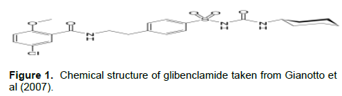
Out of the various lipid membranes used as models so far supported bilayer lipid membranes (s BLM) could be regarded as the most realistic ones in view of the facts that lipid bilayer is the key structural element of all bio membranes. Since its inception in 1960, BLM has been widely used for carrying out investigations into a variety of physical, chemical and biological phenomena including solar energy transduction (Janshoff and Steinem, 2006; Lonsdale, 1989; Tien, 1985). Glibenclamide (5-chloro-N-(2-(4-(cyclohexylcarbamoylsulfamoyl) phenyl) ethyl)-2-methoxy-benzamide) is a second generation sulfonyl urea compound used as an oral hypoglycemic or anti diabetic agent, a class of drug used to treat type-2 diabetes mellitus (Figure 1) (Bchir et al., 2004). Glibenclamide is 200 times more potent than tolbutamide in evoking pancreatic secretion of insulin. It differs from other oral hypoglycemic drugs in that it is more effective during eating than fasting (Rajkumar M et al., 2010).
Experimental work on artificial membrane using a variety of methods has demonstrated that the membrane properties may be strongly affected by the presence of membrane associated molecules. Examples of parameters that can be affected by drug-membrane interactions include the conformation of acyl groups, the membrane surface and thickness, the phase transition temperature, the membrane potential and hydration of head groups and the membrane fusion proteins (Szewczyk et al., 1993, 2006).
Literatures showed that glibenclamide was widely studied on natural cells mainly on rats. Lamprianou in his invitro studies of glibenclamide on non obese diabetic mice reported that glibenclamide prevents diabetes (Lamprianou et al., 2016). Another study also showed in vivo assessment of combined effects of glibenclamide and losartan in diabetic rats revealed the relevance of treatment of hypertensive diabetes with the combination of these two drugs (Moureq et al., 2019). Studies on formulation development, in vitro and in vivo evaluation of membrane controlled transdermal systems of glibenclamide revealed that membrane controlled transdermal systems exhibited better control of hyperglycemia and more effectively reversed the diabetes mellitus complications than oral administered glibenclamide in mice (Mutaliik and Uduga, 2005). There were also other studies on glibenclamide on rat and other animals (Wang et al., 2008; Seena et al., 2017; Ling et al., 2006; Nagamatsu et al., 2006; Maria and Reyes, 2008; Maheswari, 2001). Most of the studies conducted so far on glibenclamide and other drugs also were on animals. The use of animal is tiresome because it takes time to grow animals. On the other hand, model systems have high throughput screening than natural systems, due to this, model systems may be helpful in saving animals and could be utilized to indicate the concentration at which the drug is most effective and with least side effects. However, these model systems were not reported yet in investigation of glibenclamide drug. The objective of this study was investigation of transport of glibenclamide drug across micro-pore supported bilayer lipid membrane.
MATERIALS AND METHODS
Materials and equipments
Potentiometer (Denver instrument, model 250) and Ag/AgCl electrodes were used to measure membrane potential; conductivity meter (EUTECH instrument, syberscan CON 41) was used to measure membrane conductance. Hanas pH meter was used to measure pH of the drug solution and thermostat type GLS400 was used to regulate temperature. Electrochemical cells (each with the capacity to hold about 30 ml solutions were ordered and purchased from Addis Ababa university chemistry department and used to hold drug solutions. AXIVA, membrane filters (Nylon)CatNo.170013R, Rating: 0.2 µm, media: Nylon, diameter 13 mm, imported and processed in India by Delhi(India), used as micro-pore support for the formation of sBLM (supported bilayer lipid membrane).
Chemicals and reagents
All chemicals used for this experiment were of analytical reagent grade and were used without further treatment (L-alpha-phosphotidylcholine purchased from sigma Aldrich chemie Germany). Chloroform (99.96%, Fischer chemicals, UK) used to dissolve phosphotidylcholine.
Glibenclamide was purchased from local pharmacy. Drug solutions and KCl solution used for calibration purpose were prepared by using double distilled water.
Preparation of solutions
Fresh stock solutions of 0.0005, 0.00075, 0.001, 0.0025, 0.005, 0.0075, 0.01, 0.025, 0.05, 0.075 and 0.1 mol/lit of glibenclamide were successively prepared using double distilled water.
Preparation of supported bilayer lipid membrane
Micro-pore support BLM was prepared as follows: At the beginning 10 g of L-alpha –phosphotidylcholine was dissolved in 100 ml of chloroform to prepare lipid solution, then, the micro filter was first immersed in lipid solution then immediately immersed in water (for the purpose of proper alignment of bilayer) to get micro filter supported bilayer lipid membrane.
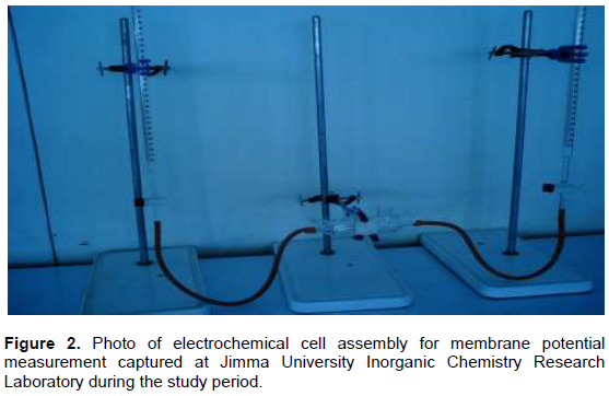
Before the experiment the micro filter was dried in air at room temperature to remove the solvent from the surface for about 20 min. The cell had two compartments. The dried micro pore support was attached on one half of the electrochemical cell and then both compartments were assembled together (Sheppard and Robinson, 1997) (Figure 2) with the introduction of burettes and electrodes and the membrane was properly adjusted and the solutions of the electrolyte poured into the two compartments through the inlets.
Measuring membrane potential
Ag/AgCl electrodes connected to potentiometer were placed in each compartment of the electrochemical cell, then, glibenclamide solutions of tenfold concentration difference were filled in both burettes and made to fill both compartment cells slowly at about equal flowing rates through the in late till overflow the membrane, then, membrane potential was measured as a function of concentration of glibenclamide used and temperature of the electrochemical cell maintained. Membrane potential was measured prior to the formation of BLM for the sake of comparison. Potentiometer was calibrated before each experiment and Ag/AgCl electrodes were placed in 3M KCl solution throughout the experiment and taken out during measurement only.
Measuring membrane conductance
The assembly was made by following the same procedure for membrane potential measurement. Membrane conductivity was measured by setting temp at 10, 15, 23, 30 and 37°C. Conductivity of different concentrations of the drug solutions were measured prior to contact with the membrane and after contact with the membrane at specified concentrations.
Measuring pH of the drug solution
pH meter was calibrated by using buffer solutions at pH 4 and 7 before measuring the pH of drug solutions and the pH of distilled water was taken as a standard.
Data processing and analysis
Selectivity in membrane permeation is usually inferred from two kinds of measurements, viz., potentiometric and conductometric for comparable concentrations of permeate species (Eisenman and Horn, 1983). The former involves measurements of difference in electric potential between two electrolyte solutions separated by a selectively permeable membrane. When the electric potential arises on account of difference in concentrations of the same solution, it is called membrane potential. Theories proposed and equations derived by Aizawa and co-workers (Kotnik et al., 2008) is useful to evaluate the transport number of anion and thermodynamic effective fixed charge density from the linear plots of (F/RT) Em against 1/C1, in accordance with the following equation.
(F/RT) Em = (2t--1) ln γ + 2(γ-1)/γ t-(1- t-) ØX /C1 (1)
Where (ØX) is thermodynamically effective fixed charge density; t- is transport number of anion; γ is concentration ratio (C2/C1); C2 is concentrated solution of the drug and C1 is dilute solution of the drug solution; while other terms have their usual thermodynamic significance.
Membrane conductance depends upon the properties of both fixed and mobile ionic species and is usually independent of membrane thickness or cross-sectional area.

Where π is membrane conductance; N is Avogadro’s number; h is Planck’s constant; R is universal gas constant; T is temperature in Kelvin; ΔH* is enthalpy of activation and ΔS* is entropy of activation.
Membrane conductance (π) data was used to evaluate (ΔH*) and (ΔS*), respectively, from the slope and intercept of linear plot of ln (πNh/RT) against 1/T in accordance with Equation 2.The evaluated activation parameters would be used to evaluate Arrhenius activation energy (Ea) and activation free energy (ΔG*) in accordance with the following relations (Kotwal and Thube, 2007; Siddiqi et al., 1988; Saddiqi and Alvi, 1989).

RESULTS
Observed values of membrane potential
The potentiometric data obtained by keeping micro-pore supported bilayer lipid membrane in between solutions of different concentrations of glibenclamide were summarized as a function of glibenclamide concentration in Table 1 and Figure 3. Each measurement was taken as the average of three readings. The data showed a decreased trend with increase in concentration.

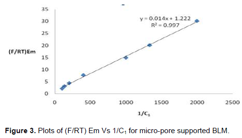
The observed value was utilized to evaluate thermodynamically effective fixed charge density ØX and transport number of anions (t-) respectively, from the slope and intercept of the linear plots of (F/RT) Em vs 1/C1, in accordance with Equation (1).
At fixed ratio, Equation (1) predicts straight line plot of (F/RT) Em against 1/C1 and allows evaluations of t- and ØX from the intercept and slope. Accordingly, the values of ØX and t evaluated from the above linear plot were 42.6 meq/lit and 0.76 respectively.
Observed values of membrane conductance
This study revealed that membrane conductance increased with increased concentration of glibenclamide solution and temperature (Table 2). Each measurement was taken as the average of three readings.

Figure 4 was used to evaluate thermodynamic activation parameters ?H* and ?S*. Table 3 indicated that the values of thermodynamic activation parameters increased with decreased concentration of the drug solution.


DISCUSSION
The values of transport number of anion indicated that glibenclamide most likely crossed the membrane in its anionic form than in its cationic form. The value of ØX indicated the presence of good effective fixed charged sites on the membrane.
Variation of membrane potential data with concentration could be explained by taking into consideration the structure of phosphotidylcholine (PC) molecule. Since H+ is very small in size it can easily react with the phosphate group of PC and neutralize it, so that, the repulsion on the anion form of the drug is minimized. In dilute range because of less viscosity of the drug solution, the positively charged group of the PC were in a better position to react with negatively charged part of the drug molecule, which being liphophilic, could cross the membrane easily and made the potential higher.
The evaluated values of anion transport number (t-) and thermodynamically effective fixed charge density (ØX) also support this idea. Study conducted in Ethiopia reported membrane potential increased with decrease in concentration (Damena et al., 2013) which was in line with the result of this study. There was also other study which supports this result (Saddiqi and Alvi, 1989).
Variation of membrane conductance with increased drug concentration and temperature might be explained by taking the following points into consideration: The number of charged particles available for the transport of current; the extent of the sorption of permeants in the membrane phase; the variation of viscosity with concentration and temperature. As concentration increased the number of charged particles crossing the membrane increased, as a result, more membrane conductance was measured at higher concentration of drug solution.
In addition to that increased temperature increases the kinetic energy of the permeants hence, permeants rapidly came close to the surface of the membrane resulted in increased membrane conductivity, besides; increased temperature lowers viscosity of the drug solution. So that, ions could easily move across supported BLM, which was again the cause for increment of membrane conductance.
The other reason might be, when the hydrated ions approached to the pores of the membrane before effective interaction with phosphotidylcholine of the lipid, certain fraction of water molecules from the hydration shell of the ion and fixed sides might be removed. The negative entropy change of activation (-ΔS*) observed indicated that an ordered water molecules become less ordered in the most diluted solutions (Saddiqi and Alvi, 1989). Study conducted in India on ion transport studies through polystyrene based model membrane conductance data and absolute reaction rate theory reported activation energy decrease with increase in concentration and the value of change of entropy were negative. It was in line with the result of this study (Ansari et al., 2012).
Increased in Gibbs free energy of activation (ΔG*) might be due to strong interaction between fixed site and permeants. Since more sites were available in dilute range, more energy was released. That was why enthalpy of activation (Δ H*) increased with decreased concentration of glibenclamide solution. In general drugs might change the permeability, fluidity, mechanical stability and structure of the membrane.
This concept was also supported by literature; Jacqueline in his review of membrane-drug interactions studied using model membrane systems reported the effect of Rifabutin and Oritavancin on the structure of model membrane (Knobloch et al., 2015).
CONCLUSIONS AND RECOMMENDATION
In this study, transport of glibenclamide across a model lipid bilayer membrane was determined. First part of the study focused on determination of membrane potential as a function of concentration. From membrane potential data, transport number of anion (t-) and thermodynamic effective fixed charge density were calculated. Higher potential was observed in dilute range. It might be due to the interaction between the drug molecule and phosphotidylcholine as discussed previously. Membrane conductance increased as a function of increased temperature and concentration of the drug solution. From membrane conductance data, thermodynamic activation parameters were evaluated and the general trend observed were, as concentration decreased the value of Ea, ΔH*, ΔG* and negative ΔS* increased. From evaluation of the data obtained in both cases, membrane was more selective when drug was in very dilute range. Glibenclamide follows passive transport mechanism [Kamp et al., 2003]. It crosses mitochondrial membrane in pancreas and initiates β-cell to secrete insulin. In most studies this drug was used at µM scale [Szewczyk et al., 1993; Zheng and Zhang, 2007; Coppack et al., 1990). This study also indicated that membrane was more selective when the drug was at very low concentration; therefore, it is recommended that if this beginning is further enriched by research it can be used as a substitute for natural systems.
CONFLICT OF INTERESTS
The authors have not declared any conflict of interests.
ACKNOWLEDGMENTS
The authors acknowledge Chemistry Department of Jimma University for allowing them conduct the study and student research project of Jimma University for financial support.
REFERENCES
|
Ansari MA, Kumar M, Singh N, Dadoriya KS, Kushwaha RS, Ayub S (2012). Ion transport studies through polystyrene based model membrane: Conductance data and absolute reaction rate theory. Journal of Advanced Applied Science Research 3(1):251-260. |
|
|
Bchir WS, Roglic G, Green A, Sicree R, King SR (2004). Global Prevalence of Diabetes. Journal of Diabetes Care 27(5):1047. |
|
|
Benhamou M (2008). Organization of Lipid Molecules within Biomembranes. Lipid Insights 1, LPI-S887. |
|
|
Coppack, SW, Lant AF, Mclntosh CS, AV Rodgers AV (1990). Pharmacokinetic and Pharmacodynamic studies of glibenclamide in non-insulin dependent diabetes mellitus. British Journal of Clinical Pharmacology. 29(6):673-684. |
|
|
Damena T, Tesema TE, Alvi NI, Siraj K (2013). Effect of pyridoxine on the transport of iron across micro filters supported lipid membrane. Journal of Surface and Interfacial Matter 1(2):2164-2169. |
|
|
Eeman M, Deleu M (2010). From Biologic membrane to Biomimetic model membranes. Biotechnologie, Agronomie, Société et Environnement 14(4):719-736. |
|
|
Eisenman G, Horn H (1983). Ionic selectivity revisited: the role of kinetic and equilibrium processes in ion permeation through channels. The Journal of membrane biology 76(3):197-225. |
|
|
Gianotto AE, Arantes PR, Lara-Filho JM, Filho CA, Fregonezi-Nery MF(2007). Dissolution test for glibenclamide tablets. Quimica Nova 30(5):1218-1221. |
|
|
Jan DS, Tsai FN (1994). Combined film and membrane diffusion-controlled transport of ions through charged membrane. Journal of membrane science, 90(1-2):109-115. |
|
|
Janshoff A, Steinem C (2006). Transport across artificial membranes an analytical perspective. Journal of Analytical and Bio analytical Chemistry 385(3):433-451. |
|
|
Kamp F, Nadeem Kizilbash N, Barbara E, Corkey Per-Olof Berggren and James AH (2003). Sulfonylureas rapidly cross phospholipid bilayer membranes by a free diffusion mechanism. Diabetes 52(10):2526-2531. |
|
|
Knobloch J, Daniel K, Suhendro Julius L, Zielenieckii Joseph G, Kopper SI (2015). Membrane- drug interactions studied using model membrane systems. Saudi Journal of Biological Sciences 22(6):714-718. |
|
|
Kotnik PM, Miklavcic TT, Kramar D, Macek-Lebar P (2008). Advance in Planar Lipid Bilayer and Liposome 6:165. |
|
|
Kotwal BK, Thube R (2007). Enhancement of iontophoretic transport of diphenhydramine hydrochloride thermosensitive gel by optimization of pH, polymer concentration, electrode design and pulse rate. Aaps Pharmscitech 8(4):320-325. |
|
|
Lamprianou S, Gysemans C, Bou Saab J, Pontes H, Mathieu C, Meda P (2016). Glibenclamide Prevents Diabetes in NOD Mice. PLoS One 11(12):e0168839. |
|
|
Ling Z, Wang Q, Stang G (2006). Glibenclamide treatment recruits β-cell subpopulation into elevated and sustained basal insulin activity. Diabetes 55(1):78-85. |
|
|
Maheswari U (2001). Lipid bilayer-methotrexate interactions: A basis for methotrexate neurotoxicity. Current Science pp. 571-574. |
|
|
Maria R, Reyes L (2008). Pharmacokinetics and pharmacodynamics of diclofenac in the presence and absence of glibenclamide in the rat. Journal of Pharmacy & Pharmaceutical Sciences 11(3):68-76. |
|
|
Moureq AR, Fatani AJ, Almnaizel AT, Ahmed MM, Abuohashish HM, Al-Rejaie SS (2019). In vivo assessment of combined effects of glibenclamide and losartan in diabetic rats. Medical Principles and Practice 28(2):178-185. |
|
|
Mutaliik S, Uduga N (2005). Formulation development, in vitro and in vivo evaluation of membrane controlled transdermal systems of glibenclamide. Journal of Pharmaceutical Science 8(1):25-38. |
|
|
Nagamatsu S, Ohara-Imaizumi M, Nakamichi Y, Kikuta T, Nishiwaki C (2006). Imaging docking and fusion of insulin granules induced by anti diabetes agents. Journal of Diabetes 55(10):2819-2825. |
|
|
Peetla C, Stine A, Labhasetwar V (2009). Biophysical interactions with model lipid membranes: applications in drug discovery and drug delivery. Molecular Pharmacy 6(5):1264-1276. |
|
|
Rajkumar AM, Kumar IK, Nagaraju T, Sowjanya L, Srikanth B, Venkateshwarulu G, Sandeep P (2010) Design and invitro evaluation of drug release and bio adhesive Properties from buco adhesive tablets of glibenclamide for systemic delivery. Journal of Chemical and Pharmaceutical Research 2(4):291-303. |
|
|
Rossi C (2007). Biomimetic tethered lipid membranes designed for membrane-protein interaction studies. European Biophysics Journal 36(8):955-965. |
|
|
Siddiqi FA, Alvi NI, Khan SA (1988). Transport studies with model membranes. Colloids and surfaces 32:57-73. |
|
|
Saddiqi FA, Alvi NI (1989). Transport studies with model membrane-II. Journal of Polymer Science 27(7):1499-1517. |
|
|
Seena T, Pandarekandy PG, Sreejesh BS, Harikumaran T, Sreekumara E (2017). Hypoglycaemic effect of Glibenclamide: A critical study on the basis of Creatinine and Lipid peroxidation status of streptozotocin- induced diabetic rat. Indian Journal of Pharmaceutical Science 79 (5):768-777. |
|
|
Sheppard DN, Robinson AK (1997). Mechanism of glibenclamide inhibition of cystic fibrosis transmembrane conductance regulator Cl-channels expressed in a murine cell line. The Journal of physiology 503(2):333. |
|
|
Szewczyk A, Mikolajek B, Pikula S, Nalecz S (1993). ATP-sensitive K+ channel in mitochondria. Acta biochimica Polonica, 40(3):329-336. |
|
|
Szewczyk A, Skalska J, Glab M, Kulawiak B, Maliska D (2006). Mitochondrial potassium channels: from pharmacology to function. Biochimica et Biophysica Acta (BBA)-Bioenergetics 1757(5-6):715-720. |
|
|
Tien HT (1985). ''Planar Bilayer" Lipid membrane in progress in surface science 19(3):169-274. Pergamon Press, New York. |
|
|
Wang QH, Heimberg H, Pipeleers D, Ling Z (2008). Glibenclamide activate translation in rat pancreatic beta cells through calcium-dependent mTOR, PKA and MEK signalling pathway. Diabetologia 51(7):1202-1212. |
|
|
Zheng J, Zhang X (2007). Antihyperglycemic activity of Prunella vulgaris L. in streptozotocin-induced diabetic mice. Asian Pacific Journal of Clinical Nutrition 16:427. |
|
Copyright © 2024 Author(s) retain the copyright of this article.
This article is published under the terms of the Creative Commons Attribution License 4.0


