Full Length Research Paper
ABSTRACT
Maerua pseudopetalosa (Gilg and Bened.) De Wolf tubers which are used traditionally as antitumor agent in Sudan were subjected to separation by column chromatography technique. Eight fractions were obtained for the ethyl acetate extract and twelve for the ethanolic extract. The ethanolic fractions F8, F9, F11 and F12, with high bioactivity were subjected to further investigations. 3-(4,5-dimethyl-2-thiazolyl)-2,5-diphenyl-2H-tetrazolium bromide (MTT) assay was used for assessment of the cytotoxicity. Remarkable cytotoxicity against Michigan Cancer Foundation-7 (MCF-7) was shown, for the first time. Actually, the results revealed that F12 is a very promising one with remarkable activity against MCF-7 cell lines (43.51 µg/g at 72 h), high antioxidant activity (91.3% by 1,1-diphenyl-2-picryl-hydrazyl (DPPH) and 97% by ABTS) and flavonoid and phenolic contents (11.75 and 20.72 mg/g). Six compounds were detected in F9 (syrngic 11.65 µg/g, sinapic 8 µg/g) and F12 (gallic 88.12 µg/g, caffic 11.1 µg/g, sinapic 38.67 µg/g) which were not recorded in any previous work in the available literature by using high performance liquid chromatography (HPLC).
Key words: Michigan Cancer Foundation-7 (MCF-7) breast cancer, Maerua pseudopetalosa, phenol and flavonoid contents, high performance liquid chromatography (HPLC) analysis, antioxidant activity.
INTRODUCTION
Sudan is rich in countless flora due to the diverse climatic and soil conditions in the different ecological regions.
Traditional medicines play a very important role in the health care system in Sudan and a high percentage of the nomads still rely on local traditional healers. As in many other countries, biodiversity in Sudan is still maintained in some parts, which are known for the presence of so many species that are characterized for their undisputed cure for a large number of diseases (Ibrahim and EL Nure, 2016).
The use of plants in medicine is not limited or restricted to any region of the world. It is an old practice in various parts of the globe for both preventive and curative purposes. Dependence on herbs as medicine in the treatment of diseases is an adopted practice by a large proportion of the rural population because of its availability and affordability (Sani et al., 2009).
Phenolic compounds are commonly found in both edible and nonedible plants, and they have been reported to have multiple biological effects, including antioxidant activity. Free radicals have been implicated in the development of a number of disorders, including cancer, neuro-degeneration and inflammation (Halliwell, 2006a, b). The presence of antioxidants such as phenolics, flavonoids, tannins and proanthocyanidins in plants may provide protection against a number of diseases; for example, ingestion of natural antioxidants has been inversely associated with morbidity and mortality from degenerative disorders (Gülcin, 2012). Medicinal plants are therefore being investigated for their antioxidant properties, and the demand for natural antioxidants and food preservatives is increasing (Peschel et al., 2006). The importance of the antioxidant constituents of plant materials in the maintenance of health and protection from coronary heart disease and cancer is also raising interest among scientists, food manufacturers, and consumers as the trend of the future is moving toward functional food with specific health effects (Lo¨liger, 1991). Flavonoids and other phenolics have been suggested to play a preventive role in the development of cancer and heart disease. Ingestion of alcohol-free red wine or a phenolic compound mixture extracted from red wine has been shown to improve the antioxidant status of plasma in humans (Serafini et al., 1998; Carbonneau et al., 1998).
The plant, Maerua pseudopetalosa (Gilg and Bened.) De Wolf (family Cappariaceae), is a Sudanese medicinal plant which is traditionally used as remedy for cough and as cure for tumors. In the Nuba Mountains and the Republic of the South Sudan, the fruits are eaten but only during famine times and after careful preparations to remove the toxic substances (Henry, 1948).
Thirty three compounds were detected in tuber fractions by using GC/MS analysis; fraction 12 showed more cytotoxic effect, this was attributed to the presence of a proline derivative (proline-N-methyl-butyl ester). This compound might be considered as the cause of the high toxicity of the fraction; since free proline was used as an inhibitor of breast cancer development. Surprisingly, M. pseudopetalosa tubers were used in the folkloric medicine in Sudan by the natives of the South Blue Nile State for the treatment of breast cancer growth without any knowledge of its chemical constituents (Ibrahim and EL Nure, 2015).
The plant kingdom represents an enormous reservoir of biologically active molecules and so far, only small fractions of plants with medicinal activity have been assayed. Nearly 50% of drugs used in medicine are of plant origin. There is therefore much current research devoted to the phytochemical investigation of higher plants that have ethnobotanical information associated with them. The phytochemicals (secondary metabolites) isolated are then screened for different types of biological activity (Harborne, 1998).
In addition, there will be need for the permanent search and development of new natural drugs. This need also arises from the advantage of certain natural products in controlling some diseases that chemicals fail to do. For example, the annonaceous acetogenins, extracted from pawpaw tree (Asimina triloba Dunal) were found to be the best effective against selected tumor type, e.g., squamtacin is selective against the human prostate carcinoma cell line (PC-3) and a series of 9-carbonyl compounds work best against the human pancreatic tumor cell line (PaCa-2) (Ahammadsahib et al., 1993).
Breast cancer is one of the leading causes of death among women in the world. At the present, using of natural compounds such as medicinal plants in cancer therapy has aroused general because of its minimal side effect, safety and efficiency (Ribereau et al., 1997; Taixiang et al., 2005).
Approximately, one-third of the women with breast cancer developed metastases and ultimately died of the disease. MCF-7 is an estrogen receptor-positive human cancer cell line, derived from a patient with metastatic breast cancer (Parkin et al., 2001). Growth of MCF-7 cells is inhibited by tumor necrosis factor (TNF alpha). Many plants claimed to induce apoptosis in MCF-7 cells such as Antrodia camphorate (Levenson and Jordan, 1997) and Gmelina asiatica. The goal of screening medicinal plants is to search for excellent anticancer agent suitable for human malignancies, therefore, the aim of this study was to screen tuber fractions with respect to their total phenolic and flavonoid content, HPLC analysis, antioxidant activity in order to find new potential sources of natural anticancer drugs. The study is a part of a larger survey with other functional properties of this plant such as their antimicrobial, secondary metabolites, toxicity and GC/MS analysis.
MATERIALS AND METHODS
Plant
The plant under investigation (M. pseudopetalosa) was collected from the South of Sudan. The plant was authenticated at the Department of Botany by Prof. Hatil H. Alkamali, Omdurman Islamic University.
Preparation of crude plant extracts
The plant material was air dried, ground into a coarse powder form and the dried ground tubers (1 kg) of the M. pseudopetalosa were soaked for 3 days in 1500 ml ethyl acetate and ethanol consecutively. Ethyl acetate extract gave 7.1 g while the ethanol extract gave 10.9 g; both with dark brown residue and were subjected to silica gel (230 - 400 mesh) column chromatography separation using stepwise gradient elution of n-hexane to chloroform, and chloroform to ethyl acetate and finally washing with pure methanol. Then, 100 ml portions were collected, concentrated and combined according to their similarity in spectrometric and thin layer chromatography (TLC) separation behaviors using suitable solvent systems. Ethyl acetate gave eight fractions while ethanol gave twelve fractions. Four fractions with bioactive effects were subjected to further investigations.
Total phenolic and flavonoid contents
The total phenolic content of the extract was determined by the Folin-Ciocalteu method (Kaur and Kapoor, 2002). Briefly, 200 L of crude extract (1 mg/mL) was made up to 3 mL with distilled water, mixed thoroughly with 0.5 mL of Folin-Ciocalteu reagent for 3 min, followed by the addition of 2 mL of 20% (w/v) sodium carbonate. The mixture was allowed to stand for a further 60 min in the dark and absorbance was measured at 650 nm. The total phenolic content was calculated from the calibration curve and the results were expressed as mg of gallic acid equivalent per g dry weight.
The total flavonoid content of crude extract was determined by the aluminium chloride colorimetric method (Chang et al., 2002). In brief, 50 L of crude extract (1 mg/mL ethanol) was made up to 1 mL with methanol, mixed with 4 mL of distilled water and then 0.3 mL of 5% NaNO2 solution; 0.3 mL of 10% AlCl3 solution was added after 5 min of incubation, and the mixture was allowed to stand for 6 min. Then, 2 mL of 1 mol/L NaOH solution was added, and the final volume of the mixture was brought to 10 mL with double-distilled water. The mixture was allowed to stand for 15 min, and absorbance was measured at 510 nm. The total flavonoid content was calculated from a calibration curve, and the result was expressed as mg rutin equivalent per g dry weight.
Antioxidant properties
1,1-Diphenyl-2-picryl-hydrazyl assay
The antioxidant activity of the extract was determined by 1,1-diphenyl-2-picryl-hydrazyl (DPPH) assay, as described earlier with some modifications (Villaño et al., 2007). Briefly, 200 L of each extract (100-500 g/mL) was mixed with 3.8 mL DPPH solution and incubated in the dark at room temperature for 1 h. The absorbance of the mixture was then measured at 517 nm. Ascorbic acid was used as a positive control. The ability of the sample to scavenge DPPH radical was determined from DPPH scavenging effect = (Control OD − Sample OD / Control OD) × 100.
ABTS+ free radical-scavenging activity
The determination of ABTS•+ radical scavenging activity was carried out as reported by Dorman and Hiltunen (2004). Briefly, the ABTS•+ radical was generated by the reaction of 7 mM ABTS aqueous solution with K2S2O8 (2.45 mM) in the dark for 16 h adjusting the absorbance at 734 nm to 0.700 at room temperature. The samples (10 μL) were added to 1490 μL ABTS•+, absorbance at 734 nm was read immediately (A0) and after 6 min (A1). Several concentrations were measured, and the percentage inhibition [(A0-A1/A0) × 100] was plotted against the phenol content and IC50 was determined (concentration of total phenol able to scavenger 50% of ABTS•+ free radical).
Chromatography
HPLC instrument employed in the study was Make Waters Analytical system (USA) with alliance 2690 pump, automatic injector, UV-dual lambda observance detector and empower-2 software. The stationary phase used is C18 column. Calibration of the system was done by accurately weighing 0.01 g of standard solution (Merck, Germany) dissolved in 100 ml of HPLC grade water. 20 μl of different concentrations mode from the standard stock solutions and samples were injected through a C18 Column. The mobile phase consisting of water: methanol (70:30 v/v) was degassed before use. Detection of stalk solution was done at 273 nm and flow rate was maintained at 1 ml/min. All the chemicals used are HPLC grade (99.8% pure). The methanol was obtained from Merck and Tri fluoro acetic acid from Finar. De-ionized water was obtained from Milli-Q (millipore, USA). The samples were run for minutes. All chromatographic data were recorded and processed using Autochro-300 software.
Anticancer activity
Cell cultures and treatments
Human breast cancer cell line (MCF-7) was obtained from the American Type Culture Collection (Rockville, MD, USA). Cells were grown in RPMI-1640 medium supplemented with 10% fetal bovine serum, 1% MEM non-essential amino acid solution, and 1% penicillin streptomycin solution (10,000 units of penicillin and 10 mg of streptomycin in 0.9% NaCl) in a humidified atmosphere of 5% CO2, 95% air at 35°C. The passage number range for cell lines was maintained between 20 and 25. The cells were cultured in 75 cm2 cell culture flasks. For experimental purposes, cells were cultured in 96 well plates (0.2 ml of cell solution/well). The optimum cell concentration as determined by the growth profile of the cell line was 2×10⁵ cells/ml (Cells were allowed to attach for 24 h before treatment with tested extracts). The stock solution was filtered with Minisart Filters (0.22 µm). Working 2 fold serially diluted test materials were prepared. Cell monolayers were washed with PBS and the addition serially diluted materials were dispensed to the pre-cultured plates for determination of test materials toxicity (Romero et al., 2003).
3-(4,5-dimethyl-2-thiazolyl)-2,5-diphenyl-2H-tetrazolium bromide (MTT) assay
The MTT assay is based on the protocol described for the first time by Mossmann (1983). The assay was optimized for the cell lines used in the experiments. Briefly, for the purposes of the experiments at the end of the incubation time, cells were incubated for 4 h with 0.8 mg/ml of MTT, dissolved in serum free medium. Washing with phosphate buffer saline (PBS 1 ml) was followed by the addition of DMSO (1 ml), gentle shaking for 10 min so that complete dissolution was achieved. Aliquots (200 μl) of the resulting solutions were transferred in 96-well plates and absorbance was recorded at 560 nm using the microplate spectrophotometer system (Spectra max190-Molecular Devices). Results were analyzed with the Soft max pro software (version 2.2.1) and are presented as percentage of the control value. The relation between surviving fraction and extract concentration is plotted to get the survival curve for cell line after the specified time. The concentration required for 50% inhibition of cell viability (IC50) was calculated (Mossmann, 1983).
RESULTS AND DISCUSSION
Total phenolic and flavonoids contents
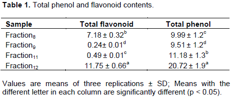
Phenolic compounds are one of the major chemical classes of plants’ secondary metabolites. They play an important role in the defense of plants against pathogens, diseases, parasites, and predators (Bhattacharyya et al., 2014). Moreover, they involve in a number of physiological mechanisms such as antioxidant activity. They also play an important role in stabilizing lipid peroxidation (Wei and Shiow, 2001). The total phenolic contents in the examined tuber fraction extracts using the Folin-Ciocalteu reagent is expressed in term of gallic acid equivalent. The values obtained for the concentration of total phenols are expressed as mg of GA/g of extract (Table 1). The highest concentration of phenols was measured for fraction 12 (20.72±1.9 mg/m) and this may increase anticancer activity of this fraction since flavonoids and phenolic compounds have been suggested to play a preventive role in the development of cancer and heart disease. Also fraction 11 represented concentration of 11.18±1.3 mg/ml, while fractions 8 and 9 exhibited low concentrations. On the other hand, the values of flavonoids content represented high concentration with fraction 12 (11.75 ± 0.660), while fraction 8 showed 7.18 ± 0.32 mg/g. The lowest flavonoid content was measured in fractions 11 and 9.
Antioxidant activity
Reactive oxygen species, such as single oxygen, superoxide ion, hydroxyl ion and hydrogen peroxide, are highly reactive toxic molecules, which are generated normally in cells during metabolism. They cause severe oxidative damage to proteins, lipids, enzymes and DNA by covalent binding and lipid peroxidation, with subsequent tissue injury. Natural antioxidant agents have attracted much interest because of their ability to scavenge free radicals (Saeed et al., 2012).
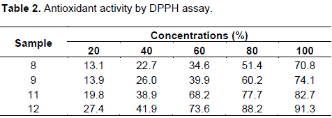
Test for antioxidant activity represented strong activity in fraction 12 at a concentration of 100 mg/ml when we used DPPH and ABTS (91.3 and 97%) (Tables 2 and 3). Also F8, F9 and F11 showed high antioxidant activity with DPPH method at a high concentration (100 mg/ml), the values were 70.8, 74.1 and 82%, respectively.
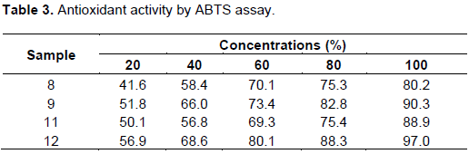
On the other hand, ABTS method exhibited high values at a concentration of 100 mg/ml, the fractions F11, F9, F8 have activity equal to 88.9, 90.3 and 80.2%, respectively.
High performance liquid chromatography (HPLC) analysis
HPLC analysis can be used for classification of herbs based upon secondary metabolites. Extract yield at optimum condition was then analyzed by HPLC for quantifying bioactive compound.
The HPLC analysis of tuber fractions showed some interesting results (Table 4). Fraction 12, which proved to be the highest cytotoxic fraction, has got 3 compounds as revealed by the HLPC analysis. One of the compounds present in fraction 12 is a gallic acid which belongs to phenolic compounds. This compound might be considered as the cause of the high anticancer activity of the fraction; since gallic acid potent high antioxidant activity is linked to anticancer agent as reported by Lo¨-liger (1991).
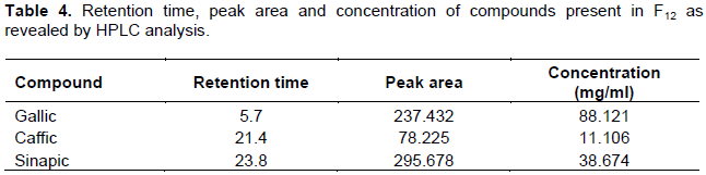
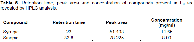
Fraction 12 represented high concentration of gallic acid with a value of 88.121 µg/ml. Gallic acid (3,4,5 trihydroxybenzoic acid) is a phenolic compound present in most plants. This metabolite is known to exhibit a range of bioactivities including antioxidant, antimicrobial, anti-inflammatory, and anticancer (Felipe and Salgado 2016). However, this molecule attracts the interest of researchers mainly for its antioxidant capacity (Kim, 2007). Other pharmacological activities described in the literature are anticancer (Chia et al., 2010). Also, sinapic acid (3,5 dimethoxy-4-hydroxycinnamic acid) which belongs to the class of phenolic acid, exhibited 38.674 µg/ml. It has been tested and reported against various pathological conditions such as cancer inflammation, diabetes and oxidative stress (Kikuzaki et al., 2002). The less concentration is shown by caffeic acid (3,4 dihydroxycinnamic acid 11.106 µg/ml). Caffeic acid is present in several medications of popular use, mainly based on propolis; moreover, it is acting as a carcinogenic inhibitor (Greenwald, 2004). On the other hand, fraction 9 (Table 5) reflected less concentration of syrngic and sinapic with values of 11.65 and 8 µg/ml, respectively.
Anticancer activity
Tuber fractions were evaluated in-vitro for their anticancer activity against MCF-7 cell lines using MTT assay. 5-flurouracil is one of the most commonly used drugs to treat cancer (positive control) and the plant extracts were used at different concentrations for 24, 48 and 72 h. The MTT assay is a sensitive, quantitative and reliable colorimetric assay that measure cell viability. The assay is based on the capacity of the cellular mitochondrial dehydrogenase enzyme in living cells to reduce the yellow water-soluble substrate 3-(4,5-dimethylthiazol-2yl)-2,5-diphenyl tetrazolium bromide (MTT) into a dark blue/purple formazan product which is insoluble in water. The amount of formazan produced is directly proportional to the cell number in a range of cells lines (Riss et al., 2016; Gerlier and Thomasset, 1986). Also, it is accurate because of its ability to describe the relationship between the amount of active cells with absorbance obtained from measuring its 50% inhibition concentration value (IC50) (Behera et al., 2003). The lesser the IC50 value, the higher the potential of the tested extract to inhibit cell proliferation. The principle of the MTT assay is to measure the activity of mitochondrial dehydrogenase in converting MTT into formazan. The concentration of formazan, which has a blue color, can be determined with a visible spectrophotometer and it has positive correlation with the number of living cells because the reduction event only exists when the mitochondrial reductase is produced (mitochondria is still active and this indicates that the cell is alive) (Chapdelaine, 2001).
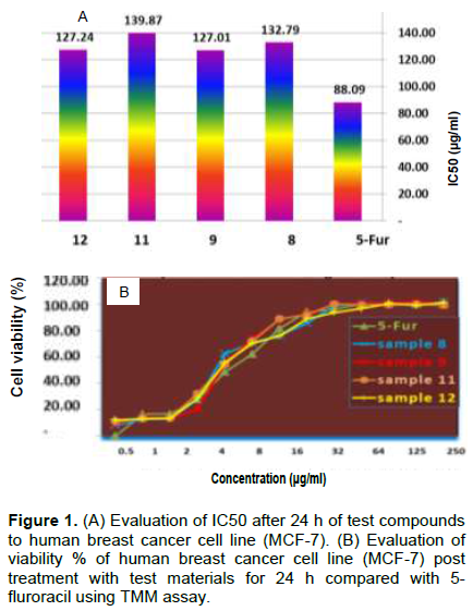
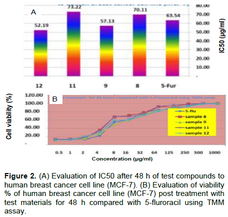
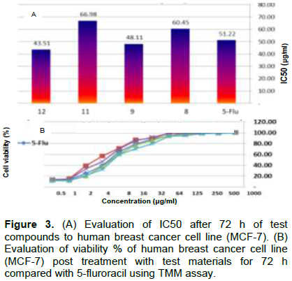
As a matter of fact, the results revealed that F12 (43.15 µg/ml) is a very promising one with remarkable IC50 against MCF-7 breast cancer cell when compared with 5-flurouracil (51.22 µg/ml) used as standard drug at 72 h (Figure 3A). The other fractions F11, F9 and F8 showed IC50 equal to 66.98, 48.11 and 60.45 µg/ml, respectively (Figure 3A). As the cell viability decreases the inhibition increase and that led to more potent drug, this is clearly shown in Figures 1, 2 and 3B.
Phytochemicals isolated from herbs have emerged as a new and promising source of anticancer remedies, or as adjuvants for chemotherapeutic drugs, to enhance their efficacy and decrease side effects (De Vita et al., 2000).
Gallic acid is a possible cause of the anticancer activity observed for fractions (F12); since Felipe and Salgado (2016) reported that the gallic acid was known to display some anticancer and antioxidant activity. Moreover, the presence of gallic acid is restricted to this fraction. This compound might be considered as the cause of the high toxicity of the fraction; since gallic acid was found to inhibit the growth of breast cancer cell MCF-7 as reported by Wang et al. (2014). He suggested it as a possible application in breast cancer therapy. However, Zheng et al. (2001) also referred to the potent antioxidant of the cinapic acid which may provide another explanation to increase antioxidant and anticancer effects of this fraction.
Furthermore, the presence of caffeic acid in fraction 12 might also be taken as another proof for the increased anticancer activity of the fraction compared to fraction 9 which lacks this acid in spite of its high IC50 value (48.11 µg/ml).
Surprisingly, M. pseudopetalosa tubers were used in the folkloric medicine of the natives of the South Blue Nile State in Sudan for the treatment of breast cancer growth without any knowledge of their chemical constituents (Ibrahim and EL Nure, 2015).
CONCLUSION
This study indicated that the tuber fractions contained high amounts of phenolic compounds and exhibited strong antioxidant activities. Gallic acid, cinapic acid, and caffeic acid are concentrated largely in fraction 12 in comparison with the other fractions and also represented anticancer effect more than 5-flurouracil which is used as anticancer chemo therapeutic drug. Hence, the plant tubers may be used as a new and promising source of breast cancer remedies, or as adjuvants for chemotherapeutic drugs to decrease side effects.
CONFLICT OF INTERESTS
The authors have not declared any conflict of interests.
ACKNOWLEDGEMENT
The authors are grateful to the management and staff of the Central Laboratory of National Research Centre, Giza, Egypt for materials and technical supports.
REFERENCES
|
Ahammadsahib KI, Hollingworth RM, McGovern PJ, Hui YH, McLaughlin JL (1993). Inhibition of NADH: ubiquinone reductase (mitochondrial complex I) by bullatacin, a potent antitumor and pesticidal Annonaceous acetogenin. Life Science (53):1113-1120. |
|
|
Behera BC, Adawadkar B, Makhija U (2003). Inhibitory activity of xanthine oxidase and superoxide-scavenging activity in some taxa of the lichen family Graphidaceae. Phytomedicine 10(7):536-543. |
|
|
Bhattacharyya P, Kumaria S, Diengdoh R, Tandon P (2014). Genetic stability and phytochemical analysis of the in vitro regenerated plants of Dendrobium nobile Lindl., an endangered medicinal orchid. Meta Gene 2:489-504. |
|
|
Carbonneau MA, Léger CL, Descomps B, Michel F, Monnier L (1998). Improvement in the antioxidants status of plasma and low-density lipoprotein in subjects receiving a red wine phenolic mixture. Journal of the American Oil Chemists Society 75:235-240 |
|
|
Chang CC, Yang MH, Wen HM, Chern JC (2002). Estimation of total flavonoid content in propolis by two complementary colorimetric methods. Journal of Food and Drug Analysis 10(13):178-182. |
|
|
Chapdelaine JM (2001). MTT reduction-a tetrazolium-based colorimetric assay for cell survival and proliferation. Pharmakon Research International, Inc. pp. 1-6. |
|
|
Chia Y, Rajbanshi R, Calhoun C, Chin RH (2010). Anti-neoplastic effects of gallic acid, a major component of Toona sinensis leaf extract, on oral squamous carcinoma cells. Molecules 15(11):8377-8389. |
|
|
De Vita V, Hellman S, Rosenberg SA (2000). Cáncer, Principios y Práctica de Oncologia. J. Ed Médica Panamericana 1(5):154-157. |
|
|
Dorman H, Hiltunen R (2004). Fe(III) reductive and free radical-scavenging properties of summer savory (Satureja hortensis L.) extract and subfractions. Food Chemistry 88:193-199. |
|
|
Felipe HA, Salgado HR (2016). Gallic Acid: Review of the methods of determination and quantification. Critical Reviews in Analytical Chemistry 46(3):257-265. |
|
|
Gerlier D, Thomasset N (1986). Use of MTT colorimetric assay to measure cell activation. Journal of Immunological Methods 94(2):57-63. |
|
|
Greenwald P (2004). Clinical trials in cancer prevention: current results and perspectives for the future. The Journal of Nutrition 134(12 Suppl):3507S-3512S. |
|
|
Gülcin I (2012). Antioxidant activity of food constituents: an overview. Archives of toxicology 86(3):345-391. |
|
|
Halliwell B (2006a). Oxidative stress and neurodegeneration: where are we now? Journal of Neurochemistry 97(6):1634-1658. |
|
|
Halliwell B (2006b). Oxidative stress and cancer: have we moved forward? Biochemical Journal 401(1):1-11. |
|
|
Harborne JB (1998). Phytochemical Methods: A Guide to Modern Techniques of Plant Analysis. 3rd Edn., Chapman and Hall, London. P 302. |
|
|
Henry AJ (1948). The toxic principle of Courbonia virgata: its isolation and identification as a tetramethylammonium salt. British Journal of Pharmacology and Chemotherapy 3(3):187. |
|
|
Ibrahim MA, El Nur EE (2015). Cytotoxicity study on Maerua pseudopetalosa (Glig and Bened.) De Wolf tuber fractions. African Journal of Plant Science 9(12):490-497. |
|
|
Ibrahim MA, El Nur EE (2016). Phytochemical analysis of Maerua pseudopetalosa: Medicinal plant with bioactive natural products. LAP Lambert Academic Publishing, Germany. P 3. |
|
|
Kaur C, Kapoor HC (2002). Antiâ€oxidant activity and total phenolic content of some Asian vegetables. International Journal of Food Science and Technology 37(2):153-161. |
|
|
Kikuzaki H, Hisamoto M, Hirose K, Akiyama K, Taniguchi, H (2002). Antioxidant properties of ferulic acid and its related compounds. Journal of Agricultural and Food Chemistry 50(7):2161-2168. |
|
|
Kim YJ (2007). Antimelanogenic and antioxidant properties of gallic acid. Biological and Pharmaceutical Bulletin 30(6):1052-1055. |
|
|
Levenson AS, Jordan VC (1997). MCF-7: the first hormone-responsive breast cancer cell line. Cancer Research 57(15):3071-3078. |
|
|
Lo¨liger J (1991). The use of antioxidants in food. In Free Radicals and Food Additives; Aruoma O. I., Halliwell B., Eds., Taylor and Francis, London. pp. 129-150. |
|
|
Mossmann T (1983). Rapid colorimetric assay for cellular growth and survival: application to proliferation and cytotoxicity assays. Journal of Immunological Methods 65:55-63. |
|
|
Parkin DM, Bray F, Ferlay J, Pisani P (2001). Estimating the world cancer burden: Globocan 2000. International Journal of Cancer 94(2):153-156. |
|
|
Peschel W, Sánchez-Rabaneda F, Diekmann W, Plescher A, Gartzía I, Jimenez D, Codina C (2006). An industrial approach in the search of natural antioxidants from vegetable and fruit wastes. Food Chemistry 97(1):137-150. |
|
|
Ribereau-Gayon G, Jung ML, Frantz M, Anton R (1997). Modulation of cytotoxicity and enhancement of cytokine release induced by Viscum album L. extracts or mistletoe lectins. Anti-Cancer Drugs 8:3-8. |
|
|
Riss TL, Moravec RA, Niles AL, Duellman S, Benink HA, Worzella TJ, Minor L (2016). Cell viability assays. Assay Guidance Manual [Internet]. P 121. |
|
|
Romero D, Gomez-Zapata M, Luna A, GarcıÌa-Fernández AJ (2003). Morphological characterization of BGM (Buffalo Green Monkey) cell line exposed to low doses of cadmium chloride. Toxicology In Vitro 17(3):293-299. |
|
|
Saeed N, Khan MR, Shabbir M (2012). Antioxidant activity, total phenolic and total flavonoid contents of whole plant extracts Torilis leptophylla L. BMC Complementary and Alternative Medicine 12(1):221. |
|
|
Sani D, Sanni S, Ngulde SI (2009).Phytochemical and antimicrobial screening of the stem aqueous extract of Anisopus mannii. Journal of Medicinal Plants Research 3(3):112-115. |
|
|
Serafini M, Maiani G, Ferro-Luzzi A (1998). Alcohol-free red wine enhances plasma antioxidant capacity in humans. Journal of Nutrition 128:1003-1007. |
|
|
Taixiang W, Munro AJ, Guanjian L,Liu GJ (2005). Chinese medical herbs for chemotherapy side effects in colorectal cancer patients. Cochrane Database of Systematic Reviews (1):4540. |
|
|
Villaño D, Fernández-Pachón MS, Moyá ML, Troncoso AM,García-Parrilla MC (2007). Radical scavenging ability of polyphenolic compounds towards DPPH free radical. Talanta 71(1):230-235. |
|
|
Wang K, Zhu X, Zhang K,Zhu L, Zhou F (2014). Investigation of gallic acid induced anticancer effect in human breast carcinoma MCFâ€7 cells. Journal of Biochemical and Molecular Toxicology 28(9):387-393. |
|
|
Zheng W, Wang SY (2001). Antioxidant activity and phenolic compounds in selected herbs. Journal of Agricultural and Food Chemistry 49(11):5165-5170. |
|
Copyright © 2024 Author(s) retain the copyright of this article.
This article is published under the terms of the Creative Commons Attribution License 4.0


