Panzeria alaschanica, family Labiate, is a widely distributed plant in Eerduosi of Inner Mongolia, China. P. alaschanica (aerial parts) are used as a characteristic medicine in Mongolian folk to treat pelvic inflammation and chronic pelvic inflammation (Li et al., 2011; Zhang et al., 2001). In previously conducted work, it was found that the highest dose (400 mg/kg) of EtOAc extract from P. alaschanica produced significant anti-inflammatory activity (Wang et al., 2015a), and phenylethanoid and acylatedflavone glycosides were isolated from P. alaschanica (Shao et al., 2015; Wang et al., 2015b). In addition, a high performance liquid chromatography (HPLC) method for the quantification of the flavonoids in
this plant was established (Wang et al., 2015c). Acylatedflavone glycosides exhibited a wide range of biological activities including anti-inflammatory, antioxidant, and hepatoprotective effects (Julião et al., 2010; Albach et al., 2003; Peng et al., 2003).
Every barber knows that the beneficial to health of acylatedflavone glycosides are due to their antioxidative and anti-inflammatory activities (Hasan et al., 2012). The EtOAc extract of P. alaschanica and the isolated compounds have widely been measured in various in vivo anti-inflammatory test systems (Wang et al., 2015a, b, d). The 5,7,4'-trihydroxyflavone-7-O-(6''-O-[E]-coumaroyl)-β-glucopyranoside (TFGN) exhibited significant in vivo anti-inflammatory activities. However, until recently, there have been few studies about the in vivo antioxidative effect of TFGN. It is necessary to study its in vivo antioxidative effect, since inflammation is closely related to oxidative stress. Therefore, focus was on the in vivo antioxidative potential of TFGN.
Oxidative stress has been connected with many chronic diseases such as atherosclerosis, diabetes and cancer. Oxidative stress in diabetes mellitus (DM) is produced through multiple mitochondrial, enzymatic and non-enzymatic pathways (Bebrevska et al., 2010). Many flavonoids from foods and herbal medicines have been tested already in vitro to determine their antioxidative effect. As these in vitro tests do not pay attention to problems of malabsorption, distribution, excretion and metabolism, it is indispensable to study the activity of a promising antioxidant in vivo. In this study, the effects of the TFGN on the plasma concentration of MDA and some fat-soluble antioxidants (co-enzyme Q9, α-and γ-tocopherol) were determined (Figure 1).
Chemicals
α-Tocopherol, α-tocopherol acetate, γ-tocopherol, co-enzymes Q9 and Q10, 1,1,3,3-tetramethoxypropane, butylated hydroxyanisole, butylated hydroxy toluene (BHT), retinol, streptozotocin, thiobarbituric acid and pentobarbital (20% solution) were purchased from Sigma. All other used reagents were of analytical grade and were purchased from Shanghai Biochemical Co. (Shanghai, China). K3E Vacutainer tubes and diabetes test kits were purchased from Becton, Dickinson and Company (BD) and Abbott, respectively.
Plant material
P. alaschanica (aerial parts) were collected in Eerduosi, Inner Mongolia, China, in July, 2015. The plant material was identified by Prof. Wuxiangjie (Inner Mongolia University for Nationalities) and a voucher specimen (NO. 20150802) was stored in the Mongolian Medicine Research Center, Inner Mongolia University for Nationalities.
Extraction and isolation
The air-dried and powdered aerial parts of P. alaschanica (6.0 kg) were extracted twice under reflux with EtOAc (25 L) after extracting with CHCl3 (10 L). The combined extracts were concentrated to a residue (510 g, yield 8.5 %) under reduced pressure. The EtOAc extract was isolated by column chromatography on silica gel and eluted by a gradient of CHCl3-CH3OH (40:1 to 5:1) to give seven fractions (Fractions 1-7). Fraction 5 (4.0 g) was further eluted on a Sephadex LH-20 column with MeOH yielding TFGN (405 mg) (Wang et al., 2015b). The purity of TFGN was determined to be above 98.0% by normalization of the peak areas detected by HPLC.
TFGN: Yellow needles; UV (MeOH) λmax nm (log ε): 269 (4.35), 327 (4.23). IR (neat) ν (cm-1): 3447, 3365, 1688, 1654, 1639, 1602, 1588, 1510, 1487, 1361, 1278, 1167, 1074 cm-1. 1H NMR (500 MHz, in DMSO- d6) δH: 6.89 (1H, s, H-3), 6.47 (1H, d, J = 1.5 Hz, H-6), 6.81 (1H, d, J = 1.5 Hz, H-8), 7.93 (2H, d, J = 8.0 Hz, H-2′, 6′), 6.93 (2H, d, J = 8.0 Hz, H-3′, 5′), 5.17 (1H, d, J = 7.0 Hz, H-1′′), 7.36 (2H, d, J = 8.0 Hz, H-2′′′, 6′′′), 6.66 (2H, d, J = 8.0 Hz, H-3′′′, 5′′′), 7.51 (1H, d, J = 16.0 Hz, H-7′′′), 6.34 (1H, d, J = 16.0 Hz, H-8′′′). 13C-NMR (125MHz, in DMSO- d6) δC: 164.6 (C-2), 103.3 (C-3), 182.5 (C-4), 161.7 (C-5), 99.8 (C-6), 163.0 (C-7), 95.5 (C-8), 157.3 (C-9), 105.6 (C-10), 121.3 (C-1′), 129.1 (C-2′), 116.6 (C-3′), 161.3 (C-4′), 116.6 (C-5′), 129.1 (C-6′), 100.3 (C-1′′), 73.4 (C-2′′), 76.7 (C-3′′), 70.1 (C-4′′), 74.1 (C-5′′), 63.9 (C-6′′), 125.3 (C-1′′′), 130.6 (C-2′′′), 116.0 (C-3′′′), 160.2 (C-4′′′), 116.0 (C-5′′′), 130.6 (C-6′′′), 145.5 (C-7′′′), 114.1 (C-8′′′), 166.9 (C-9′′′); HR-ESI-MS: m/z 723.1705 [M-H]- (calcd for 723.1708).
Animals and experimental design
Male Wistar rats at the age of 3 months were provided by Changchun Yisheng Laboratory Animal Technology Co., Ltd. (Changchun, China). The 50 animals were housed in an air-conditioned room with 12/12h-light/dark cycles and provided with standard laboratory food (Rat sterile granulated feed, product executive standard: GB14924–2001, license: SCXK–(JI) 2010–0001) and water ad libitum. All animals received humane care in compliance with local regulations of laboratory animal care and institutional guidelines. At day 7, after an acclimatisation period of 1 week, rats were randomly assigned to five experimental groups (n = 10) as described later. Streptozotocin was administered intraperitoneally (i.p.) at a single dose of 60 mg/kg, prepared with citrate buffer (10.01 mg/ml) to animals in groups 3 to 5.
Group 1 (G1): Healthy control, received 2.0 ml/kg i.p. of citrate buffer.
Group 2 (G2): Toxicity group, received 2.0 ml/kg i.p. of citrate buffer.
Group 3 (G3): Negative control, received 60 mg/kg i.p. of streptozotocin solution.
Group 4 (G4): Treatment group, for 7 weeks (streptozotocin solution, 60 mg/kg, i.p.).
Group 5 (G5): Positive group, for 7 weeks (streptozotocin solution, 60 mg/kg, i.p.).
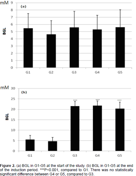
The animals were treated with the TFGN after an induction period of 7 weeks. G2 received a high dose of 500 mg/kg TFGN solution (10.0 mg/mL); G4 received the TFGN solution (50 mg/kg); G5 received a dose of 50 mg/kg α-tocopherol acetate solution (10.0 mg/mL). G1 and G3 received water during the treatment period. Treatment was given once at approximately the same time interval of 24 h every day. The weights of animals were determined every 7 days. The presence of diabetes was confirmed by measuring blood glucose levels using a portable blood sugar monitor. During the induction period, the blood glucose level (BGL) was tested at equal intervals of 14 days. Rats with BGL less than 10 mM were excluded at the end of the induction period of 7 weeks. The BGL was measured at equal intervals of 4 days during the treatment period.
Sample preparation
At the end of the treatment period, plasma samples were collected from the arteria carotis in 1 ml tubes (Eppendorf) containing potassium EDTA (30 µl 7.5% EDTA/ml blood sample) and immediately centrifuged at 1000 × g for 12.5 min. The plasma samples were stored at -70°C.
Lipid peroxidation assay
A HPLC-fluorimetric detection method was used to determine the oxidative damage due to lipid peroxidation (Hermans et al., 2005). Briefly, this method quantifies MDA after reaction with thiobarbituric (TBA) in acid and heat conditions, and the resulting pink fluorescent complex is analysed by HPLC fluorescence detection.
Determination of fat-soluble antioxidants
For the determination of α- and γ-tocopherol and coenzyme Q plasma levels, an optimized and validated HPLC-coulometric detection method was used as described by the published method (Hermans et al., 2005).
Statistical analysis
SPSS 15.0 and GraphPad Prism 5 software packages was used to evaluate the data. The difference between the means was estimated using an appropriate test. All data are shown as mean ± standard error of mean (SEM). The Levene’s test was used to test the homogeneity of variances. If variances were unequal data were mathematically transformed (logarithmic or power transformation). A value of P < 0.05 was considered significant.
The BGL in G3 significantly increased at the end of this induction period, confirming the successful development of DM (Figure 2b). Moreover, the characteristic symptoms of DM such as the increased appetite, polydipsy, polyuria, and loss of weight were observed. From Figure 3, all animals which received oxidative stress displayed a significant decrease in body weight compared to G1, even after treatment with G4 or G5. There were no significant differences between the body weight of G4 or G5, with G3. As shown in Figure 4, the level of MDA-TBA complex in G3 was significantly promoted as compared to the G1 (P<0.001), which indicated the suitability of the chosen system for in vivo lipid peroxidation inhibitory activity evaluation.
Comparing to G3, there was a significant reduction in the oxidative stress damage in G4 and G5. However, the damage of lipid peroxidation was not decreased to G1. The level of lipid damage in G4 and G5 was similar. Thus, the G4 and G5 had similar effect on the in vivo oxidative stress status of the diabetic animals. The potent lipid peroxidation chain breaking activity in G3 compound with G2 or G4 produced a similar effect. From Figure 5, it is found that the level of plasma α-tocopherol in G3 significantly reduced compared to G1. However, there was no significant difference between G4 and G1 or G3. In addition, no significant difference was observed between G5 and G3.
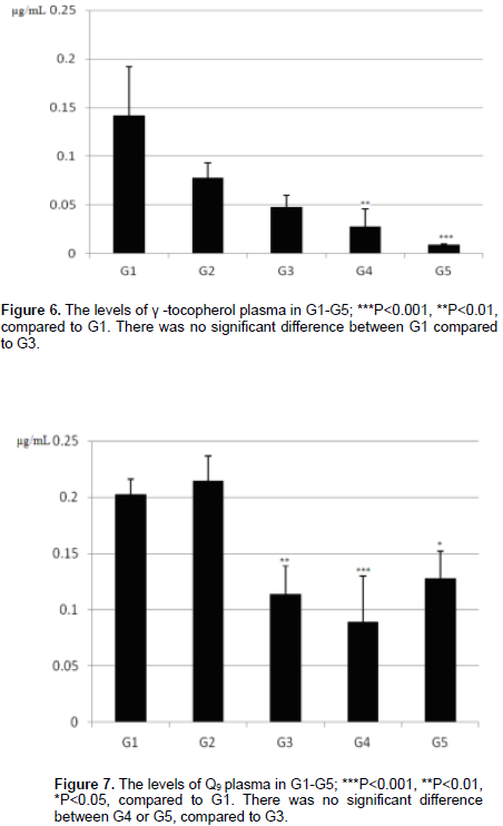
The G1 and G2 showed similar α-tocopherol levels. Concerning γ-tocopherol, there was no significant difference between G1 and G3 (Figure 6). It is surprising that the levels of γ-tocopherol in G4 and G5 were significantly reduced with the G1, and the observed levels were not different from G3. It should be noted that the levels of γ-tocopherol were very low. The discoveries showed that in G3, G4 and G5 where oxidative stress was induced almost complete depletion of γ-tocopherol had happened. Any positive influence of the treatment with TFGN on the level of γ-tocopherol under the condition of oxidative stress could not be observed. From Figure 7, the level of Q9 was higher in the G1 as compared to the G3.
The plasma level of Q9 in the G4 or G5 was different from the G1 but not from the G3. The diabetic animals in G3 and G4 displayed a significant decrease at this phase of the experiment. This might be an after effect of its depletion as a factor in the antioxidant defence against the induced oxidative stress, which was not equalized by upregulation of its synthesis in this case. There was no significant difference between G4 or G5 on the Q9 level compared to G3. Thus, the dynamics of the up-regulation and depletion of this molecule are needed to further investigate. For detecting potential toxic effects of TFGN, the animals in G2 received 10× the treatment dose (500 mg/kg). The animals in G2 were not injected with streptozotocin. With regard to the level of BGL, MDA-TBA complex, the fat-soluble antioxidants and body weight measured, no significant differences were observed between G1 and G2.