Full Length Research Paper
ABSTRACT
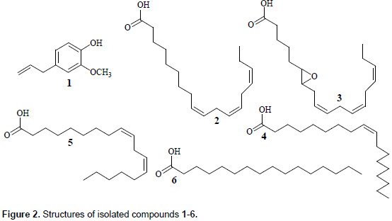
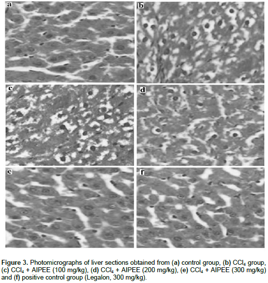
The aim of this study was to investigate the chemical constituents of petroleum ether extract from Artemisia integrifolia L (AIPEE) and to evaluate its hepatoprotective potential and in vivo antioxidant effects. Six compounds, namely eugenol (1), linolenic acid (2), 6,7-epoxy-linolenic acid (3), linoleic acid (4), oleic acid (5) and hexadecanoic acid (6) were isolated from the AIPEE. Oral administration of AIPEE significantly reduced carbon tetrachloride-induced elevations in the levels of plasma markers of hepatic damage and lipid peroxidation. It also rescued histopathologic alterations observed in the liver and levels of oxidative stress markers. AIPEE exhibited antioxidant and hepatoprotective activities in vivo, which may be attributable to its chemical constituents such as five fatty acids and eugenol.
Key words: Artemisia integrifolia L., fatty acids, eugenol, antioxidant, hepatoprotection.
INTRODUCTION
MATERIALS AND METHODS
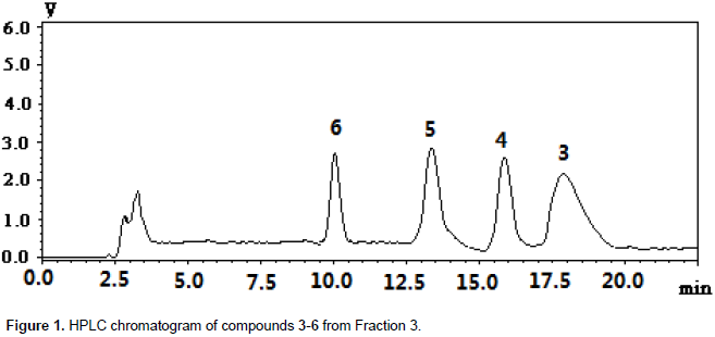
RESULTS
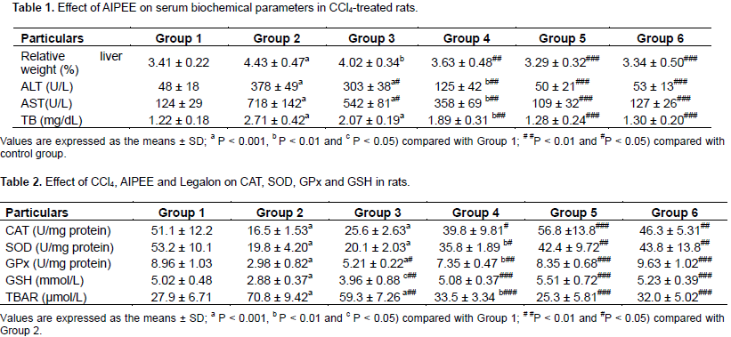
DISCUSSION
CONFLICT OF INTERESTS
ACKNOWLEDGEMENTS
REFERENCES
|
Bao-zhong Z, Lin L (2008). Influence of alpha-linoleic acid plus anti-oxidant agent on drosophila life and mouse antioxidation ability. ZHONGGUO ZUZHI GONGCHENG YANJIU YU LINCHUANG KANGFU, 12(7): 1264-1267. |
|
|
Das S, Vasishat S, Snehlata R, Das N, Srivastava LM (2000). Correlation between total antioxidant status and lipid peroxidation in hypercholesterolemia. Curr Sci. Bangalore 78(4):486-487. |
|
|
Deng XH, Zhang CJ, Wu Y, Qin LP (2013). Chemical Constituents of Whole Plant of Veronicastrum axillare (Sieb. et Zucc.) Yamazaki. Chin. Pharm. J. 48:777-781. |
|
|
Halliwell B (2012). Free radicals and antioxidants: Updating a personal view. Nutr. Rev. 70:257-265. |
|
|
Hu JF, Feng XZ (1999). New guaianolides from Artemisia selengensis. J. Asia. Nat. Prod. 1:169-176. |
|
|
Khan SA, Priyamvada S, Arivarasu NA, Khan S, Yusufi AN (2007). Influence of green tea on enzymes of carbohydrate metabolism, antioxidant defense, and plasma membrane in rat tissues. Nutrition 23:687-695. |
|
|
Liu JY, Chen CC, Wang WH, Hsu J, Yang M, Wang C (2006). The protective effects of Hibiscus sabdariffa extract on CCl4-induced liver fibrosis in rats. Food. Chem. Toxicol. 44:336-343. |
|
|
Liu R, Dong Q, Wang XH (2010). Study on the antioxidant activity of flavonoid in Artemisia selengensis. J. Sci. Technol. Food. Ind. 31:319-21. |
|
|
Megahed HA, Zahran HG, Arbid MS, Osman A, Kandll SM (2010). Comparative study on the protective effect of biphenyl dimethyl dicarboxylate (DDB) and silymarin in hepatitis induced by carbon tetrachloride (CCl4) in rats. N. Y. Sci. J. 3:1-11. |
|
|
Milena K, Carlos ABG, Gabriel AS, Naira JNB, Jorge AL, Sibele OT, Maria GA, Marilis DM, Didier S, Obdulio GM (2014). Chemical composition, antioxidant activity and hepatoprotective potential of Rourea induta Planch. (Connaraceae) against CCl4-induced liver injury in female rats. Nutrition 30:713-718. |
|
|
Mistry S, Dutt KR, Jena J (2013). Protective effect of Sida cordata leaf extract against CCl4 induced acute liver toxicity in rats. Asia. Pacif. J. Trop. Biom. 3:280-284. |
|
|
Setty SR, Quereshi AA, Swamy AV, Patil T, Prakash T, Prabhu K, Gouda AV (2007). Hepatoprotective activity of Calotropis procera flowers against paracetamol-induced hepatic injury in rats. Fitoterapia 78:451-454. |
|
|
Sharma P, Bodhankar SL, Thakurdeasi PA (2012). Protective effect of aqueous extract of Feronia elephantum correa leaves on thioacetamide induced liver necrosis in diabetic rats. Asian Pac. J. Trop. Biomed. 2:691-695. |
|
|
Silva GN, Martins FR, Matheus ME (2005). Investigation of anti-inflammatory and antinociceptive activities of Lantana trifolia. J. Ethnopharmacol. 100(3):254-259. |
|
|
Singh N, Kamath V, Narasimhamurthy K, Rajini PS (2008). Protective effect of potato peel extract against carbon tetrachloride-induced liver injury in rats. Environ. Toxicol. Pharmacol. 26(2):241-246. |
|
|
Srivastrivastava A, Shivanandappa T (2010). Hepatoprotective effect of the root extract of Decalepis hamiltonii against carbon tetrachloride induced oxidative stress in rats. Food Chem. 118(2):411-417. |
|
|
Talwar S, Jagani HV, Nayak PG, Kumar N, Kishore A, Bansal P, Shenoy RR, Nandakumar K (2013). Toxi-cological evaluation of Terminalia paniculata bark extract and its protective effect against CCl4-induced liver injury in rodents. BMC Complement. Altern. Med. 13:127-135. |
|
|
Tu Y, Liu XH (2007). The Daur people eating-Artemisia integrifolia. Chin. Folk. Ther. 15:10-11. |
|
|
Wang BH, Zuzel KA, Rahman K, Billington D (1998). Protective effects of aged garlic extract against bromobenzene toxicity to precision cut rat liver slices. Toxicology 126:213-222. |
|
|
Wang JL, Zhang GF, Dong LW, Zhao M, Zhang SJ (2010). Chemical constituents in seed crust of Syringa oblate. Chin. Trad. Herb. Drug. 41: 1598-1601. |
|
|
Wang MX, Huang FH, Liu CS, Wang JW, Huang QJ (2007). Study on antioxidative effects of natural antioxidants toα-ethyl linolenate. Chin. J. Oil Crop Sci. 29:466-469. |
|
|
Yang Q, Wang SW, Wang JB, XIE YH, LIU Z, SUN JY, MIAO S, MIAO Q (2008). Study on extraction and identification of α-linoleicacid from Semen Zanthoxyli Bungeani. Chin. New Drug J. 17:318-120. |
|
|
Yeum KJ, Beretta G, Krinsky NI, Russell RM, Aldini G (2009). Synergistic interactions of antioxidant nutrients in a biological model system. Nutrition 25:839-846. |
|
|
Zan LF, Bu T, Bao HY, Li Y (2008). Chemical composition in fruiting body of Cortinarius rufo-olivaceus. Mycosystema 27:284-288. |
|
|
Zhang GP, Yang JX, Zhu YA (2007). Study on antioxidant activity of Lithospermum erythrorhizon in vitro. J. Wuhan Bot. Res. 25:490-493. |
|
|
Zhang J, Li SJ, Xiang BP, He JG (2008). Modulation effects of Artemisia integrifolia on immue function of mice. Food. Sci. 29:405-408. |
|
Copyright © 2024 Author(s) retain the copyright of this article.
This article is published under the terms of the Creative Commons Attribution License 4.0


