Full Length Research Paper

ABSTRACT
The aim of this research was to explore the in vitro antioxidant, antibacterial and anticancer activities of flavonoid rich fraction from the leaves of Enicostemma axillare. The total phenolic and flavonoid contents were assessed by Folin–Ciocalteu and Aluminium chloride method respectively. Antioxidant activities of this plant was confirmed on the source of ABTS (2,2'-azino-bis-3-ethyl benzthiazoline-6-sulphonic acid), Radical scavenging assay, Inhibition of lipid peroxidation, Super oxide radical scavenging activity, Nitric oxide radical scavenging activity and Metal chelating activity. Correspondingly, antibacterial activities were accomplished by disc diffusion method and MIC (Minimum Inhibitory Concentration) against Staphylococcus aureus, Pseudomonas aeruginosa, Klebsiella pnemoniae, Escherichia coli and Enterococcus faecalis and anticancer activity was performed against human breast cancer cell MCF-7. The total phenolic and flavonoid content in flavonoid rich fraction of E. axillare was 136.8±13.00 mg GAE/g and 75.2±1.23 μg RE/g respectively. The flavonoid rich fraction of the leaves of E. axillare exhibited significant antioxidant activity. Similarly, the study on antibacterial activity of flavonoid rich fraction of E. axillare exposed inhibitory activity. However flavonoid rich fraction of E. axillare showed high inhibitory zone against E. coli, S. aureus, K. pneumoniae (18, 15 and 14 mm). The flavonoid rich fraction of E. axillare also unveiled strong cytotoxic effect with IC50 values of 15.39 μg/ml against MCF-7. This research work has made it clear that E. axillare possess excellent antioxidant, antibacterial and cytotoxic activity and the extracts can be more extensively used in developing countries for the prevention and treatment of ageing and infective associated diseases and may be considered as good source for drug discovery.
Key words: E. axillare, flavonoid rich fraction, antioxidant, antibacterial, cytotoxic activity.
INTRODUCTION
Plants have been used in ancient times as medicine to treat several diseases and medical complaints by most, if not all civilizations. Herbal-therapy in India is predominantly extensive and documented. Similarly plant based medicinal system remain to be the primary therapeutic system in various parts of India. Recently, consumer’s aspect to decrease the risk or accomplish a specific health ailment through improved food diet. Plants and fruits have diverse phytochemicals and enzymes as antioxidant defense to sustain growth and metabolism system (Pandhair and Sekhon, 2006). The research on antioxidant activity has been improved due to the apprehension about health enhancement involving agricultural products with their probable benefits (Moore et al., 2005). Antioxidants that are chiefly supplied as dietary ingestions can obstruct carcinogenesis by scavenging free radicals or interfering with binding of carcinogens to deoxyribonucleic acid. Several reports have exposed that the majority of the antioxidant activity may be from phytochemicals such as flavonoids, isoflavones, flavones, anthocyanins, catechins and other phenolics (Alothman et al., 2009; Isabelle et al., 2010).The antioxidative abilities of phenolic compounds can be accredited to their strong capacity to transfer electrons to reactive oxygen species or free radicals, chelating metal ions by stimulating antioxidant enzymes and inhibitory oxidases (Choi and Lee, 2009). In addition, free radicals and reactive oxygen species are continuously produced in-vivo and cause oxidative injury to biomolecules, a process held to check only by the presence of multiple antioxidants or repair systems as well as the replacement of injured lipids or proteins (Bapjpai et al., 2009).
As a consequence, many irredeemable human diseases including cancer, cardio and cerebro-vascular diseases have been recognized (Hossain et al., 2015). The possible ways to fight these irredeemable diseases is to progress our body’s transformation due to antioxidant defenses. High ingestion of plants, fruits and vegetables has lowered the incidence of such deteriorating or irredeemable diseases (Hossain et al., 2006). Plants can contribute in this area chiefly due to the antioxidant activity of polyphenolic compounds (Feng et al., 2006). Flavonoids that mainly exists as colouring pigments in plants too function as potent antioxidants at innumerable levels. Some reports exhibited that flavonoids could shield membrane lipids from oxidation (Cos et al., 1998). Microbial contamination is one of the major cause responsible to induce oxidative reactions which intern lead to cell damage (Bhargava et al., 2010; Cabiscol et al., 2000). Although many antimicrobials have been effectively used but remarkable flexibility and the development of resistance are major problems (Grant and Hung, 2013). Similarly, Cancer is also one of the world's shocking diseases leading to disease-related impermanence and anomalous growth of cells and tissues. Its treatment includes surgical, radiation, chemotherapeutic drugs which often destroys healthy cells and cause toxicity to humans. Drug designs for cancers are well evolving due to the overview of plant molecules. Among this, breast cancer is one of the most
common cancer and the prominent cause of cancer-associated deaths in females (Sorlie et al., 2001). It has been categorized into several subgroups based on the pathology and gene expression profiles, which subsidize to several responses to the same therapeutic strategies (Stockler et al., 2000).
Enicostemma axillare (Lam.) Raynal (Syn Enicostemma littorale Blume, Gentianaceae) is a perennial herb widely distributed all over India and common in coastal areas. According to Ethnobotanical information this plant is used to treat diabetes mellitus, rheumatism, abdominal ulcers, hernia, swelling, itching and insect poisoning. In pharmacological research, it’s been reported that this plant possess anti-inflammatory, hypoglycemic and anticancer activities (Roy et al., 2010). Swertiamarin, alkaloids, steroids, triterpenoids, saponins, flavonoids, xanthones, phenolic acids, etc. were isolated from the E. axillare (Ghosal et al., 1974). Keeping this in view, there has been considerable interest by the industry and a growing trend in consumer preferences for natural antioxidants and antimicrobial compounds and elimination of synthetics food applications has given more impetus to explore natural sources. Up to date, the search for new natural antioxidant, antimicrobial and anticancer agents from plants remain a potential area of investigation. Thus, they are of interest to both food and health scientists and there has been a convergence of interest among researchers in these fields and their impact on human health has come under attention. The study is therefore designed to determine antioxidant, antimicrobial and anticancer activity of flavonoid rich fractions from the leaves of E. axillare.
MATERIALS AND METHODS
Plant materials
Leaves of E. axillare were collected from Government Siddha Medical College, Herbal Garden, Chennai 600 106, Tami Nadu, India. Plant was authenticated by Dr. S. Sankaranarayanan, Head, Department of Medicinal Botany, Government Siddha Medical College, Arumbakkam, Chennai-600 106, Tamilnadu.
Extraction of flavonoid rich fraction
The Leaves of E. axillare were dried in hot air oven at (40°C) for 1h, after which it was pulverized to uniform powder with house hold mixer grinder. The aqueous extract was prepared by soaking 100 g of the dry powdered plant materials in 500 ml of aqueous at 4°C for 24 h. The extract was filtered first through a What mann filter paper No. 42 (125 mm) and then centrifuged at 5000 rpm for 10 min (Remi-R-8C, India). The clear solution was separated with petroleum ether and chloroform for exclusion of other than flavonoid compound and finally aqueous extracts were partitioned with butanol. It was condensed using a rotary evaporator with the water bath set at 40°C. The percentage yield of extracts extended from 7 to 19% w/w.
Determination of total phenolics
The concentration of total phenolics in the flavonoid extract of E. axillare was resolute by using Folin-Ciocalteu reagent and standardized externally with gallic acid. Briefly, 0.2 ml of flavonoid rich fraction and 0.2 ml of Folin-Ciocalteu reagent were added and mixed strongly. After shaking for 4 minutes, 1 ml of 15% Na2CO3 was added, and finally the combination was allowed to stand for 2 h at room temperature. The absorbance was measured at 760 nm using Deep Vision 1371 spectrophotometer. The concentration of the total phenolic was assessed as mg of gallic acid comparable by using an equation obtained from gallic acid calibration curve. The quantification of phenolic content was carried out in triplicate and the results were averaged (Singleton et al., 1999).
Determination of flavonoid content
The amount of total flavonoids in the extract was measured according to Quettier-Deleu et al. (2000). This method is based on the formation of a complex flavonoid-Aluminium, with the absorbance maximum at 430 nm. Rutin was used to brand a calibration curve. To 1 ml of flavonoid rich fraction, added 1 ml of 2% AlCl3 and it was incubated at room temperature for 15 min. Then absorbance was measured at 430 nm using Deep Vision 1371 spectrophotometer.
Invitro antioxidants properties
ABTS (2,2'-azino-bis-3-ethyl benzthiazoline-6-sulphonic acid) radical scavenging assay
ABTS radical scavenging activity of flavonoid rich fraction of E. axillare was followed by Re et al. (1999). ABTS radical was newly prepared by addition 5 ml of 4.9 mM potassium persulfate solution to 5 ml of 14 mM ABTS solution and kept for 16 h in dark. This solution was diluted with distilled water to produce an absorbance of 0.70 at 734 nm and the same was used for the antioxidant activity. The final solution of standard group was made up to 1 ml with 950 μl of ABTS solution and 50 μl of Ascorbic acid. Correspondingly, in the experiment group, 1 ml reaction mixture encompassed 950 μl of ABTS solution and 50 μl of different concentration of flavonoid rich fraction. The reaction mixture was vortexed for 10 s and after 6 min, absorbance was recorded at 734 nm against distilled water by using a Deep Vision (1371) UV–Vis Spectrophotometer and compared with the control ABTS solution. Ascorbic acid was used as reference antioxidant compound. ABTS Scavenging Effect (%) = [(A0- A1/A0) ×100] Where A0 is the absorbance of the control reaction and A1 is the absorbance of flavonoid rich fraction.
Inhibition of lipid peroxidation activity
Lipid peroxidation induced by Fe2+ascarbate system in egg yolk was assessed as thiobarbituric acid reacting substances (TBARS) by the method of Ohkawa et al. (1979). The experimental mixture contained 0.1 ml of egg yolk (25% w/v) in Tris-HCl buffer (20 mM, pH 7.0); KCl (30 mM); FeSO4 (NH4)2SO4.7H2O (0.06 mM); and different concentrations of flavonoid rich fraction of E. axillare in a final volume of 0.5 ml. The experimental mixture was incubated at 37°C for 1 h. After the incubation period, 0.4 ml was collected and treated with 0.2 ml sodium dodecyl sulphate (SDS) (1.1%); 1.5 ml thiobarbituric acid (TBA) (0.8%); and 1.5 ml acetic acid (20%, pH 3.5). The final volume was made up to 4.0 ml with distilled water and then kept in a water bath at 95 to 100 °C for 1 hour. After cooling, 1.0 ml of distilled water and 5.0 ml of n-butanol and pyridine mixture (15:1 v/v) were added to the reaction mixture, shaken vigorously and centrifuged at 4000 rpm for 10 min. The absorbance of butanol-pyridine layer was recorded at 532 nm in Deep Vision (1371) UV–Vis Spectrophotometer) to quantify TBARS. Inhibition of lipid peroxidation was determined by comparing the optical density (OD) of test sample with control. Ascorbic acid was used as standard.
Inhibition of lipid peroxidation (%) by the flavonoid rich fraction was calculated according to 1-(E/C) × 100, where C is the absorbance value of the fully oxidized control and E is absorbance of the test sample (Abs532+TBA–Abs532+TBA).
Superoxide radical scavenging assay
This assay was based on the capacity of the flavonoid rich fraction to inhibit the photochemical reduction of Nitroblue tetrazolium (NBT) in the presence of the riboflavin-light-NBT system (Tripathi and Pandey Ekta, 1999; Tripathi and Sharma, 1999). Each 3 ml reaction solution contained 50 mM phosphate buffer (pH 7.8), 13 mM methionine, 2 μM riboflavin, 100 μM Ethylene diamine tetra acetic acid (EDTA), NBT (75 μM) and different concentration of flavonoid rich fraction. It was kept visible in fluorescent light and absorbance was taken after 6 min at 560 nm by using a Deep Vision (1371) UV–Vis Spectrophotometer. Identical tubes with reaction mixture were kept in the dark served as blanks. The percentage inhibition of superoxide radical activity was measured by comparing the absorbance of the control with test sample solution:
% Super oxide radical scavenging capacity= [(A0-A1)/A0] ×100 Where A0 was the absorbance of control and A1 was the absorbance of flavonoid rich fraction.
Nitric oxide radical scavenging activity
Nitric oxide scavenging ability of flavonoid rich fraction of E. axillare was measured according to the method described by Olabinri et al. (2010). 0.1 ml of sodium nitroprusside (10 mM) in phosphate buffer (0.2 M, pH 7.8) was mixed with different concentration of flavonoid rich fraction and incubated at room temperature for 150 min. After treated period, 0.2 ml of Griess reagent (1% Sulfanilamide, 2% Phosphoric acid and 0.1% N-(1-Naphthyl) ethylene diamine dihydrochloride) was added. The absorbance of the experimental sample was read at 546 nm against blank. All readings were taken in triplicate and ascorbic acid was used as standard. The percentage of inhibition was calculated by following equation:
% Nitric oxide radical scavenging capacity= [(A0-A1)/A0] ×100
Where A0 was the absorbance of control and A1 was the absorbance of flavonoid rich fraction.
Metal chelating activity
Metal chelating capacity of flavonoid rich fraction of E. axillare was measured according to Iihami et al., (2003). 1 ml of different concentrations of flavonoid rich fraction was added to 0.05 ml of 2 mM ferric chloride solution. The reaction was initiated by the addition of 0.2 ml of 5 mM Ferrozine and the mixture was shaken vigorously. After 10 min, the absorbance was measured at 562 nm against blank. All readings were taken in triplicate and ascorbic acid was used as standard. The % inhibition of ferrozoine-Fe2+complex was calculated by following equation.
% Inhibition of ferrozoine-Fe2+complex = [(A0-A1)/A0] ×100
Where A0 was the absorbance of control and A1 was the absorbance of flavonoid rich fraction.
GC–MS analysis
The flavonoid fraction was analyzed by GC–MS (QP-2010, Shimadzu Co., Kyoto, Japan) equipped with 30- mX 0.25 mm DB-5MS column (Agilent Technologies, J&W Scientific Products, Folsom, CA). The carrier gas was helium. The temperature program was set as follows: 100°C hold for 5 min, raised at 4°C/min to 280°C, and hold for 5 min. The injector and detector temperatures were set at 250 and 280°C, respectively. The ion source and interface temperatures were set at 200 and 250°C, correspondingly. The mass range was scanned from 50 to 900 amu. The control of the GC–MS system and the data peak processing were controlled by Shimadzu’s GC–MS solution software, version 2.4. Compound identification was confirmed based on the relative retention time and mass fragmentation pattern spectra with those of standards and the NIST147. LIB. Database of the GC–MS system (Shimadzu).
Antibacterial properties
Bacterial strains
Bacteria used for the assessing antibacterial activities were Gram positive viz; Staphylococcus aureus MTCC 29213, Klebsiella pneumoniae MTCC 1771 and Enterococcus faecalis MTCC 439 and gram negative viz; Pseudomonas aeruginosa MTCC 2488, and Escherichia coli MTCC 25922. The bacterial strains were obtained from Microbial Type Culture Collection and Gene Bank, Institute of Microbial Technology Sector 39-A, Chandigarh-160036, India. All bacterial strains were sub cultured on nutrient agar medium, incubated at 37°C for 24 h and stored at 4°C in refrigerator to maintain stock culture.
Antibacterial assay
Antibacterial activity was assessed using disc diffusion method (Velickovic et al., 2003). Petriplates were prepared with 20 ml of sterile nutrient agar (HIMEDIA). The tested cultures were swabbed on top of the solidified media and allowed to dry for 10 min. The test was conducted in four different concentrations of the flavonoid rich fraction (5, 10, 15 and 20 μl/ml) and treated discs (Whatman No.1 filter paper was used to prepare discs) were air dried. The treated discs were placed on the surface of the medium and incubated at room temperature for 24 h. The relative inhibition of organisms to the flavonoid rich fraction was indicated by a clear zone of inhibition around the discs. It was then detected, measured and documented in millimeters with three replicates.
Cytotoxicity screening
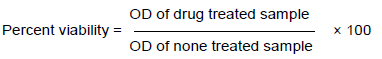
The cytotoxicity of flavonoid rich fraction of E. axillare was determined by Methyl tetrazolium (MTT) assay (Selvakumaran et al., 2003). Cells (2×10³/well) were placed in 100 μl of medium/well in 96-well plates. After overnight incubation, flavonoid rich fraction was added in 5 wells for each concentration. After treating with flavonoid rich fraction for 1st, 2nd, 3rd , 4th , and 5th day, 20 μl of 5 mg/ml MTT (pH 4.7) was added to each well and cultured for another 4 h. The supernatant was eliminated and 100 μl DMSO was added per well. After shaking the samples for 15 min. the absorbance was measured at 570 nm with microplate reader (Bio-Rad), using wells without cells as blanks. All experiments were performed in triplicate. The treated cell line was expressed relatively on its cell viability, using the following formula.
Statistical analysis
The impact of the flavonoid rich fraction of E. axillare on its antioxidant activity was measured by the ABTS assay, lipid peroxidation, superoxide scavenging, metal chelating and nitric oxide radical were determined using one-way analysis of variance (ANOVA). Likewise, Duncan’s post hoc test was applied, so as to determine the statistically significant different values. All statistical handling was performed using SPSS software, v. 14.0 (SPSS, Chicago, Ill., U. S. A.).
RESULTS AND DISCUSSION
Total phenolic and flavonoid content
E. axillare exhibited the highest amount of total phenolic (172.08±13.00 mg/g) and flavonoid contents (75.2±1.23 μg RE/g). Flavonoids has extensive health benefits, as anti-allergic, anti-inflammatory, analgesic, and cardio protective, hepatoprotective, anticancer, antiviral activities. These diverse properties of Flavonoids are due to its varied structures (Agrawal, 2011). The above data in accordance with previous researches (Maisuthisakul et al., 2007), had revealed that the high total polyphenols content increases antioxidant activity and there was a linear correlation between phenolic and flavonoid content for antioxidant activity.
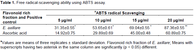
Free radical-scavenging ability using ABTS assay
The radical scavenging ability was measured by ABTS assay as per given in table 2. The inhibition percentage of the ABTS radical activity was assessed on average and high free radical-scavenging values were found in flavonoid rich fraction of E. axillare. In ABTS assay, the EC50 of the pure ascorbic acid (29.97 µg/ml) was lower than E. axillare, (18.97 µg/ml) (Table 1). Nevertheless, in our study, it is showed that these activities were mainly due to phenolics and flavonoids. It is known that vitamin C (ascorbic acid) and carotenoids are chief source of discrepancy of antioxidant/ antiradical activities in plant materials. Although these components were not explored, their contributions towards antioxidant/ antiradical activities of these studied medicinal plants may be insignificant. The presence of phenolic compounds in plants shows a synergic effect of antioxidants in association with phytochemicals (Kumar and Pandey, 2013; Teow et al., 2007).
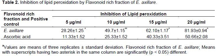
Inhibition of lipid peroxidation
Flavonoid rich fraction of E. axillare also inhibited the lipid peroxidation induced by ferrous sulfate in egg yolk homogenates. Maximum inhibition was recorded in flavonoid rich fraction of E. axillare with EC50 value at 9.28 μg/ml and ascorbic acid 32.98 μg/ml. As it is identified that lipid peroxidation is the net result of any free radical attack on membrane and other lipid components present in the system, the lipid peroxidation may be enzymatic (Fe/NADPH) or non-enzymatic (Fe/ascorbic acid). In the present study, egg yolk was used as substrate for free radical mediated lipid peroxidation, which is a non-enzymatic method. Significantly, E. axillare suppressed the degree of lipid peroxidation than positive control (Table 2). Normally, the mechanism of phenolic compounds for antioxidant activity includes neutralizing lipid free radicals and preventing decomposition of hydroperoxides into free radicals (Prior et al., 2005). In previous studies it is recommended that the Pasteurization of blackberry juice preserves polyphenol-dependent inhibition for lipid peroxidation and intracellular radical (Kim et al., 2002; Gabriela et al., 2015).
Superoxide scavenging assay activity
Flavonoid rich fraction of E. axillare exhibited powerful scavenging activity for superoxide radicals in a concentration dependent process than positive control. Flavonoid rich fraction of E. axillare showed EC50 values of 19.39 μg/ml and the positive control with an EC50 value of 20.50 μg/ml (Table 3). One of the standard method to produce Superoxide radicals is through photochemical reduction of nitro blue tetrazolium (NBT) in the presence of a riboflavin-light-NBT system. These superoxide radicals are extremely toxic and may be produced either through xanthine activity or through mitochondrial reaction. Superoxide radicals are reasonably a weak oxidant may decompose to form stronger reactive oxidative species, such as singlet oxygen and hydroxyl radicals (Dirk et al., 2003). The present study recommended that E. axillare possessed strong scavenging activity for superoxide radicals in a concentration dependent process than positive control. The presence of secondary metabolites like Flavonoids, carotenoids and triterpenes in higher plants have excellent antioxidant activity by scavenging reactive oxygen species which prevent possible damage to cellular components such as DNA, proteins and lipids (Ksouri et al., 2013).

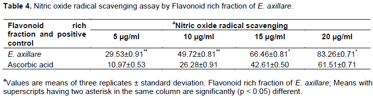
Nitric oxide radical scavenging assay
Nitric oxide radical quenching activity of the Flavonoid rich fraction of E. axillare was identified and compared with the standard ascorbic acid. The flavonoid rich fraction of E. axillare displayed the maximum inhibition of 68% at a concentration of 20 μg/ml with an EC50 value of 29.42 μg/ml, in a concentration-dependent process when compared to ascorbic acid with EC50 value of 59.48 μg/ml (Table 4). In the current study, nitrite was produced by incubation of sodium nitroprusside in standard phosphate saline buffer at 25°C was reduced by flavonoid rich fraction. Significant scavenging activity may be due to the antioxidant property of flavonoid, which compete with oxygen to react with nitric oxide, leading to less production of nitric oxide (Alasalvar et al., 2006).
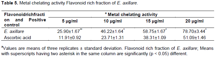
Metal chelating activity
The metal chelating property of Flavonoid rich fraction of E. axillare was displayed as per Table 5. The Flavonoid rich fraction of E. axillare was evaluated for their ability to compete with ferrozine for ferrous iron in the solution. In this evaluation, the leaf extract of E. axillare hindered the formation of ferrous and ferrozine complex, signifying that they have chelating activity and are capable of capturing ferrous iron before ferrozine. The Flavonoid rich fraction of E. axillare reduced the red color complex immediately and showed the highest chelating activity (EC507.59 μg/ml) than positive control (EC5027.11 μg/ml). It’s concluded that flavonoid compounds in extract would be responsible for antioxidant activities and reducing ferric ions might not be directly involved in ferrous ion chelation. This result was in accordance with the data that chelating agents, that form σ-bonds with a metal, are effective as secondary antioxidants as they reduce the redox potential, there by stabilizing the oxidized form of the metal ion (Kumaran and Karunakaran, 2006).
Anti-bacterial activity of Flavonoid rich fraction of E. axillare tested against pathogenic bacteria
Antibacterial activities of the Flavonoid rich fraction of E. axillare was tested against pathogenic organisms as per Table 6. The plants vary in their activities against the micro-organisms tested. Flavonoid rich fraction of E. axillare exhibited maximum antibacterial activity against Klebsiella pneumoniae, Escherichia coli, Enterococcus faecalis, Staphylococcus aureus than Pseudomonas aeruginosa. Highest antibacterial activity was recorded with flavonoid rich fraction of E. axillare against E. coli, S. aureus, K. pneumoniae (18, 15 and 14 mm) respectively while lowest activity was recorded against E. faecalis, and P. aeruginosa with the inhibition zone of 13.9 and 13.4 mm. The current investigation revealed that Flavonoid rich fraction has potential antibacterial activity against entire tested organisms. Similar investigation was reported in methanol extract exhibiting strongest and broadest spectrum of anti-bacterial activity (Chan et al., 2007). Previous research reveals that plant extracts are less active in Gram Negative bacteria than Gram Positive bacteria as it has an outer membrane consisting of lipoprotein and lipopolysaccharide, which is selectively permeable and thus regulates access to the underlying structures (Ghadir et al., 2014).

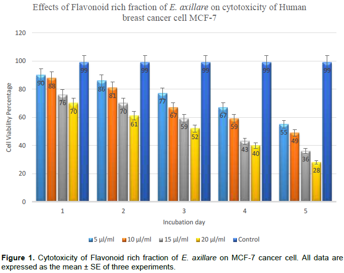
Effects of cytotoxicity on human breast cancer cell MCF-7
Cytotoxic effects of Flavonoid rich fraction of E. axillare was observed by using MTT assay against human breast cancer cell lines MCF-7 for daily treatment of 24, 48, 72, 96 and 120h, respectively. As shown in Figure 1, Flavonoid rich fraction of E. axillare displayed strong cytotoxic effect with IC50 values of 15.39 μg/ml against MCF-7. Cell morphological variations too indicated that the treatment of breast cancer cells at various doses of flavonoid rich fraction reflected its significant cytotoxicity to human breast cancer cells at doses of 5 μg/ml and above with high cytotoxic effect. Current investigation recommended that the flavonoid rich fraction of E. axillare, with a high phenolic content, possesses stronger anticancer activity. Natural compounds copious in plants are capable of arresting cell growth in tumor cells and modulating cell signaling pathways associated to cell death (Zou and Chang, 2011). The present study was in accordance with the previous findings which revealed that poly-phenolic extracts from several plants exhibited high efficiency in antitumor activity (Pan et al., 2008).
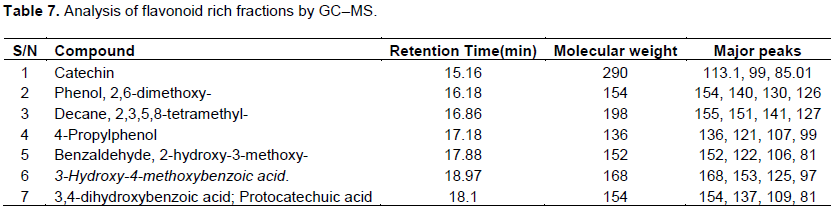
Analysis of flavonoid rich fractions by GC–MS
The flavonoid rich fraction of the E. axillare ensured highest flavonoid content and exhibited the strongest antioxidant activity (Table 7). It was therefore analyzed by GC–MS to determine its chemical composition that may contribute to this activity. The GC–MS analysis showed a variety of phenolic compounds (Figure 2).

CONCLUSION
The flavonoid rich fraction of E. axillare can protect the body from oxidative stress may be due to the existence of phytochemicals in the form of flavonoids compounds. These phytochemicals are present in this plant and it contributes to their medicinal properties that can be used in nutraceuticals and pharmaceutical industry. Nevertheless, additional studies are essential to develop a technique for the fractionation and identification of most active antioxidant, antibacterial and anticancer molecules and thus can be used in the prevention and treatment of ageing and infective related diseases and can be considered as good source for drug discovery.
CONFLICT OF INTERESTS
The authors have not declared any conflict of interests.
ACKNOWLEDGEMENTS
Authors take great pleasure in expressing their heartfelt thanks to the Directors of Gloris Biomed Research Centre (P) Ltd, No. 171, Fourth St, Kannigapuram, K.K. Nagar Chennai-78 for the invaluable guidance and supervision throughout the work and permission to use the facilities available in the laboratory. We are deeply indebted to the Principal, Guru Nanak College for the constant support and guidance to carry out the work.
REFERENCES
|
Agrawal AD (2011). Pharmacological activities of flavonoids: a review. Int. J. Pharm. Sci. Nanotechnol. 4(2):1394-1398. |
|
|
Alasalvar C, Karamac M, Amarowicz R, Shahidi F (2006). Antioxidant and antiradical activities in extracts of hazelnut kernel (Corylusa vellana L.) and hazelnut green leafy cover. J. Agric. Food Chem. 54:4826-4832. |
|
|
Alothman M, Bhat P, Karim AA (2009). Antioxidant capacity and phenolic content of selected tropical fruits from Malaysia, extracted with different solvents. Food Chem. 115:785-788. |
|
|
Bapjpai VK, Yoon JI, Kang SC (2009). Antioxidant and antidermatophytic activities of essential oil and extracts of Metasequoia glyptostroboides Miki ex Hu. Food Chem. Toxicol. 47:1355-1361. |
|
|
Bhargava A, Khan S, Panwar H, Pathak N, Punde RP, Varshney PK, Mishra PK (2010). Occult hepatitis B virus infection with low viremia induces DNA damage, apoptosis and oxidative stress in peripheral blood lymphocytes. Virus Res. 53:143-50. |
|
|
Cabiscol E, Tamarit J, Ros J (2000). Oxidative stress in bacteria and protein damage by reactive oxygen species. Int. Microbiol. 3:3-8. |
|
|
Chan EWC, Lim YY. Mohammed Omar (2007). Antioxidant and antibacterial activity of leaves of Etlingera species (Zingiberaceae) in Peninsular Malaysia. Food Chem. 104:1586-1593. |
|
|
Choi Y, Lee J (2009). Antioxidant and antiproliferative properties of a tocotrienol rich fraction from grape seeds. Food Chem. 114:1386-1390. |
|
|
Cos P, Ying L, Calomme M, Hu JP, Cimanga K, Van Poel B, Pieters L, Vlietinck AJ, Vanden Berghe D (1998). Structure activity relationship and classification of flavonoids as inhibitors of xanthine oxidase and superoxide scavengers. J. Nat. Prod. 61:71-76. |
|
|
Dirk T, Thomas B, Andreas L, Petra C, Steffi H, Reinhard B, Wolfgang K, Renate R (2003). Reaction rate constants of superoxide scavenging by plant antioxidants. Free Rad. Biol. Med. 35(12):1599-607. |
|
|
Feng ZQ, Ni KF, He Y, Ding ZS, Zhu F, Wu LG, Shen MH (2006). Experimental study on effect of Tetrastigma hemsleyanum Diels et Gilg Flavone on apoptosis of SGC-7901 cell line in vitro. Chin. J. Clin. Pharmacol. Ther. 11(6):669-672. |
|
|
Gabriela A, Silvia Q, Ana MP, Fabrice V, Alain M (2015). Pasteurization of blackberry juice preserves polyphenol-dependent inhibition for lipid peroxidation and intracellular radicals. J. Food Composition Anal. 42:56-62. |
|
|
Ghadir AEl-C, Abeer FA, Eman SR (2014). Evaluation of the antioxidant and antibacterial properties of various solvents extracts of Annona squamosa L. leaves. Arab J. Chem. 7:227–233. |
|
|
Ghosal SS, Sharma AK, Chaudhuri PV (1974). Chemical constituents of Gentianaceae IX: natural occurrence of Erythrocentaurin in Enicostemma hissopifolium and Swertia lawii. J. Pharm. Sci. 63:944-945. |
|
|
Grant SS, Hung DT (2013). Persistent bacterial infections, antibiotic tolerance, and the oxidative stress response. Virulence 4(4):273-283. |
|
|
Hossain MA, Salehuddin SM, Ismail Z (2006). Rosemarinic acid and methyl rosmarinate from Orthosiphon stamineus, Benth. J. Bangl. Acad. Sci. 30(2):167-171. |
|
|
Hossain MA, Shah MD (2015). A study on the total phenols content and antioxidant activity of essential oils and different solvent extracts of endemic plant Merremia borneensis. Ara. J. Chem. 8: 66-71. |
|
|
Iihami G, Emin BM, Munir O, Irfan KO (2003). Antioxidant and analgesic activities of turpentine of Pinusnigra arnsubsppallsian A (Lamb) Holmboe. J. Ethnopharmacol. 86:51-88. |
|
|
Isabelle M, Lee BL, Lim MT, Koh MT, Huang D, Nam C (2010). Antioxidant activity and profiles of common fruits in Singapore. Food Chem. 123:77-84. |
|
|
Kim DO, Lee KW, Lee HJ, Lee CY (2002). Vitamin C equivalent antioxidant capacity (VCEAC) of phenolic phytochemicals. J. Agric. Food Chem. 50:3713-3717. |
|
|
Ksouri WM, Medini F, Mkadmini K, Legault J, Magne C, Abdelly C (2013). LC-ESI-TOF-MS identification of bioactive secondary metabolites involved in the antioxidant, anti-inflammatory and anticancer activities of the edible halophyte Zygophyllum album Desf. Food Chem. 139:1073-1080. |
|
|
Kumar S, Pandey AK (2013). Chemistry and biological activities of flavonoids: an overview. Sci. World J. pp. 1-16. |
|
|
Kumaran A, Karunakaran RJ (2006). Antioxidant and free radical scavenging activity of an aqueous extract of Coleus aromaticus. Food Chem. 97:109-114. |
|
|
Maisuthisakul P, Suttajit M, Pongsawatmanit R (2007). Assessment of phenolic content and free radical-scavenging capacity of some Thai indigenous plants. Food Chem. 100:1409-1418. |
|
|
Moore J, Hao Z, Zhou Z, Luther Z, Costa JYL (2005). Phenolic content and antioxidant activity of extracts from whole buckwheat (Fagopyrum esculentum Moench) with or without microwave irradiation. J. Agri. Food Chem. 53:6649-6657. |
|
|
Ohkawa H, Ohisi N, Yagi K (1979). Assay for lipid peroxides in animals tissue by thiobarbituric acid reaction. Anal. Biochem. 95:351-358. |
|
|
Olabinri BM, Odedire OO, Olaleye MT, Adekunle AS, Ehigie LO, Olabinri PF (2010). In vitro evaluation of hydroxyl and nitric oxide radical scavenging activities of artemether. Res. J. Biol. Sci. 5: 102-105. |
|
|
Pan MH, Ghai G, Ho CT (2008). Food bioactives, apoptosis, and cancer. Mol. Nutri. Food Res. 52:43-52. |
|
|
Pandhair V, Sekhon BS (2006). Reactive oxygen species and antioxidants in plants. An overview. J. Plant Biochem. 15: 71-80. |
|
|
Prior RL, Wu XL, Schaich K (2005). Standardized methods for the determination of antioxidant capacity and phenolics in foods and dietary supplements. J. Agric. Food Chem. 53:4290-4302. |
|
|
Quettier-Deleu C, Gressier B, Vasseur J, Dine T, Brunet C, Luyckx M (2000). Phenolic compounds and antioxidant activities of buckwheat (Fagopyrum sculentum Moench) hulls and flour. J. Ethnopharmacol. 72:35-42. |
|
|
Re R, Pellegrini N, Proteggente A, Pannala A, Yang M, Rice-Evans C (1999). Antioxidant activity applying an improved ABTS radical cation decolorizing assay. Free Rad. Biol. Med. 26:1231-1237. |
|
|
Roy SP, Niranjan CM, Jyothi TM, Shankrayya MM, Vishawanath KM, Prabhu K (2010). Antiulcer and anti-inflammatory activity of aerial parts of Enicostemma littorale Blume. J. Young Pharm. 2(4):369-373. |
|
|
Selvakumaran M, Pisarcik DA, Bao R, Yeung AT, Hamilton TC (2003). Enhanced cisplatin cytotoxicity by disturbing the nucleotide excision repair pathway in ovarian cancer cell lines. Cancer Res. 63: 1311. |
|
|
Singleton VL, Orthofer R, Lamuela-Raventos RM (1999). Analysis of total phenols and other oxidation substrates and antioxidants by means of Folin–Ciocalteu reagent. Methods Enzymol. 299:152-178. |
|
|
Sorlie T, Perou CM, Tibshirani R, Aas T, Geisler S, Johnsen H, Hastie T, Eisen MB, van de Rijn M, Jeffrey SS, Thorsen T, Quist H, Matese JC, Brown PO, Botstein D, Lønning PE, Børresen-Dale AL (2001). Gene expression patterns of breast carcinomas distinguish tumor subclasses with clinical implications. Proc. Natl. Acad. Sci. U. S. A. 98:10869-10874. |
|
|
Stockler M, Wilcken NR, Ghersi D, Simes RJ (2000). Systematic reviews of chemotherapy and endocrine therapy in metastatic breast cancer. Cancer Treat. Rev. 26:151-168. |
|
|
Teow CC, Truong VD, Mc Feeters RF, Thompson RL, Pecota KV, Yencho GC (2007). Antioxidant activities, phenolic and b-carotene contents of sweet potato genotypes with varying flesh colours. Food Chem. 103:829-838. |
|
|
Tripathi YB, Pandey Ekta (1999). Role of alcoholic extract of shoot of H. perforatum (Lim) on LPO and various species of free radicals in Rats. Indian J. Exp. Biol. 37:567-571. |
|
|
Tripathi YB, Sharma M (1999). The Interaction of R. cordifolia with iron redox status: mechanistic aspects in FR reactions. Phytomedicine 6: 51-57. |
|
|
Velickovic AS, Smelcerovic AA (2003). Chemical constituents and antimicrobial activity of the ethanol extracts obtained from the flower, leaf and stem of Salvia officinalis L. J. Serb. Chem. Soc. 68:17-24. |
|
|
Zou Y, Chang SKC (2011). Effect of black soybean extract on the suppression of the proliferation of human AGS gastric cancer cells via the induction of apoptosis. J. Agric. Food Chem. 59:4597-605. |
|
Copyright © 2024 Author(s) retain the copyright of this article.
This article is published under the terms of the Creative Commons Attribution License 4.0


