Full Length Research Paper

ABSTRACT
The present work is focused on the evaluation of the Maytenus senegalensis roots effect on bronchial hyperleukocytosis and the search of its antioxidant potential both in vitro and in vivo. The roots of M. senegalensis (Celastraceae) are traditionally used for the treatment of cough and asthma. This study evaluates the effect of the hydroalcoholic extract of the roots of M. senegalensis on the bronchial hyperleukocytosis that occur during airway inflammation and determine its antioxidant capacity. In an eight days mouse asthma model sensitized to ovalbumin, the effect of the hydroethanolic extract of M. senegalensis on infiltration of leukocytes in general and eosinophils particularly in the airways was studied. The antioxidant activity of the extract was evaluated in vitro by the tests of DPPH, nitric oxide and AAPH then in vivo by the malondialdehyde dosage. The extract significantly inhibited bronchial infiltration of leukocytes in general, and eosinophils particularly (p<0.01; p<0.001). In vitro and in vivo, antioxidant tests revealed the reducing effect and the inhibitory of membrane lipoperoxidation potential of the extract. Phytochemical tests have shown that the extract contains polyphenols such as flavonoids. These compounds would be partly responsible for the antioxidant and anti-inflammatory activities of the extract. This study suggest that M. senegalensis roots would have an anti-inflammatory effect in asthma, which would be partially related to its antioxidant potential.
Key words: Maytenus senegalensis, asthma, bronchial inflammation, antioxidant.
INTRODUCTION
Asthma is one of the most common chronic respiratory diseases, which affect people of all ages around the world (Cukic et al., 2012). It is a major public health problem since it is one of the leading causes of hospitalization and death in the world. Approximately 339 million people worldwide develop this disease (The Global asthma report, 2018).
Asthma is defined as a heterogeneous disease characterized by chronic bronchial inflammation, airway hyper responsiveness leading to exaggerated bronchial smooth muscle contraction and mucus hypersecretion followed by enlarged mucus glands and reversible obstruction of bronchi (Pynn et al., 2012). According to Holgate et al. (2010), airway hyper responsiveness and bronchial obstruction are consequences of bronchial inflammation. In allergic asthma, inflammation is mainly orchestrated by Th2 lymphocytes and is characterized by an infiltration into the airways of leucocytes mainly eosinophils (Lambrecht et al., 2015). Eosinophils are the main effector cells in asthma. By releasing the contents of their granules, they cause epithelial damage, bronchial hyperreactivity, bronchospasm, activation of mast cells and increased vascular permeability (Gleich, 2000; Kay, 2005).
During inflammation in asthma, activated inflammatory cells of the airway excessively produce free radicals in response to the allergen. These free radicals cause in the airways several deleterious effects such as lipid peroxidation (Henricks and Nijkamp, ​​2001). They also enhance the expression of pro-inflammatory mediators and cytokines and thus the recruitment of leukocytes (Rangasamy et al., 2005); increase of contractile response of bronchial smooth muscle to acetylcholine (Barnes, 1990), bronchial hyperreactivity and mucus hypersecretion (Wood et al., 2003). Damage caused by theses radicals can lead to denaturation of bronchial epithelium and amplification of airway inflammation.
The existing asthma treatments (anti-inflammatories, bronchodilators) control only the symptoms of the disease and this conducts to long-term treatment. The high cost of the treatments weakening the economic situation and their side effects lead to the research of new therapies by people. Nowadays, using medicinal plants has become an attractive option, which populations are turning towards (Park et al., 2011).
Maytenus senegalensis (Lam.) Exell (Celastraceae) is a plant widely used in traditional medicine. Synonym of Gymnosporia senegalensis (Lam.) Loes, the plant is commonly called Confetti tree in English. It is known for its antiplasmodial (El Tahir et al., 1999; Malebo et al., 2015); analgesic (Sanogo et al., 2006); anthelmintic (Zangueu et al., 2018) and antibacterial properties (Lindsey et al., 2006, Jain et al., 2008). Its leaves and roots have been shown to possess anti-inflammatory activity in vitro and in eodema models induced by carrageenan or croton oil (Sosa et al., 2007; da Silva et al., 2011; Makgatho et al., 2018). Missebukpo et al. (2007) have also demonstrated its antitussive effect. There are no studies performed to evaluate the effect of M. senegalensis on airways inflammation that occurs in asthma.
The present study investigated the effect of the hydroetanolic extract of Maytenus senegalensis on bronchial inflammation exactly leukocyte infiltration in a murine model of asthma and then analyzed the antioxidant power of the extract.
MATERIALS AND METHODS
Plant material
The roots of M. senegalensis were harvested in August 2016 in Tsévié-Boloumondji, an area situated at 35 km at the northern part of Lomé. A specimen is authenticated by the Department of Ecology and Botany of the University of Lome. It is then deposited in the herbarium of the University of Lome (TG 15182). The extraction process is the one used by Metowogo et al. (2011). The roots were washed, cut, dried and crushed. The powder was macerated in a mixture of distilled water / ethanol (1: 1) for 72 h with intermittent manual stirring. The macerate obtained is filtered and then evaporated under vacuum at 45°C using a rotary evaporator (Buchi R120) until a dry extract (8.55% of yield).
Animals
Thirty ICR mice of both sexes weighing between 20 and 25 g from the animal house of the Department of Animal Physiology at the University of Lomé were used for this study. These mice were kept in chamber under a 12 h light / 12 h dark cycle and had free access to food and water. All the tests were carried out following the rules of WHO Guidelines for the care and use of human blood and laboratory animals and were approved by the national bioethics committee of University of Lomé-Togo.
EXPERIMENTAL PROTOCOL
Sensitization, challenge and treatment procedure
The sensitization of the mice was performed according to the method used by Metowogo (2010). Six groups of 5 mice were constituted. On days 0, 1 and 2, all the mice were sensitized by an intraperitoneal injection of 100 µl of a mixture of ovalbumin and aluminum hydroxide dissolved in 0.9% NaCl. On days 5, 6 and 7, the mice received by intranasal route ovalbumin (12,5 µl) for challenge except the control group who received NaCl 0.9%. Two hours earlier, the mice were treated in the following manner.
(i) Groups 1 (control) and 2 (untreated sensitized): the animals received orally distilled water.
(ii) Groups 3, 4 and 5 (sensitized and treated with the extract): the mice of these lots received the extract by single dose gavage respectively at 125, 250 and 500 mg/kg.
(iii) Group 6 (treated with the reference drug): this batch received cetirizine at 10 mg / kg.
Bronchoalveolar lavage and cell count
On day 8, 18 to 24 h after the last ovalbumin administration, all the mice were sacrificed and their trachea intubated. Using a syringe connected to the catheter, 4 ml of 0.9% NaCl was introduced into the lungs and then aspirated back into the syringe. For each mouse, a part of the resulting liquid (bronchoalveolar fluid, BAL) is taken under the microscope for counting total leukocytes using the Malassez cell; the rest is centrifuged at 700g for 10 min and the cells stained with May-Grunwald-Giemsa. The percentage of eosinophils was established after reading the stained slides based on morphological criteria (Missebukpo et al., 2011).
Evaluation of the antioxidant activity of the extract of M. senegalensis
DPPH test
The test has been done according to the method used by missebukpo et al., (2013) with some modifications. To DPPH (100 μmol/L), extract’s dilutions (62.5 to 500 μg / ml) were added for search its DPPH reduction effect. Ascorbic acid was used as standard control. For each concentration 3 trials were made. The control consists of a mixture of DPPH and methanol. Absorbances were read at 517 nm After 15 min incubation. The percentage of inhibition (PI = (A. control - A. sample / A. control) × 100) of each sample was calculated A = absorbance.
Nitric oxide assay
The nitric oxide scavenging effect of the extract was searched with the method used by Jagetia and Baliga (2004). A solution of sodium nitroprusside 5mM was added to dilutions of the extract and ascorbic acid (50, 100, 200 and 400 μg / ml). After 150 min of incubation, the Greiss reagent was added and the absorbance read at 546 nm. The percentages of inhibition were calculated like described above.
AAPH induced hemolysis assay
Blood from rats was centrifuged for separate erythrocytes from plasma. Dilutions of the extract and ascorbic acid with PBS were prepared (100, 150, 200, 250 and 500 µg/ml). To the erythrocyte suspension, was added the extract / ascorbic acid and AAPH (200 mM). For each concentration, 3 trials were made. The control was made up of the erythrocyte suspension, PBS and AAPH. All mixtures were incubated at 37°C for 3 h. After incubation, the degree of hemolysis was determined by measurement of the absorbance of each mixture with spectrophotometer at 540 nm. The percentages of inhibition were calculated (Missebukpo et al., 2013).
In vivo antioxidant activity: malondialdehyde (MDA) assay
At day 8 of the manipulation, two mice were selected respectively in groups 1, 2, 3, 4 and 5 and their lungs were removed. The level of MDA in these lungs was measured out as described previously (Missebukpo et al., 2013). To 175 µl of lung homogenate or MDA dilutions, were added successively 250 µl of 1M HCl; 100μl sodium dodecyl sulfate 9.8%; 1ml of thio-barbituric acid 0.67% and 330μl of distilled water. After incubation at 90°C, 2.5 ml of n-butanol was added and the whole was centrifuged at 1006.2 g for 10 minutes. The absorbance was read at 535nm and the concentration of MDA present in each lung was expressed in ng/mg lung tissue.
Phytochemical tests
Phytochemical screening
Staining tests were carried out on the extract in order to determine the large chemical groups it contains. Alkaloids, flavonoids, tannins, saponins, carbohydrate and reducing compounds were searched in the extract with the methods used by Karumi et al. (2004) and Edeoga et al. (2005).
Quantitative determination of phenols and total flavonoids
Determination of total phenols content: Five ml of folin-ciocalteu diluted to 10% and 5 ml of sodium carbonate were added to 500 μl of extract for determine total phenols it contains. The control and gallic acid dilutions were prepared in the same way. Absorbance was read at 765 nm after 15 min of incubation. The gallic acid curve serves to the extract absorbance calibration. This amount is expressed in mg gallic acid equivalent per gram of extract (Pourmorad et al., 2006).
Total flavonoids determination: 1.5 ml of methanol was added to 500 μl of the extract. Then 100 μl of aluminum chloride, 100μl of potassium acetate and 2.8ml of distilled water were successively added. The standard range was made with quercetin. Absorbance was read at 415nm after 30min of incubation. The absorbance of the extract calibrate with quercetin curve allow the determination of total flavonoids it contains. This quantity is expressed in mg quercetin equivalent per gram of extract (Pourmorad et al., 2006).
Statistical analysis
The data obtained were expressed as mean ± SEM. One way ANOVA followed by the bonferonni test for multiple comparisons serve to the results analysis. A p- value < 0.05 is considered as significant. The statistical software used is the version 6.01 of Graphpad prism (USA).
RESULTS AND DISCUSSION
Evaluation of the effect of the extract on airway inflammation
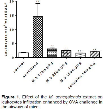
Figures 1 and 2 show the effect of the extract on the leukocytes (eosinophils) infiltration into the airways. The OVA-sensitization provoked in mice’s BALF a significant enhancement of the total leukocytes number and the percentage of eosinophils when compared to the control. Treatment with the extract and cetirizine provoked a significant reduction of these leukocytes number in BALF. The larger reduction’s activity is observed at the dose of 500 mg/kg. For both figures, each value represents the mean of the counted cells ± SEM with n = 5. ##p <0.001 (sensitized vs control); ** p <0.01; *** p <0.001 (treated vs sensitized). Control= saline group; sensitized= OVA group; M.S = groups treated with the extract.

Evaluation of the antioxidant activity of the hydro-ethanolic extract of M. senegalensis
In vitro tests
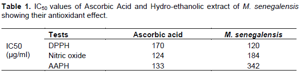
The percentages of inhibition of the extract obtained from the DPPH, nitric oxide and AAPH tests were used to determine the IC50 value of the extract in each test. This value represent respectively the half maximal DPPH reduction concentration, the 50% nitric oxide scavenging concentration and the half maximal inhibitory concentration of hemolysis induced by AAPH. The extract effect was compared to that of ascorbic acid (Table 1).
Malondialdehyde dosage
This test is carried out in order to evaluate oxidative stress, particularly lipid peroxidation in our asthma model and the effect of the M. senegalensis extract on the stress. In sensitized mice, a significant increase (p <0.01) of the MDA concentration is observed when compared to the control group. The extract induced a significant reduction (p <0.05, p <0.01) of this concentration at doses of 250 (38.91%) and 500 mg / kg (55.15%) (Figure 3).

Phytochemical tests
Phytochemical screening: The results obtained from the staining tests realized on the M. senegalensis hydro-ethanolic extract are summarized in the Table 2.

Total phenols and flavonoids determination: Gallic acid and quercetin served to determine the quantity of total phenols and total flavonoids present in the hydro-ethanolic extract of M.senegalensis. Total phenol amount was 45, 26 mg equivalent of gallic acid/g of extract and flavonoid was 29,59 mg equivalent of quercetin/g.
DISCUSSION
Bronchial inflammation is the main feature of asthma and its treatment constitute the basis of the disease treatment (Holgate, 2012). This inflammation is characterized by infiltration into the airways of leukocytes mainly mast cells, T lymphocytes and especially eosinophils. In allergic asthma, eosinophils are the foremost effector cells responsible for epithelial damage, chronicity of inflammation, and bronchial hyperreactivity (Gleich, 2000; Kay, 2005). Therefore, substances that limit the accumulation of leukocytes and especially eosinophilia in the airways are therapeutic interest for asthma.
Investigation was done on the effect of the hydro-ethanolic extract of M. senegalensis roots on the migration of leucocytes in general and particulary eosinophils in the airways of mice sensitized to ovalbumin. The results obtained showed that the extract significantly reduced the influx of leucocytes and particularly the eosinophilia caused by ovalbumin. The extract of M. senegalensis therefore inhibited the recruitment of these leucocytes into the airways. Yang et al.(2008) and Mahajan and Mehta (2011) have shown respectively on guinea-pig and mouse asthma models that the inhibition of leukocyte recruitment in the airways is due to a reduction of the OVA specific IgE number and the inhibition of the expression of Th2-like chemokines and cytokines such as TNF-α, IL-4, 5 and 13 in the airways. The cetirizine is an antiallergic antagonist of the H1 receptor. The extract of M. senegalensis would acted by controlling one or more of these parameters. In sum, the M. senegalensis hydro-ethanolic extract have an inhibitory effect on airways allergic inflammation in our murine model. However, the mechanisms by which this extract acts remain to be more investigated.
During bronchial inflammation, free radicals produced in the bronchi contribute to the pathogenesis of asthma. These oxidants especially ROS, nitric oxide cause epithelial damage and lead to more recruitment of leukocytes into the bronchi. Leukocytes in their actions against allergen also produce more radicals and this increase the inflammation (Rangasamy et al., 2005). The use of antioxidants or substances able to restore the oxidant-antioxidant balance would contribute to the treatment of bronchial inflammation (Kirkham and Rahman, 2006). For this, the research analyzed the antioxidant effect of the M. senegalensis hydro-ethanolic extract by in vitro and in vivo tests.
The DPPH test showed that the extract has capacity to reduce DPPH; Bhimrao et al. (2015) also showed this activity of the plant by working on methanolic extracts of the leaves and seeds of M. senegalensis. Nitric oxide is involved in the asthmatic reaction. In stress conditions, its reaction with the superoxide anion produces the peroxynitrite responsible for lipid peroxidation on the cells of the bronchial epithelium (Henricks and Nijkamp, ​​2001). It further tilts the inflammatory reaction on the Th2 side by inhibiting Th1 (Barnes and Liew, 1995). The extract showed an important inhibition of the nitric oxide production. By this effect, the extract would therefore limit the epithelial lesions and the amplification of the Th2 inflammatory response. The AAPH test shows that the extract inhibited AAPH-induced hemolysis. This inhibition of hemolysis would be linked to a protection of the red blood cell membrane due to the trapping of free radicals produced by AAPH. The M. senegalensis extract could therefore protect in vivo bronchial cells from membrane lipoperoxidation during bronchial inflammation in asthma. To inspect this hypothesis, the malondialdehyde assay was performed.
MDA is a marker of lipid peroxidation whose high concentration in an organ indicates oxidative stress and gives an idea on ​​the ROS effects on this organ (Wood et al., 2003; Michel et al., 2008). The MDA dosage showed that its level increased significantly in the lungs of sensitized mice and that the extract at doses of 250 and 500 mg/g significantly reduced this level of MDA. These results suggest that lipoperoxidation of bronchial cells occurred in our asthma model and that the extract of M. senegalensis would have an inhibitory effect on this process in vivo. The hydro-ethanolic extract of M. senegalensis would therefore have a protective effect of bronchial cells against ROS and their deleterious effects in asthma. Since free radicals are a source of worsening inflammation in asthma, the antioxidant effect of the extract would have contribute to the reduction of bronchial inflammation.
Moreover, a link has been made between the presence of polyphenolic compounds in a plant and its antioxidant and anti-inflammatory activities. Studies have shown that polyphenols, mainly tannins and flavonoids, are potentially important antioxidants and anti-inflammatories (Hagerman et al., 1998; Kim et al., 2004). Phytochemical tests were performed on the extract to see if these compounds are present. The results of the phytochemical screening showed that the hydro-ethanolic extract of M. senegalensis contains flavonoids, tannins, saponins, carbohydrates and reducing compounds. As phytochemical screening is a qualitative analysis, one cannot affirm alkaloids absence in the extract. Moreover, this test carried out in Nigeria revealed the presence of alkaloids in the methanolic extracts of the roots and the ethanolic leaves extract of the plant (Tor-Anyiin and Anyam, 2013; Kafu and Adebisi, 2015). Phytochemical screening revealed the presence of polyphenols in the extract; the determination of total phenols and flavonoids has confirmed the presence of these compounds in the plant. Polyphenols present in would probably have contributed to the reduction of oxidative stress and bronchial inflammation in our asthma model.
The results revealed that the M. senegalensis hydro-ethanolic extract prevent bronchial eosinophilia, have antioxidant potential in murine asthma model and possess phenolic compounds. It suggested that the plant would be beneficial in asthma treatment. However, this work remain partial because all the parameters of bronchial inflammation have not been explored yet. Thus, the study must be continued in order to deepen the extract effect on airway inflammation and find the mechanisms by which it acts; to search the effect of the extract on the others asthma parameters like airway hyper-responsiveness, bronchoconstriction and bronchial tissue damage.
CONCLUSION
The results obtained from this study show that the hydro-ethanolic extract of M. senegalensis acts on bronchial inflammation in asthma preventively. The mechanisms by which the extract would have inhibited this inflammation await elucidation. The antioxidant power of the extract both in vitro and in vivo is also confirmed in this work. This antioxidant activity would be partly involved in the inhibition of the inflammation exerted by the extract. Phytochemical studies of the extract revealed the presence of phenolic compounds known for their anti-inflammatory and antioxidant properties.
CONFLICT OF INTERESTS
The authors have not declared any conflict of interests.
REFERENCES
|
Barnes PJ (1990). Reactive oxygen species and airway inflammation. Free Radical Biology and Medicine 9(3):235-243. |
|
|
Barnes PJ, Liew FY (1995). Nitric oxide and asthmatic inflammation. Immunology today. 16(3):128-130. |
|
|
Bhimrao VJ, Vasim SS, Abhijit AK, Rajesh DT (2015). Assessment of Total Phenolics and Antioxidant Activity from Different Solvent extracts of Maytenus senegalensis(Lam) Excell: Purification and Partial Characterization of Antioxidant. International Journal of Current Research in Biosciences and Plant Biology 2(6):144-152. |
|
|
Cukic V, Lovre V, Dragisic D, Ustamujic A (2012). Asthma and chronic obstructive pulmonary disease (COPD)–differences and similarities. Materia socio-medica 24(2):100-105. |
|
|
Da Silva G, Taniça M, Rocha J, Serrano R, Gomes ET, Sepodes B, Silva O (2011). In vivo anti-inflammatory effect and toxicological screening of Maytenus heterophylla and Maytenus senegalensis extracts. Human and experimental toxicology 30(7):693-700. |
|
|
Edeoga HO, Okwu DE, Mbaebie BO (2005). Phytochemical constituents of some Nigerian medicinal plants. African journal of biotechnology 4(7):685-688. |
|
|
El Tahir A, Satti GM, Khalid SA (1999). Antiplasmodial activity of selected Sudanese medicinal plants with emphasis on Maytenus senegalensis (Lam.) Exell. Journal of Ethnopharmacology 64(3):227-233. |
|
|
Gleich GJ (2000). Mechanisms of eosinophil-associated inflammation. Journal of Allergy and Clinical Immunology 105(4):651-663. |
|
|
Hagerman AE, Riedl KM, Jones GA, Sovik KN, Ritchard NT, Hartzfeld PW, Riechel TL (1998). High molecular weight plant polyphenolics (tannins) as biological antioxidants. Journal of Agricultural and Food Chemistry 46(5):1887-1892. |
|
|
Henricks PA, Nijkamp FP (2001). Reactive oxygen species as mediators in asthma. Pulmonary pharmacology and therapeutics. 14(6):409-421. |
|
|
Holgate ST (2012). Innate and adaptive immune responses in asthma. Nature medicine 18(5):673-683. |
|
|
Holgate ST, Arshad HS, Roberts GC, Howarth PH, Thurner P, Davies DE (2010). A new look at the pathogenesis of asthma. Clinical Science 118(7):439-450. |
|
|
Jagetia GC, Baliga MS (2004). The evaluation of nitric oxide scavenging activity of certain Indian medicinal plants in vitro: a preliminary study. Journal of Medicinal Food 7(3):343-348. |
|
|
Jain N, Light ME, Van Staden J (2008). Antibacterial activity of hairy-root cultures of Maytenus senegalensis. South African Journal of Botany 74(1):163-166. |
|
|
Kafu ES, Adebisi O (2015). Phytochemical and Antibacterial Acivity of Three Medicinal Plants Found in Nasarawa State. International Journal of Scientific and Research Publications 5(1):164-168. |
|
|
Karumi Y, Onyeyili PA, Ogugbuaja VO (2004). Identification of active principles of M. balsamina (Balsam Apple) leaf extract. Journal of Medical Sciences 4(3):179-182. |
|
|
Kay AB (2005). The role of eosinophils in the pathogenesis of asthma. Trends in molecular medicine. 11(4):148-152. |
|
|
Kim HP, Son KH, Chang HW, Kang SS (2004). Anti-inflammatory plant flavonoids and cellular action mechanisms. Journal of Pharmacological Sciences 96(3):229-245 |
|
|
Kirkham P, Rahman I (2006). Oxidative stress in asthma and COPD: antioxidants as a therapeutic strategy. Pharmacology and Therapeutics 111(2):476-494. |
|
|
Lambrecht BN, Hammad H (2015). The immunology of asthma. Nature immunology. 16(1):45-56. |
|
|
Lindsey KL, Budesinsky M, Kohout L, Van Staden J (2006). Antibacterial activity of maytenonic acid isolated from the root-bark of Maytenus senegalensis. South African Journal of Botany 72(3):473-477. |
|
|
Mahajan SG, Mehta AA (2011). Suppression of ovalbumin-induced Th2-driven airway inflammation by β-sitosterol in a guinea pig model of asthma. European Journal of Pharmacology 650(1):458-464. |
|
|
Makgatho ME, Nxumalo W, Raphoko LA (2018). Anti-mycobacterial,-oxidative,-proliferative and-inflammatory activities of dichloromethane leaf extracts of Gymnosporia senegalensis (Lam.) Loes. South African Journal of Botany 114:217-222. |
|
|
Malebo HM, Wiketye V, Katani SJ, Kitufe NA, Nyigo VA, Imeda C P, Mammuya B (2015). In vivo antiplasmodial and toxicological effect of Maytenus senegalensis traditionally used in the treatment of malaria in Tanzania. Malaria Journal 14(1):79-85. |
|
|
Metowogo K, Eklu-Gadegbeku K, Agbonon A, Aklikokou KA, Gbeassor M (2011). Gastroprotective effect of hydroalcoholic extract of Aloe buettneri. Iranian Journal of Pharmaceutical Research 10(1):69-74. |
|
|
Metowogo K (2010) Effets d'un extrait de feuilles d'Aloe buettneri sur l'inflammation bronchique, l'ulcère gastrique et la réparation tissulaire. Thesis pp. 56-57. |
|
|
Michel F, Bonnefont-Rousselot D, Mas E, Drai J, Thérond P (2008). Biomarqueurs de la peroxydation lipidique: aspects analytiques. Annales de Biologie Clinique. 66(6):605-620. |
|
|
Missebukpo A, Metowogo K, Agbonon A, Eklu-Gadegbeku K, Aklikokou KA, Gbeassor M (2011). Evaluation of antiasthmastic activities of Ixora coccinea L. (Rubiaceae). Journal of Pharmacology and Toxicology 6(6):559-570 |
|
|
Missebukpo A, Agbonon A, Eklu-Gadegbeku K, Aklikokou K, Gbeassor M (2007). Etude de l'activité biologique d'Ixora coccinea L. (Rubiaceae) et de Maytenus senegalensis (Lam) Exell (celastraceae); plantes à propriétés antitussive. Annale de l'Université de Lomé. Tome 16:73-84. |
|
|
Missebukpo A, Metowogo K, Diallo A, Lawson-Evi P, Eklu-Gadegbeku K, Aklikokou KA, Messanvi G (2013). Antioxidant effects of Ixora coccinea Linn. in a rat model of ovalbumin-induced asthma. African Journal of Pharmacy and Pharmacology 7(42):2794-2800. |
|
|
Park YN, Lee YJ, Choi JH, Jin M, Yang JH, Li Y, Chang HW (2011). Alleviation of OVA-induced airway inflammation by flowers of Inula japonica in a murine model of asthma. Bioscience, Biotechnology, and Biochemistry 75(5):871-876. |
|
|
Pourmorad F, Hosseinimehr SJ, Shahabimajd N (2006). Antioxidant activity, phenol and flavonoid contents of some selected Iranian medicinal plants. African Journal of Biotechnology 5(11):1142-1145. |
|
|
Pynn MC, Thorthon CA, Davies GA (2012). Asthma pathogenesis. Pulmão RJ, 21(2):11-17. |
|
|
Rangasamy T, Guo J, Mitzner WA, Roman J, Singh A, Fryer AD, Biswal S (2005). Disruption of Nrf2 enhances susceptibility to severe airway inflammation and asthma in mice. Journal of Experimental Medicine 202(1):47-59. |
|
|
Sanogo R, Maiga A, Diallo D (2006). Activités analgésique et anti-inflammatoire des extraits de maytenus senegalensis, stereospermum kuntrianum et tricrilia emetica utilisées dans le traitement traditionnel des dysménorrhées au mali. Pharmacopée et Médecine Traditionelle Africaine 14:123-36. |
|
|
Sosa S, Morelli CF, Tubaro A, Cairoli P, Speranza G, Manitto P (2007). Anti-inflammatory activity of Maytenus senegalensis root extracts and of maytenoic acid. Phytomedicine 14(2):109-114. |
|
|
The Global Asthma Report (2018). Auckland, New Zealand: Global Asthma Network, 2018. |
|
|
Tor-Anyiin TA, Anyam JV (2013). Phytochemical evaluation and antibacterial activity: A comparison of various extracts from some Nigerian trees. Peak Journal of Medicinal Plant Research 1(2):13-18. |
|
|
Wood LG, Gibson PG, Garg ML (2003). Biomarkers of lipid peroxidation, airway inflammation and asthma. European Respiratory Journal 21(1):177-186. |
|
|
Yang EJ, Lee JS, Yun CY, Kim JH, Kim JS, Kim DH, Kim IS (2008). Inhibitory effects of Duchesnea chrysantha extract on ovalbumin-induced lung inflammation in a mouse model of asthma. Journal of Ethnopharmacology 118(1):102-107. |
|
|
Zangueu CB, Olounlade AP, Ossokomack M, Djouatsa YNN, Alowanou GG, Azebaze AGB, Hounzangbe-Adote MS (2018). In vitro effects of aqueous extract from Maytenus senegalensis (Lam.) Exell stem bark on egg hatching, larval migration and adult worms of Haemonchus contortus. BMC Veterinary Research 14(1):147-157. |
|
Copyright © 2024 Author(s) retain the copyright of this article.
This article is published under the terms of the Creative Commons Attribution License 4.0


