African Journal of
Pharmacy and Pharmacology

  • Abbreviation: Afr. J. Pharm. Pharmacol.
  • Language: English
  • ISSN: 1996-0816
  • DOI: 10.5897/AJPP
  • Start Year: 2007
  • Published Articles: 2287

Full Length Research Paper

Omega 3-fatty acids, atorvastatin as modulators for inflammatory pattern versus diclofenac in osteoarthritis induced in experimental rats

Mohammad M. El-Seweidy
  • Mohammad M. El-Seweidy
  • Biochemistry Department, Faculty of Pharmacy, Zagazig University, Zagazig 44519 Egypt.
  • Google Scholar
Sousou I. A.
  • Sousou I. A.
  • Biochemistry Department, Faculty of Pharmacy, Zagazig University, Zagazig 44519 Egypt.
  • Google Scholar
Sahar E. Elswefy
  • Sahar E. Elswefy
  • Biochemistry Department, Faculty of Pharmacy, Zagazig University, Zagazig 44519 Egypt.
  • Google Scholar
Mai M. Mashhour
  • Mai M. Mashhour
  • Biochemistry Department, Faculty of Pharmacy, Zagazig University, Zagazig 44519 Egypt.
  • Google Scholar


  •  Received: 08 March 2016
  •  Accepted: 25 May 2016
  •  Published: 15 June 2016

 ABSTRACT

Antiinflammatory properties of statins and omega-3 fatty acids are well known and documented before. Present work aimed mainly to demonstrate their effects on inflammatory pattern of osteoarthritis (OA) induced in rats. Osteoarthritis was induced by single intraarticular injection of monosodium iodoacetate (MIA) in the right knee joints in a dose level of 24.6 mg/kg body weight. Omega 3 fatty acids and atorvastatin were applied topically(cream form) in a dose levels 1 g/kg and 10 mg/kg body weight respectively either individually or in combination  versus diclofenac sodium  in a dose level 5 mg/kg body weight for comparison. The treatment started after 24 h of OA induction, daily for 3 weeks. Collective results indicated that the drugs under study significantly decreased serum interleukine-6 (IL-6), tumor necrosis factor-α (TNF-α), C-reactive protein (CRP) and total cholesterol (TC). Joint tissue contents showed significant decrease in myeloperoxidase (MPO), matrix metalloproteinase2 (MMP2) along with an increase in tissue inhibitor metalloproteinase2 (TIMP2). Combined form of atorvastatin and omega 3 fatty acids demonstrated marked effects than their individual use as compared to Diclofenac.

Key words: Osteoarthritis, monosodium iodoacetate, atorvastatin, omega-3 fatty acids, diclofenac.


 INTRODUCTION

Osteoarthritis (OA) is a chronic joint disease, widely distributed all over the world. Joint is composed of articular cartilage and its mechanical properties are due to the integrity of extracellular matrix, which consists of proteoglycan and collagens. Degeneration of the joint cartilage is the main picture of OA, beside other features like changes in synovial and subchondral bone metabolism (Martel -Pelletier et al., 2008).
The clinical features of OA include joint pain, swelling, stiffness and loss of mobility (Goldring and Goldring, 2006) which may be progressed later to characteristic pathological form (Lee, 2003). Matrix metalloproteinases (MMPs) in general facilitate the breakdown of extracellular matrix of the connective tissue. On the other hand tissue inhibitor metalloproteinases (TIMPs) act as inhibitor for MMPs. Therefore during OA pathogenesis, TIMP shows significant decrease (Martel-Pelletier et al., 2008).
 
Proinflammatory cytokines are mediators of OA where interleukin (IL-1B) and tumor necrosis factor α (TNF-α) potentiate chondrocytes to induce matrix degradation factors and activate catabolic condition (Goldring, 2000). Monosodium iodoacetate (MIA) is a glycolytic pathway inhibitor which blocks the activity of glyceraldehydes 3-phosphate dehydrogenase in chondrocytes leading to disruption of metabolism and subsequent chondrocyte death (Grossin et al., 2006).
 
Diclofenac is an anti-inflammatory drug and has been used in treatment of OA (Burke et al., 2006). Its long term use is commonly associated with potential risk, however, testing other medications of marked therapeutic use and free of side effects may be better clinically (Farid et al., 2010). Last years, many studies referred to the anti-inflammatory effect of statins in chronic disease beside its potential as hypocholesterolemic agent (Youssef et al., 2002). Omega-3 fatty acids (FAs) are long chain polyunsaturated FAs which must be supplemented with diet, since the human body is unable to synthesize it in significant amount (Hussein et al., 2005), and its potential as anti-inflammatory agent is documented before (Simopoulos, 2002).
 
Docosahexaenoic acid and eicosapentaenoic acid represent the main components of omega-3 FAs (Kris-Etherton et al., 2002). Eicosapentaenoic acid can activate the eicosanoid production in turn have anti-inflammatory and antiarteriosclerotic effect (Dwyer et al., 2004), as inhibitor of pro-inflammatory cytokines (Caughey et al., 1996). Systemic administration of these agents is well known (Raatz et al., 2009), however their application topically (with the except of diclofenac) is not reported before. The present work aimed mainly to study the therapeutic potential of the drugs under study versus Diclofenac (topically) on the inflammatory pattern of Osteoarthritis induced in joints of experimental rats. 


 MATERIALS AND METHODS
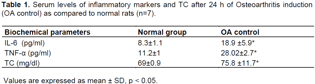
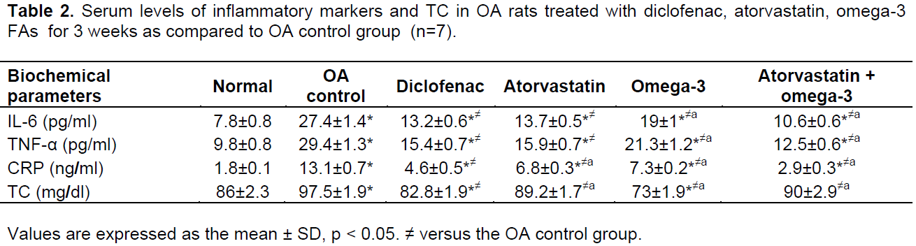
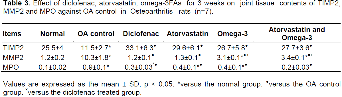
Animals
 
Adult male albino rats (150 to 200 g) were used in the present study. Animals were kept in a plastic cages at room temperature and on 12 h light-dark-cycle, were fed commercially available standard chow diet and water ad libitum. Experimental design, protocol of the study and animal handling were performed according to the guidelines of the Ethical Committee of the Faculty of Pharmacy, Zagazig University, Zagazig, Egypt.
 
Induction of osteoarthritis
 
Forty two rats as supplied from Egyptian organization for biological products and vaccine (Cairo, Egypt), were divided into 6 groups (n = 7). First group was kept as normal, the remaining groups from (2 to 6) were anesthetized by thiopental 40 mg/kg. Right knees were shaved and disinfected, received a single intra-articular injection through the patellar  ligament, of  24.6  mg/kg  monosodium iodoacetate (MIA) in 0.6 ml saline (Sigma Aldrich).  
 
Experimental design
 
Osteoarthritic rats were divided into five groups: The first one received no drugs and kept as OA control.  Four groups received specific doses in the form of topical cream application of the following drugs separately for 3 weeks, diclofenac, (Novartis Pharma AG Basel, Switzerland) 5 mg/kg body weight, atorvastatin, (MUP pharmaceutical CO Isamelia, Egypt) in a dose level 10 mg/kg body weight, omega-3 fatty acids (eicosapentaenoic acid 54.5%,docosahexaenoic acid 45.5% ,Arab Co. for Gelatin and Pharmaceutical Products, Egypt) in a dose level 1 g/kg body weight . The last group received a combination of atorvastatin and omega-3 FAs using the above mentioned doses.
 
Cream preparation for atorvastatin and omega-3 FAs
 
Atorvastatin powder was finely ground in a glass morter to form very fine powder. Cream base was added in portions to atorvastatin and mixed thoroughly.Omega-3 FA and diclofenac sodium were similarly prepared like atorvastatin and were freshly prepared before their application.
 
Cream base formula
 
Cetyl alcohol 5 g, cetomacrogol 5 g and emulsifying wax, mineral oil 20 g, glycerin 10 g,methyl paraben 0.18 g, propyl  paraben 0.02 g and purified water 59.8 g.
 
Dose incorporation
 
Diclofenac, atorvastatin, omega-3 FAs, in cream form were applied daily for 3 weeks in a constant weight 0.5 g cream/200 g body weight of the rat, corresponding to 5 and 10 mg, 1 g/kg body weight respectively.
 
Blood and tissue sampling
 
At the end of 1 day (after OA induction) and 3 weeks of treatment, 2 ml blood from retro-orbital vein were collected and centrifuged for serum preparation. Serum IL-6, C-reactive protein(CRP), and TNFα were evaluated by ELISA technique, following the instruction of their corresponding kits Ray Biotech Inc., DRG International Inc., USA and Koma Biotech Inc. Korea, respectively (Banerjee et al., 2003). Serum total cholesterol (TC) was determined colorimetrically using commercially available kit, xpress Bio and Biochain, CA, USA according to Allian et al. (1974).  At the end of 3 weeks, right knee joints were isolated and rinsed in ice cold saline, divided into 2 parts; the first one was stored at -80°C for subsequent measurements of myeloperoxidase (MPO), TIMP2 and MMP2 using RT PCR technique to (Pfaffl, 2001). The second part was used for histopathological examination.
 
Histopathological study
 
Each knee joint was kept in 10% formalin, 1% HNO3 for 24 h or more till they became soft, rinsed with running water, dehydrated in alcohol series, kept in xylene, paraffin 45°C, and lastly frozen. Five-micron tissue sections were cut by Leica Microtome, stained with haematoxylin and eosin (H&E) and subjected to histopathological examination.
 
 
Statistical analysis
 
Statistical analyses of data were done by Prism 5, Graph Pad, CA, USA. Results were presented as mean ± Standard Deviation (SD). Statistical differences were compared using student's t-test or one-way Analysis Of Variance (ANOVA), followed by Tukey test, considering p < 0.05 as statistically significant.


 RESULTS

Effect of MIA after 24 h
 
Rats which received MIA injection demonstrated  after 24 h significant increase in serum IL-6, TNFα and TC as compared to normal rats (p<0.05, Table 1).
 
 
Effect of atrovastatin, Omega-3 FAs and diclofenac after 3 weeks
 
Topical application of diclofenac induced a significant decrease of all the inflammatory markers.  Omega 3  FAs and atorvastatin application showed similar decrease, while their combination demonstrated marked anti-inflammatory effect. Serum IL-6 demonstrated significant decrease following diclofenac (54.8%), atorvastatin (59.1%), omega 3 FA (45.8%), combination of the last two achieved a marked decrease (69.4%), total cholesterol illustrated also significant decrease (Table 2). Matrix metalloproteinase 2 and MPO showed significant increase along with TIMP2 decrease in OA group. Drugs application induced the reverse effects (Table 3, Figures 1 to 3).


Histopathological results
 
Joint tissues of normal rats exhibited normal articular surface, bone, synovium and chondrocytes (lesion score 0+) (Figure 4a). Osteoarthritic control group demonstrated intense pathological alteration in articular surfaces components (lesion score 3+) pyknotic chondrocytes, debris in articualr cavity, thickened synovial membrane by edema and inflammatory cells.
 
Moreover fragmentation of bony trabeculae with partial replacement by fibrous tissues accompanied by numerous osteoclasts (Figure 4b, c). Diclofenac treated group showed slight improvement in pathological changes (lesion score 2+). The articular surface showed loosing of chondrocytes from their lacunae, mild spindle cells proliferation together with normal synovium and articular cartilage (Figure 4d). Atorvastatin treated group had moderate improvement in OA (lesion score2+) and usually showed inflammatory cells aggregation inside the joint cavity, mild thickening in synovial membrane and early necrotic changes in bony tissues (malacia) (Figure 4e). Omega-3 FA group illustrated great improvement in OA changes of joints (lesion score 1+), softening and disorganised bone trabeculae, disorganized lacunae and absence of inflammatory cells (Figure 4f). The combination treatment demonstrated great amelioration in the pathological alteration in the articular surfaces (lesion score 1+). Mild changes characterized by disorganized cartilageneous lacunae and articular cartilage, with a few pyknotic chondrocytes and normal synovial without inflammatory changes (Figure 4g).
 
 
3+------intense pathological changes in articular surfaces.
2+------moderate pathological changes in articular surfaces.
1+-----mild pathological changes in articular surfaces.
0+-----no pathological changes in articular surfaces.


 DISCUSSION

Present study demonstrated that topical application of omega-3 FAs and atorvastatin either individually or in combination induced marked anti-inflammatory effect as compared to diclofenac. Histopathological examination of the knee joints of OA treated rats showed evident correlation with the biochemical findings. It was previously reported that
MIA produces a rapid, technically, straight forward OA model which mimics the pathological and pharmacological
 
features associated with human OA (Guzman et al., 2003).
 
In addition, it induces chemical injury and inflammation of chondrocytes or synovial membrane or both. Mechanical loading that induces OA causes the synovial cells to induce signals which can mediate the production of pro-inflammatory cytokines and cartilage degradation (Goldring and Goldring, 2007). Our histopathological findings present clear evidence of this and are in agreement with previous studies (Al-saffar et al., 2009). Induction of OA is associated with flactuation of TC. Gierman et al. (2014) indicated that hypercholesterolemia may have a role in the development of OA. 
 
Tissue inhibitor metalloproteinase is an endogenous protease inhibitor which bind to active MMPs. MMPs derived from chondrocytes, synovium and polymorpho-nuclear leukocytes, play a major role in cartilage degradation in OA. The balance between TIMPs and MMPs is completely controlled in healthy joint, however in OA, MMP levels exceeds TIMPs leading to degradation of cartilage extracellular matrix (Alam et al., 2011).
 
Significant increase in MMP2 along with TIMP2 decrease in synovial fluid as observed in control group may suggest a disturbance in the balance of the enzymes, leading to high rate turnover in articular cartilage (Lee et al., 2008). Joint tissue of osteoarthritic rats demonstrated high levels of MPO. Presence of the later in syonvial fluid and neutrophils and/or macrophages within the affected joint can exaggerate the inflammatory response (Benito et al., 2005). Tissue MPO and MMP2 showed significant decrease after diclofenac treatment along with TIMP2 increase, as compared to OA control group.
Mahdy et al. (2002) demonstrated that decreased IL-6 may be attributed to reduced cyclic adenosine monophosphate and prostaglandin (PG) production. Interleukine-10 represents a responsive anti-inflammatory agent to PG effect and subsequent suppression of the later resulted in IL-10 increase (Mitchell and Warner, 2006).
 
Non-steroidal anti-inflammatory drugs as diclofenac exhibit their action through inhibition of cyclo-oxygenase (COX) enzymes, and PG production (Barrios-Rodiles et al., 1999). COX-1 and COX-2 enzymes can be induced by cytokines as TNFα (Wahane and Kumar, 2010), although COX-1 and COX-2 were not measured. In the present study, it seems likely that they were involved and so, reduced TNF-α following diclofenac treatment may have involved COX metabolites. The current study demonstrated that topical application of atorvastatin inhibited the tested inflammatory cytokines (IL-6 and TNFα), confirming the anti-inflammatory propereties of statins that have been reported before (Maher et al., 2009).
 
The mechanism of action of statins in arthritis may be generated from their ability to suppress 3-hydroxy 3-methyl glutaryl coenzyme A reductase (HMG-CoA) reductase enzyme, and subsequent inhibition  of isoprenoid intermediates synthesis which control many inflammatory pathways (Kwak et al., 2003). In turn cholesterol level determination may be relevant to inflammatory pattern of OA in the present study. Reduced TC here may be attributed to an inhibition of HMG-CoA and cholesterol biosynthesis (McCarey et al., 2004).
 
McCarey et al. (2004) demonstrated that statin can inhibit the levels of IL-6 and ameliorate endothelial dysfunction in rheumatoid arthritis. Previous study indicated that atorvastatin can shift the balance of cytokines milieu in the joints towards the production of anti-inflammatory cytokine IL-10, away from pro-inflammatory cytokines IL-6 and TNFα (Barsante et al., 2005). Topical application of omega-3 FAs significantly reduced serum IL-6, TNFα, and CRP as compared to OA control group. Omega-3 FAs can also reduce arachidonic acid metabolites and decrease the formation of pro-inflammatory compounds like leukotrienes and PGs (Chapkin et al., 1992; Joe and Lokesh, 1997).
 
Pischon et al. (2003) demonstrated an inverse relationship between omega-3 FAs intake and plasma level of soluble TNF receptors 1 and 2. The later encourage formation of complexes, which preserve the active trimeric form of TNF, preventing TNFα turn out into inactive monomeric forms. The receptors represent a binding protein and/or a slow release reservoir for TNF-α, indeed prolonging its half life.
 
Topical omega-3 FAs application resulted in hypocholesterolemic effect after 3 weeks of treatment in comparison to osteoarthritic group. Cell membrane FAs play a critical role in signal transduction where omega 3 FAs is able to modify gene expression, and change lipid level via this mechanism (Lapillonne et al., 2004). Omega-3 FAs modulate the function of sterol regulatory binding protein and peroxisome proliferation-activated receptors, both of which are involved in lipid homeostasis (Xu et al., 1999).
 
Yang et al. (2011) postulated that diclofenac down regulates MMP2 and MMP9 expression and their upstream enzymes of plasminogen activator urokinase and plasminogen inhibitor, both are associated with destruction of articular cartilage. Present study demonstrated also that atorvastatin significantly decreased MPO, due to an inhibition of neutrophil migration exerted Okouchi et al. (2003), and neutrophil influx to the joint of arthritic rats. This reflects a modification of tissue destruction (Joe and Lokesh, 1997). The current histopathology may present significant support to the biochemical one. Dalcico et al. (2012) reported that IL-6, IL-1 and TNFα activate the expression of metalloproteinase, so that cytokines inhibition by atorvastatin is associated with MMP2 reduction. Omega -3 fatty acids  suppresses also MMP and increases TIMP2 production via reduction of TNF-α and PGE2 (Curtis et al., 2002), while  its association  with atorvastatin showed significant results  as compared to diclofenac treatment.


 CONCLUSION

We conclude that topical application of omega 3 FAs and atorvastatin either individually or in combination induces anti-inflammatory and hypocholesterolemic effect in OA rats as compared to Diclofenac.  This  represents  a  new topical candidate for OA treatment in experimental rats but clinical trials for long term use may be recommended to confirm the present findings.


 CONFLICT OF INTERESTS

The authors have not declared any conflict of interests.


 ACKNOWLEDGEMENTS

We wish to thank Dr. Heba Khalil Mohammed, Theodor Belhars Research Institute, Egypt and Prof. Dr. Abd Al-Monem Ahmed Ali Mohamed, Head of Pathology Department, Veterinary Medicine College, Zagazig University, Egypt for their scientific help and performance of the histopathological study of the current work.


 REFERENCES

Alam R, Ji JR, Kim MS, Kim NS (2011). Biomarkers for identifying the early phases of osteoarthritis secondary to medial patellar luxation in dogs. J. Vet. Sci. 12(3):273-280.
Crossref


Allian CC, Roon LS, Chan CS (1974). Enzymatic determination of total serum cholesterol. Clin. Chem. 20:470.


Al-Saffar FJ, Ganabadi S, Yaakub H, Fakurazi S (2009). Collagenase & sodium iodoacetate-indeuced experimental osteoarthritis model in sprague Dawley rat. Asian J. Sci. Res. 2(4):167-179.
Crossref


Banerjee M, Tripathi LM, Srivastava VM, Puri A, Shukla R (2003). Modulation of inflammatory mediators by ibuprofen and curcumin treatment during chronic inflammation in rat. Immunopharmacol. Immunotoxicol. 25(2):213-224.
Crossref


Barrios-Rodiles MG, Tiraloche MG, Chadee K (1999). Lipopolysaccharide modulates cyclooxygenase-2 transcriptionally and posttranscriptionally in human macrophages independently from endogenous IL-1 beta and TNF-alpha. J. Immunol. 163:963-969.


Barsante MM, Roffe E, Yokoro CM, Tafuri WL, Souza DG, Pinho V, Castro MS, Teixeira MM (2005). Antiinflammatory and analgesic effects of atorvastatin in a rat model of adjuvant-induced arthritis. Eur. J. Pharmacol. 516:282-289.
Crossref


Benito MJ, Veale DJ, Fitzgerald O, Vanden Berg WB, Bresnihan B (2005). Synovial tissue inflammation in early and late osteoarthritis. Ann. Rheum. Dis. 64:1263-1267.
Crossref


Burke A, Smyth E, Fitzgerald GA (2006). Analgesic antipyretic agents pharmacotherapy of goat. In: Brunton LL, Lazo JS, Parker KL, editors. Good man & Gilman's the Pharmacological Basis of Therapeutics, 11th edition New York, Ny: McGraw-Hill. pp. 673-715.


Caughey GE, Mantzioris E, Gibson RA, Cleland LG, James MJ (1996). The effect on human tumor necrosis factor alpha and interleukin 1 beta production of diets enriched in n-3 fatty acids from vegetable oil or fish oil. Am. J. Clin. Nutr. 63:116-122.


Chapkin RS, AKoh CC, Lewis RE (1992). Dietary fish oil modulation of in vivo peritoneal macrophage leukotriene production and phagocytosis. J. Nutr. Biochem. 3:599-604.
Crossref


Curtis CL, Rees SG, Little CB, Flannery CR, Hughes CE, Wilson C, Dent CM, Otterness IG, Harwood JL, Caterson B (2002). Pathologic indicators of degradation and inflammation in human osteoarthritic cartilage are abrogated by exposure to n-3 fatty acids. Arthritis Rheum. 46(6):1544-53.
Crossref


Dalcico R, de Menezes AM, Deocleciano OB, Oria RB, Vale ML, Ribeiro RA, Brito GA (2012). Protective mechanisms of simvastatin in experimental periodontal disease. J. Periodontol. 84(8):1145-1157.
Crossref


Dwyer JH, Allayee H, Dwyer KM, Fan J, Wu H, Mar R, Lusis AJ, Mehrabian M (2004). Arachidonate 5-lipoxygenase promoter genotype, dietary arachidonic acid, and atherosclerosis. N. Engl. J. Med. 350:29-37.
Crossref


Kris-Etherton PM, Harris WS, Appel LJ (2002). Fish consumption, fish oil, omega-3 fatty acids, and cardiovascular disease. Circulation. 106: 2747-2757.
Crossref


Farid R, Rezaieyazdi Z, Mirfeizi Z, Hatef MR, Mirheidari M, Mansouri H, Esmaelli H, Bentley G, Foo Y, Watson RR, Lu Y (2010). Oral intake of purple passion fruit peel extract reduces pain and stiffness and improves physical function in adult patients with knee osteoarthritis. Nutr. Res. 30:601-606.
Crossref


Gierman LM, Kuhnast S, Koudijs A, Pieterman EJ, Kloppenburg M, van Osch GJ, Stojanovic-Susulic V, Huizinag TW, Princen HM, Zuurmond AM (2014). Osteoarthritis development is induced by increased dietary cholesterol and can be inhibited by atorvastatin in APOE*3Leiden.CETP mice: A translational model for atherosclerosis. Ann. Rheum. Dis. 73(5):921-7.
Crossref


Goldring MB (2000). Osteoarthritis and cartilage: The role of cytokines. Curr. Rheumatol. Rep. 2:459-465.
Crossref


Goldring MB, Goldring SR (2007). Osteoarthritis. J. Cell Physiol. 213:626-634.
Crossref


Goldring SR, Goldring MB (2006). Clinical aspects, pathology and pathophysiology of osteoarthritis. J. Musculoskelet. Neuronal. Interact. 6(4):376-378.


Grossin L, Cournil-Henrionnet C, Pinzano I, Gaborit N, Dumas D, Etienne S, Stoltz JF, Terlain B, Netter P, Mir LM, Gillet P (2006). Gene transfer with HSP 70 in rat chondrocytes confers cytoprotection in vitro and during experimental osteoarthritis. FASEB J. 20:65-75.
Crossref


Guzman RE, Evans MG, Bove S, Morenko B, Kilgore K (2003). Monoiodoacetate-induced histologic changes in subchondral bone and articular cartilage of rat femorotibial joints: An animal model of osteoarthritis. Toxicol. Pathol. 31:619-624.
Crossref


Hussein N, Ah-Sing E, Wilkinson P, Leach C, Griffin BA, Millward DJ (2005). Long-chain conversion of [13C]linoleic acid and alpha-linolenic acid in response to marked changes in their dietary intake in men. J. Lipid Res. 46:269-280.
Crossref


Joe B, Lokesh BR (1997). Prophylactic and therapeutic effects of n-3 polyunsaturated fatty acids, capsaicin, and curcumin on adjuvant-induced arthritis in rats. J. Nutr. Biochem. 8:397-407.
Crossref


Kwak BR, Mulhaupt F, March F (2003). Atherosclerosis: Anti-inflammatory and immunomodulatory activities of statins. Autoimmune. Rev. 2:332-338.
Crossref


Lapillonne A, Clarke SD, Heird WC (2004). Polyunsaturated fatty acids and gene expression. Curr. Opin. Clin. Nutr. Metab. Care 7:151-156.
Crossref


Lee HB, Alam MR, Seol JW, Kim NS (2008). Tartrate-resistant acid phosphatase, matrix metalloproteinase-2 and tissue inhibitor of metalloproteinase-2 in early stages of canine osteoarthritis. Vet. Med. (Praha). 53:214-220.


Lees P (2003). Pharmacology of drugs used to treat osteoarthritis in veterinary practice. Inflammopharmacol. 11:385-399.
Crossref


Mahdy AM, Galley HF, Abdel Wahed MA, Korny KF, Sheta SA, Webster NR (2002). Differential modulation of interleukin-6 and interleukin-10 by diclofenac in patients undergoing major surgery. Br. J. Anaesth. 88(6):797-802.
Crossref


Maher BM, Dhonnchu TN, Burke JP, Soo A, Wood AE, Watson RW (2009). Statins alter neutorphil migration by modulating cellular Rho activity: A potential mechanism for statins-mediated pleotropic effects. J. Leukoc. Biol. 85:186-93.
Crossref


Martel-Pelletier J, Boileau C, Pelletier JP, ROughley PJ (2008). Cartialge in normal and osteoarthritis conditions. Best Pract. Res. Clin. Rheumatol. 22:351-84.
Crossref


McCarey DW, McInnes IB, Madhok R, Hampson R, Scherbakov O, Ford I, Capell HA, Sattar N (2004). Trial of atorvastatin in rheumatoid arthritis (TARA): Double-blind, randomized placebo-controlled trial. Lancet 363:2015-21.
Crossref


Mitchell JA, Warner TD (2006). COX isoforms in the cardiovcular system: Understanding the activities of non-steroidal anti-inflammatory drugs. Nat. Rev. Drug Discov. 5:75-86.
Crossref


Okouchi M, Okayama N, Omi H, Imaeda K, Shimizu M, Fukutomi T, Itoh M (2003). Cerivastatin ameliorates high insulin-enhanced neutrophil-endothelial cell adhesion and endothelial intercellular adhesion molecule 1 expression by inhibiting mitogen-activated protein kinase activation. J. Diabetes Complicat. 17:380-386.
Crossref


Pfaffl MW (2001). A new mathematical model for relative quantification in real-time RT-PCR. Nucleic Acids Res. 29:e45.
Crossref


Pischon T, Hankinson SE, Hotamisligil GS, Rifai N, Willett WC, RImm EB (2003). Habitual dietary intake of n-3 and n-6 fatty acids in relation to inflammatory markers among US men and women. Circulation 108:155-160.
Crossref


Raatz SK, Redmon JB, Wimmergren N, Donadio JV, Bibus DM (2009). Enhanced absorption of omega-3 fatty acids from emulsified compared with eucapsulated fish oil. J. Am. Diet. Assoc. 109(6):1076-1081.
Crossref


Simopoulos AP (2002). Omega-3 fatty acids in inflammation and autoimmune diseases. J. Am. College Nutr. 2(6):495-505.
Crossref


Wahane VD, Kumar VL (2010). Atorvastatin ameliorates inflammatory hyperalgesia in rat model of moon-articular arthritis. Pharmacol. Res. 51:329-333.
Crossref


Xu J, Nakamura MT, Cho HP, Clarke SD (1999). Sterol regulatory element binding protein-1 expression is suppressed by dietary polyunsaturated fatty acids: A mechanism for the coordinate suppression of lipogenic genes by polyunsaturated fats. J. Biol. Chem. 274:23577-2583.
Crossref


Yang S, Hsieh Y, Lue K, Chu S, Chang I, Lu K (2011). Effects of non-steroidal anti-inflammatory drugs on the expression of urokinase plasminogen activator and inhibitor and gelatinases in the early osteoarthritic knee of humans. Clin. Biochem. 41:109-116.
Crossref


Youssef S, Stuve O, Patarroyo JC, Ruiz PJ, Radosevich JL, Hur EM, Bravo M, Mitchell DJ, Sobel RA, Steinman L, Zamvil SS (2002). The HMG-CoA reductase inhibitors, atorvastatin, promotes a Th2 bias and reverse paralysis in central nervous system autoimmune disease. Nature 420:78-84.
Crossref


          */?>