ABSTRACT
Mangroves are plant communities growing in the intertidal zone of tropical to subtropical coastal rivers. Some endophytic fungi which live in the tissues of mangrove plants produce some biologically active substances. By screening these biologically active substances some researchers have found that these substances have antimicrobial activity. This research is aimed to determine the antibacterial activity of endophytic fungi isolated from leaves of mangrove plants Excoecaria agallocha, Avicennia marina, Rhizophora mucronata and Lumnitzera racemosa in Sarasalai area in Jaffna Peninsula in Sri Lanka. Various species of endophytic fungi were isolated from the leaves of mangrove plants and identified based on morphological characteristics. Five fungal species were isolated from E. agallocha four from R. mucronata, A. marina and two from L. racemosa. Fifteen endophytic fungi were tested against six selected bacteria for their antagonistic effect. Antibacterial activity was tested against Escherichia coli, Bacillus sp, Klebsiella sp, Pseudomonas sp, Staphylococcus sp. and Proteus sp. using disc diffusion assay. Almost all endophytic fungi inhibited the growth of bacteria. Aspergillus flavus had the highest amount of inhibition against E. coli, Pseudomonas and Staphylococcus sp. Aspergillus tamari had higher amount of inhibition against Klebsiella sp. Few other species of Aspergillus also showed higher inhibitory activity against different bacteria when compared to other endophytic fungi.
Key words: Mangrove, endophytic fungi, bacteria.
Mangrove plants grow well in between sea and terrestrial ecosystem that contain brackish water. Mangroves live in wide range of salinities, tidal amplitudes, changes in sea level, winds, high temperatures, muddy and anaerobic soil conditions. They are well adapted for their extreme environmental conditions. In addition, most mangrove species are used as medicinal plants and also they have antimicrobial properties. These mangroves contain bioactive compounds that have potential antimicrobial, antiviral, anticancer, antidiabetic, antimalarial and antioxidant compounds (Zhang et al., 2009). Previous studies showed that most of the bioactive compounds were derived from the interaction between plants and microbes such as bacteria and endophytic fungi (Rossiana et al., 2016). Endophytic microorganisms grow within tissues of higher plants as facultative saprophytic, parasitic, mutualistic and commensalistic symbioses. These microorganisms grow intracellularly or intercelullarly in the tissues of higher plants without causing any symptoms on the host plants in which they live (Molina et al., 2012). Endophytic microorganisms are generally capable of producing bioactive compounds similar to their host plants (Nurhajati, 2011). Several studies have found that the endophytic fungi are one of the main sources of producing new antibiotics (Zhang et al., 2009). Endophytic fungi have been widely investigated as source of bioactive compounds (Bills et al., 1991). Most of these bioactive compounds have antimicrobial activity. The objective of this study was to isolate and identify endophytic fungi from selected mangroves and to test the biological activity of fungal isolates.
The mangroves which are common in Sarasalai area in the Northern part of Sri Lanka were selected for this study. Leaves were collected from mangroves namely Excoecaria agallocha, Avicennia marina, Rhizophora mucronata and Lumnitzera racemosa at five random sites in the area during July before the rainy season. Three plants were selected from each mangrove species. The identification was based on the herbarium specimens (M 23/1900) available in the Department of Botany University of Jaffna Sri Lanka and assistance from a taxonomist. The mangrove specimens collected for this study were preserved as herbarium and maintained in the laboratory for future reference.
Mature leaves (3-4) from the selected mangrove plants were collected into sterile polythene bags in the field. After reaching the laboratory, the leaves were immersed in Sodium hypochloride for 1-2 min for sterilization. The leaves were washed with sterile water 3 times. Four small segments (about 4 mm x 4 mm size) from each leaf in between the mid rib and periphery were cut using a sterile razor. Potato Dextrose Agar (PDA 39 g/L) medium was prepared in Petri dishes with the addition of streptomycin to prevent the growth of bacteria. Leaf segments were placed on the surface of PDA. The plates were incubated at room temperature for 2-3 days to observe fungal growth. The individual fungi were sub cultured until pure fungal isolates were obtained. Microscopic morphology and macroscopic characters were used for the examination of fungal cultures in the laboratory. Fungi were identified using standard keys available for fungi (Pitt and Hocking, 1997).
Discs (5 mm diameter) of 5 days old fungal culture were inoculated into 250-mL conical flasks containing 50 mL of Potato Dextrose Broth medium (PDB). The conical flasks were placed on a thermostatic shaker at 180 rpm at 28°C for 7 days for fermentation.
The cultured fungal mats were filtered through cheese cloth. The filtrate (50 mL) was transferred into separating flask. The crude metabolites were extracted using ethyl acetate at room temperature. The extracted ethyl acetate fractions were pooled into a conical flask, dried over anhydrous MgSO4 and evaporated by using rotary evaporator. The crude extract was dissolved in Dimethyl Sulphoxide (DMSO).
The bacterial cultures used in this study were obtained from the bacterial culture collection available in the laboratory in the Department of Botany University of Jaffna. The choice of bacteria was based on the previous studies made on the antibacterial activity. Bacteria (Escherichia coli, Bacillus sp, Klebsiella sp, Pseudomonas sp, Staphylococcus sp and Proteus sp) were streaked on the surface of the Nutrient Agar (NA) (28 g/L) medium. The plates were incubated at 37°C for overnight. After the incubation, a loop full of young bacterial culture (16-24 h old) from the isolated colony was transferred into the universal bottle containing sterile distilled water. It was stirred well by using vortex stirrer. Suspension was prepared which contain 105 CFU/ml from each bacterial culture.
Nutrient agar (NA) medium was prepared. 0.1 ml of bacterial suspension was transferred into the centre of the NA plates separately by using sterile pipette. Thereafter, the suspension was spread all over the surface of the NA medium by using sterile glass spreader.
A 50 µL of fungal metabolite extract was added into a sterile paper disc (5 mm diameter, Whatman No. 1). The paper disc was placed on NA plates which were surface inoculated with bacterial cultures. The antibacterial agent ampicillin (60 µg/mL) was used as a positive control and DMSO was used as a negative control. The plates were incubated at 37°C for 24-48 h and inhibition zones were measured. The experiment was carried out in triplicates.
Fifteen endophytic fungi were tested against six selected bacteria for their antagonistic effect. Positive and negative controls were also maintained. The obtained results were analyzed by 2-way ANOVA.
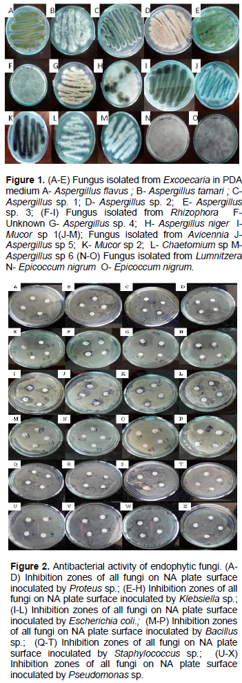
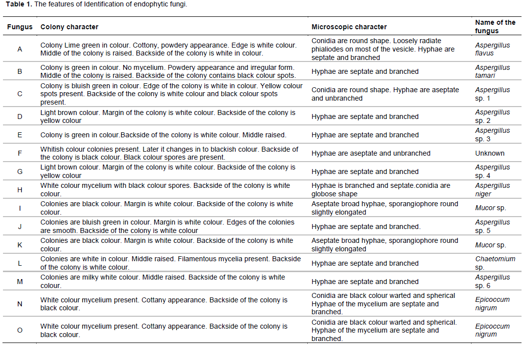
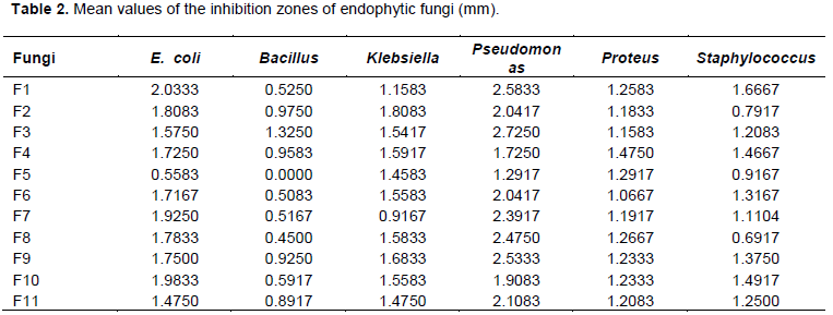
A total of 15 different species of endophytic fungi were isolated from the leaves of mangrove plants. Five different fungal species were isolated from Excoecaria sp., four from Rhizophora sp and Avicennia sp and two from Lumnitzera sp (Figures 1 and 2). The features of identification are given in Table 1. The mean values of the inhibition zones are given in the Table 2.
Almost all fungi inhibited the growth of E. coli. Endophytic fungus Aspergillus flavus (F1) has the higher amount of inhibition against E. coli. Endophytic fungus Aspergillus sp (F5) has the lowest amount of inhibition against E. coli. Most of the fungus inhibited the growth of Bacillus sp. Endophytic fungus Aspergillus sp (F3) has the higher amount of inhibition against Bacillus sp. Endophytic fungus Aspergillus sp (F5) and Epicoccum nigrum (F14) had the lowest amount of inhibition against Bacillus sp. Almost all fungus inhibited the growth of Klebsiella sp. Endophytic fungus Aspergillus tamarii (F2) has the higher amount of inhibition against Klebsiella sp. Endophytic fungus Chaetomium sp (F7) has the lowest amount of inhibition against Klebsiella sp. Almost all fungus inhibited the growth of Proteus sp. Endophytic fungus Aspergillus sp (F4) has the higher amount of inhibition against Proteus sp. Endophytic fungus Mucor sp (F6) has the lowest amount of inhibition against Proteus sp. Almost all fungus inhibited the growth of Pseudomoas sp. Endophytic fungus A. flavus (F1) has the higher amount of inhibition against Pseudomoas sp. Endophytic fungus E. nigrum (F15) has the lowest amount of inhibition against Pseudomoas sp. Almost all fungus inhibited the growth of Staphylococcus sp. Endophytic fungus A. flavus (F1) has the higher amount of inhibition against Staphylococcus sp. Endophytic fungus E. nigrum (F14) has the lowest amount of inhibition against Staphylococcus sp.
At P=0.05, statistical analysis showed that, there is no interaction between mean values of diameter of fungus. Also, there is no significant difference (P=0.05) in the diameters of clear zones of different fungi tested in this study. Highest antagonistic activity was shown by Aspergillus sp. (F3) against Pseudomonas sp. Whereas lowest antagonistic activity was shown by E. nigrum (F14) against Bacillus sp. The interaction between fungi and bacteria is shown in Figures 1 and 2.
Different mangroves used in this research study had different endophytic fungi or different strains of the same endophytic fungus. This study was done during the dry period or just before the rainy season. Future studies should be carried out to correlate the seasonal variations and the presence of endophytic fungi. It can be concluded that endophytes are rich sources of bioactive natural products with promising applications in development of pharmaceutical and industrial compounds. The fungal metabolites in the crude form have been used in this study. Further research should be carried out with the purified extracts. The follow up study would be the identification of the endophytic fungi at the molecular level to confirm the species.
The authors have not declared any conflict of interests.
REFERENCES
|
Bills GF and Polishook.JD (1991). Microfungi from Carpinus caroliniana. Canadian Journal of Botany 69:1477-1482.
Crossref
|
|
|
|
Molina G, Pimentel MR, Bertucci TCP, Pastore GM (2012). Application of fungal endophytes in biotechnological processes. Chemical Engineering Transaction 27:289-294.
|
|
|
|
|
Nurhajati J (2011). Salmonella dan Salmonellosis. Universitas Padjajaran Press, Bandung, pp. 14-36.
|
|
|
|
|
Pitt AJ, Hocking AD (1997). Fungi and food spoilage. Second edition. ISBN 0412554607, 9780412554605, Blackie Academic & Professional Publishers.
Crossref
|
|
|
|
|
Rossiana N, Miranti M, Rahmawati R (2016). Antibacterial activities of endophytic fungi from mangrove plants Rhizophora apiculata L. and Bruguiera gymnorrhiza (L.) Lamk. on Salmonella typhi. In AIP Conference Proceedings, 1744(1):020040. AIP Publishing\LLC.
Crossref
|
|
|
|
|
Zhang Y, Mu J, Feng Y, Kang Y, Zhang J, Gu PJ, Wang Y, Ma LF, Zhu YH (2009). Broad spectrum antimicrobial epiphytic and endophytic jamur from marine organism: isolation, bioassay and taxonomy. Marine Drugs 7(2):97-112.
Crossref
|
|