International Journal of
Genetics and Molecular Biology

  • Abbreviation: Int. J. Genet. Mol. Biol.
  • Language: English
  • ISSN: 2006-9863
  • DOI: 10.5897/IJGMB
  • Start Year: 2009
  • Published Articles: 139

Full Length Research Paper

Bael, Aegle marmelos (L.) Correa, an Indian medicinal plant protects V79 cells against the genotoxic effect of doxorubicin

Ganesh Chandra Jagetia
  • Ganesh Chandra Jagetia
  • Department of Zoology, Mizoram University, Tanhril, Aizawl-796 0094, India.
  • Google Scholar
Ponemone Venkatesh
  • Ponemone Venkatesh
  • Department of Zoology, Mizoram University, Tanhril, Aizawl-796 0094, India.
  • Google Scholar


  •  Received: 28 June 2011
  •  Accepted: 25 June 2015
  •  Published: 30 July 2015

 ABSTRACT

Alleviation of doxorubicin (DOX)-induced cytotoxicity and genotoxicity by leaf extract of bael also known as Aegle marmelos (AME) was studied in cultured Chinese hamster V79 cells. The optimum protective dose of AME was determined by treating V79 cells with different concentrations of AME before exposure to 10 µg/ml DOX and then by evaluating the cell survival and micronuclei frequency in the cytokinesis blocked V79 cells. MTT assay results revealed that AME pretreatment resulted in a concentration dependent elevation in the cell survival up to 25 μg/ml, whereas a further increase in AME concentration reduced the cell survival. Assessment of DNA damage by micronuclei assay showed that 25 µg/ml AME reduced the micronuclei frequency to a maximum extent. Therefore, 25 μg/ml AME was considered as an optimum chemo-protective concentration and further studies were carried out using this concentration, where V79 cells were treated with 25 μg/ml AME before exposure to different concentrations of DOX. The results of MTT assay at various post-DOX treatment times showed a time and concentration dependent decline in the cell survival with a maximum decline at 72 h post-DOX treatment. The IC50 values of 122, 108, 88 and 47 µg/ml DOX was observed at 12, 24, 48 and 72 h post-DOX treatment, respectively. Treatment of V79 cells with 25 μg/ml AME before DOX exposure to different concentrations of resulted in a rise in the IC50 by 60, 24, 44 and 41 µg/ml at 12, 24, 48 and 72 h, respectively. These results were corroborated by clonogenic assay where DOX-treatment caused a concentration dependent decline in the cell survival; whereas treatment of V79 cells with 25 µg/ml AME before DOX exposure arrested the DOX-induced decline in the cell survival. The micronuclei frequency increased in a concentration dependent manner in cells exposed to DOX, whereas AME pretreatment significantly reduced the DOX-induced micronuclei formation. Our results suggest that AME did reduce the cytotoxic and genotoxic effects of DOX and may be useful in clinical setup to reduce DOX-induced toxicity.
 
Key words: V79 cells, Aegle marmelos, doxorubicin, MTT, cell survival, micronuclei.


 INTRODUCTION

Doxorubicin (DOX, Adriamycin) is an anthracycline antibiotic derived from Streptomyces peucetius. It is a clinically important antitumor agent widely used in the chemotherapy of hematological malignancies and solid tumors and is one of the most important anticancer agents (Carter, 1975). DOX is a valuable component of various chemotherapeutic regimens including breast carcinoma, small-cell lung carcinomas and metastatic thyroid carcinoma. DOX is also an important agent for the successful treatment of Hodgkin’s and non-Hodgkin’s lymphomas. It has been reported to inhibit neoplastic proliferation in mouse and rat tumors in vitro and in vivo (Martin and McNally, 1980).
 
Chemotherapy is a major treatment modality for cancer therapy and is used to control advanced stages of malignancies in clinical settings (Kinghorn, 2003). However, most of these chemotherapeutic agents exhibit severe normal toxicity, and cause undesirable side effects. The clinical use of anthracyclines like DOX is a double-edged sword, on the one hand anthracyclines play a crucial role in the treatment of many neoplastic disorders, while on the other hand, their chronic administration induce severe cardiomyopathy and congestive heart failure (Minotti et al., 2004; Tacar et al., 2013), a major problem that limits their clinical application.
 
Since DOX is an important antineoplastic agent, it is necessary to screen other agents that can reduce the DOX-induced toxicity. Recently, intensive researches on biological function of natural antioxidants have been carried out with numerous botanicals worldwide, including those used as foods (Rice-Evans et al., 1995; López-Alarcón and Denicola, 2013). The natural antioxidants including phenolic or thiolic compounds could protect against damages caused by reactive oxidants by various biological mechanisms in living cells (Rice-Evans et al., 1995; van Acker et al., 1996; Sugamura, Keaney, 2011). The natural products and botanicals have been used since time immemorial to treat various disorders and offer an alternative to the synthetic drugs, as they have been considered either nontoxic or less toxic than their synthetic counterparts. Plants are complex mixtures of many biomolecules compounds and it is likely that their presence in this form in botanicals may provide better protective effects against any toxicant than a single molecule. The presence of many molecules in plants may be advantageous, as some of them may counteract the toxicity of other and as a result net effect may be beneficial for therapeutic purposes (Jagetia and Venkatesha, 2005).
 
Aegle marmelos, commonly known as bael, is a spinous tree belonging to family Rutaceae. The leaves, roots, bark, seeds and fruits of Aegle marmelos are edible. The medicinal properties of bael are well documented in the Ayurvedic texts of India. Its stem, bark, root, leaves and fruits have been valued for their, medicinal properties and bael has a long history of use as an herbal medicine to treat various ailments. The leaves of bael are bitter, astringent, laxative, febrifuge and expectorant. The leaves are used as a remedy for ophthalmic, ulcers, dropsy, cholera and beriberi associated with weakness of heart. They are also useful in ophthalmia, deafness, inflammations, catarrh, diabetes and asthmatic complaints. The unripe fruits are bitter, acrid, sour, astringent, digestive and stomachic, and are useful in diarrhea, dysentery and stomachalgia. Fresh aqueous and alcoholic leaf extracts of bael are reported to have a cardiotonic effect like digitalis and decrease the requirement of circulatory stimulants (Nadkarni, 1976). Bael has also been reported to possess a cardioprotective effect in mice (Jagetia, 2008; Jagetia and Venkatesh, 2015). Recently, bael has been reported to be radioprotective in vivo and in vitro (Jagetia et al., 2003, 2004; Jagetia and Venkatesh, 2005, 2007). Bael has shown antidiarrhoeal, antimicrobial, antiviral, anticancer, chemopreventive, antipyretic, ulcer healing, antigenotoxic, diuretic, antifertility and anti-inflammatory activites (Rahaman and Parvin, 2014). The chemotherapeutic agents not only injury cancer cells but also damage normal cells, which is the major cause of toxicity and side effects and doxorubicin is no exception. Therefore it is necessary to study toxic and genotoxic potential of doxorubicin using normal cells.
 
The diverse medicinal properties attributed to the Aegle marmelos stimulated us to investigate its chemopro-tective activity by assessing cell survival and micro-nucleus formation in the cultured V79 cells exposed to different concentrations of doxorubicin hydrochloride (DOX).


 MATERIALS AND METHODS
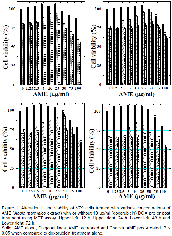
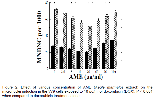
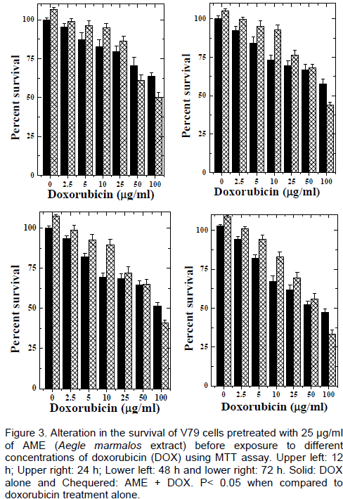
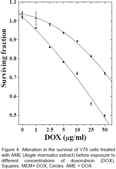
Preparation of extract
 
The identification of bael (Aegle marmelos (L.) Correa), family, Rutaceae and other aspects including collection extraction etc. have been described elsewhere (Jagetia et al., 2003, 2004)  Briefly, the mature leaves of bael were collected, shade dried, powdered and extracted in 50% ethanol using a Soxhlet apparatus. The leaf extract was freeze-dried and stored at -80°C until further use. Henceforth, the leaf extract will be denoted as AME.
 
Drug and chemicals
 
Doxorubicin hydrochloride (Adriamycin) was obtained from the Biochem Pharmaceutical Industries, Mumbai, India. Cytochalasin-B, eagle’s minimum essential medium (MEM), L-glutamine, gentamycin sulfate, fetal calf serum, tetrazolium dye, 3-(4, 5-dimethylthiazol-2-yl)-2,5- diphenyltetrazolium bromide (MTT) and dimethyl sulfoxide (DMSO) were procured from Sigma Chemical Co. St. Louis, USA. DOX and AME were dissolved in MEM as required, whereas cytochalasin-B was dissolved in DMSO at a concentration of 10 mg/ml, stored at -80°C and diluted with sterile MEM immediately before use.
 
Cell line and culture
 
The entire study was carried out using V79 (Chinese hamster lung fibroblasts) cells procured from the National Centre for Cell Sciences Pune, India. The cells were routinely grown in 75 cm2 flasks (Falcon, Becton Dickinson, USA) with loosened caps, containing MEM supplemented with 10% fetal calf serum, 1% L-glutamine and 50 µg/ml gentamycin sulfate at 37°C in a CO2 incubator (NuAire, Plymouth, MN, USA) in an atmosphere of humidified 5% COin 95% air.
 
Experimental design
 
A fixed number (5 × 10⁵) of exponentially growing V79 cells were inoculated into several individual culture flasks and allowed to grow until plateau phase.
 
Optimum dose selection
 
Several individual cell cultures were setup for the determination of optimum chemoprotective dose of AME, where plateau phase cell cultures were divided into various groups as follows:
 
AME group
 
The cells of this group were treated with 0, 2.5, 5, 10, 25, 50, 75 or 100 mg/ml of AME only.
 
AME + DOX group
 
The cells of this group were treated with different concentrations of AME for 1 h, thereafter the AME containing media was replaced with media containing 10 µg/ml DOX.
 
DOX + AME group
 
This group of cultures was exposed to 10 µg/ml DOX for 1 h and then drug containing media was replaced with media containing various concentrations of AME.
 
MTT assay
 
The effect of pretreatment and post-treatment of AME on the doxorubicin-induced cytotoxicity was studied to understand whether AME is able to protect even if it is administered after doxorubicin treatment. The MTT assay was performed to determine cell viability of V79 cells under the influence of AME or DOX alone or in combination of both as described previously (Mosmann, 1983). The yellow MTT (3-(4,5-dimethythiazol-2-yl)-2,5-diphenyl tetrazolium bromide) is reduced to dark purple coloured formazan by mitochondrial succinate dehydrogenase in living cells but not dead cells which are unable to reduce MTT, and this reaction is used as the end point in a rapid drug-screening assay. Approximately 5 × 10³ V79 cells/well were plated into 96-well plates (Nunc, Rosklide, Denmark) and kept in the CO2 incubator overnight at 37°C as described above (see 2.3). Next day, the cells were fed with a fresh medium containing or not 0, 2.5, 5, 10, 25, 50, 75 or 100 µg/ml AME for 1h. Thereafter the AME containing media was replaced with media containing 0, 1, 2.5, 5, 10, 25 or 50 mg/ml DOX and the cells were incubated for 1 h at 37°C.
 
The microplates were removed and DOX-containing medium was replaced with a fresh drug free MEM, immediately transferred into a CO2 incubator and allowed to grow for 12, 24, 48 or 72 h. The cell viability was assessed by the MTT assay at different post-treatment times. Briefly, at stipulated time following treatment/s, the medium was aspirated, MTT (50 µl of a 5 mg/ml stock solution in PBS) added into each well of the 96-well plate and the microplates were incubated for 2 h at 37°C. During this period the living cells produced blue insoluble formazan from the yellow soluble MTT. The plates were spun, supernatants were discarded and purple colored precipitates of formazan were dissolved in 150 µl of dimethylsulfoxide. The absorbance was recorded at 540 nm of each aliquot using a microplate reader with a reference at 650 nm serving as a blank. The obtained values were calculated as the percentage of cell survival in comparison with the non-drug/s treated controls taken as 100% survival. The data of eight replicates were collected for each concentration in each group at different assessment times.
 
The group DOX+AME was not incorporated in further experiment as there was no significant difference between the pre and post AME-treatment by MTT assay.
 
Micronucleus assay
 
A separate experiment was conducted to assess the effect of AME on DOX-induced micronuclei formation, where groupings and other conditions were essentially similar to that described for optimum dose selection, except that the plateau phase cells were grown in 25 cm2 culture dishes. The AME or DOX containing medium was removed and the cells were dislodged by trypsin-EDTA treatment. The cells from each culture were replated into quadruplicates and micronuclei were prepared as described earlier (Fenech and Morley, 1985) with minor modifications. Briefly, the cells from both experiments were allowed to attach for 6 h and incubated with 3 µg/ml of cytochalasin-B to inhibit cytokinesis (Jagetia and Adiga, 1998). The cells were left undisturbed and allowed to grow for another 16 h. Thereafter, the medium containing cytochalasin-B was discarded and cells were washed twice with PBS. Finally, cells were dislodged with trypsin EDTA treatment, centrifuged, subjected to mild hypotonic treatment (0.7% ammonium oxalate) for 5 min at 37°C, centrifuged again and the resultant cell pellet was fixed in Carnoy's fixative (3:1 methanol, acetic acid). The cells were centrifuged again, resuspended in a small volume of fixative and spread on to precleaned coded slides to avoid observer's bias. The slides containing cells were stained with 0.25% acridine orange (BDH, England, Gurr Cat. No. 34001 9704640E) in Sorensen's buffer (pH 6.8), and washed twice in this buffer. The buffer mounted slides were observed under a fluorescence microscope equipped with 450 to 490 nm BP filter set with excitation at 453 nm (Carl Zeiss Photomicroscope III, Oberkochen, Germany) using a 40 X Neofluar objective for the presence of micronuclei (MN) in the binucleate cells (BNC). A minimum of thousand BNC with well-preserved cytoplasm was scored from each culture and the frequency of micronucleated binucleate cells (MNBNC) was scored. The micronuclei in BNC were identified as described earlier (Kirsch-Volders et al., 2003).
 
Chemoprotection
 
A separate experiment was carried out to ascertain the chemoprotective potential of AME in V79 cells. The plateau phase cell cultures were divided into the following groups:
 
 
MEM+DOX
 
The cell cultures were incubated with 0, 1, 2.5, 5, 10, 25 or 50 mg/ml of DOX for 1 h, thereafter the drug containing media was replaced with fresh drug-free MEM.
 
AME + DOX
 
The cell cultures of this group were treated with 25 mg/ml of AME for 1 h after which the AME containing media was replaced with MEM containing various concentrations of DOX for another 1 h. After 1 h of DOX treatment the drug containing media was discarded, the cells were washed with PBS, dislodged by trypsin EDTA treatment and clonogenic and micronucleus assays were carried out from the same stock of cells. A separate experiment was carried out for MTT assay, where grouping and the conditions were essentially similar to that described above. The details of MTT and micronucleus assays have been described in optimum dose selection section.
 
Clonogenic assay
 
Clonogenicity of cells was measured using colony-forming assay of Puck and Marcus (1955). The plateau phase cells were pretreated with 25 µg/ml AME for 1 h before exposure to different concentrations of DOX. One hour after the DOX treatment, the drug containing medium was removed and the cells were washed twice with sterile PBS. The cells from each group of flasks were dislodged by trypsin EDTA treatment. Usually, 200 to 300 cells were plated on to 25 cm2 culture dishes (Nunc, Rosklide, Denmark) containing 5 ml medium in triplicate for each drug concentration for each group. The cells were allowed to grow for 9 days. The resultant colonies were stained with 1% crystal violet in methanol and clusters containing 50 or more cells were scored as a colony. The plating efficiency of cells was determined and the surviving fraction was fitted on to non-linear polynomial functions. The left over cells were used for micronucleus assay. The results of each experiment were confirmed by repetition of the experiment. The test of homogeneity was applied to compare the data of both experiments. Since no significant differences were observed, the data of both experiments were combined and presented as results.
 
Statistical analyses
 
The   significance  between  the  treatments  was  analyzed using student’s‘t’ test for MTT assay, one-way ANOVA. The Bonferroni’s post-hoc test was used wherever necessary. The Fisher’s exact test was applied for micronucleus assay. The data were fitted on to linear quadratic model SF = exp – (αD + βD2) for survival or Y = C + D + bD2, for micronucleus assay. Where C is control MNBNC frequency, D is drug dose and α and β are the constants.
 
The Solo 4 statistical package ((BMDP Statistical software Inc, Los Angeles, CA, USA) was used for statistical analyses. The protection factor (PF) for micronuclei was calculated using the following formula:


 RESULTS

The results expressed are mean ± Standard error of the mean in Table 1 to 2 and Figures 1 to 6 for MTT, micronuclei and clonogenic assays.


Optimum dose selection
 
MTT assay
 
Exposure of V79 cells to different concentrations of AME did not alter the cell viability significantly (Figure 1). Treatment of V79 cells with various concentrations of AME before DOX-treatment resulted in a gradual rise in the cell survival (MTT assay) and a maximum survival of 84, 80, 75 or 70% was observed at a concentration of 25 µg/ml AME for 12, 24, 48 and 72 h post-DOX treatment (PDT). A similar effect was discernible when the cells were treated with various concentrations of AME immediately after DOX-treatment except that the effect was less pronounced when compared to AME pretreatment (Figure 1).
 
 
Micronuclei assay
 
The chemo-protective effect of AME against DOX-induced genotoxicity was studied by micronucleus assay. AME treatment alone did not alter the spontaneous frequency of micronuclei in binucleate V79 cells. However, a marginal but non-significant decline in MNBNC was observed up to a concentration of 50 µg/ml AME treatment alone (Figure 2). Exposure of V79 cells to 10 µg/ml DOX resulted in a significant rise in the MNBNC frequency that was 2.6 folds higher than that of spontaneous MNBNC frequency. Treatment of V79 cells with different concentrations of AME before exposure to 10 µg/ml DOX caused a significant decline in the DOX-induced micronuclei formation and a maximum reduction in MNBNCs was observed for 25 µg/ml AME (Table 1). A similar trend was discernible for BNC bearing one, two and multiple micronuclei. Since 25 µg/ml AME caused the maximum rise in the cell survival accompanied by a maximum reduction in micronuclei frequency, it was considered as the optimum concentration for chemo-protection when compared to the other concentrations of AME, therefore, further studies were carried out using this AME concentration.
 
 
Chemoprotection
 
MTT assay
 
Chemoprotective activity of 25 µg/ml AME was studied by exposing V79 cells to different concentrations of DOX. Exposure of V79 cells to different concentrations of DOX resulted in a concentration dependent reduction in the cell survival (MTT formazan assay) in MEM+DOX group. This reduction in cell survival depended on the post-DOX treatment assay time. The cell survival showed a time dependent decline and a highest decline was observed for 100 µg/ml DOX at 72 h post-DOX treatment in MEM+DOX group (Figure 3). The IC50 was found to be 122, 108, 88 and 47 µg/ml DOX for 12, 24, 48 and 72 h post-DOX treatment in MEM+DOX group. Treatment of V79 cells with AME before exposure to different concentrations of DOX arrested the DOX-induced decline in the cell survival. The elevation in cell survival was significantly greater in AME+DOX group when compared to MEM+DOX group (Figure 3). The IC50 concentrations were 183 (á1.5), 132 (á1.2), 122 (á1.4) and 88 (á1.9) µg/ml for 12, 24, 48 and 72 h post-DOX treatment, respectively in AME+DOX group indicating a time dependent elevation in the IC50.
 
 
Clonogenic assay
 
Since clonogenic assay is the gold standard for determining the cytotoxic activity, the results of MTT assay were further confirmed by clonogenic assay. Treatment of V79 cells with different concentrations of DOX resulted in a concentration dependent decline in the cell survival in MEM+DOX group as evidenced by a reduction in the surviving fraction, which was lowest for 50 µg/ml DOX (Figure 4). Treatment of V79 cells with 25 µg/ml AME before exposure to different concentrations of DOX in AME+DOX group resulted in a rise in the cell survival when compared with the MEM+DOX group. One interesting fact was that the chemo-protective effect of AME increased with increasing concentration of DOX, where surviving fraction increased by 0.25 and 0.22 for 25 and 50 µg/ml DOX in AME + DOX group when compared with the concurrent MEM+DOX group.
 
 
Micronuclei assay
 
The frequency of micronuclei increased with increasing concentration of DOX up to a concentration of 25 mg/ml in MEM+DOX group, thereafter the frequency of MNBNC remained unaltered (Figure 5). Pretreatment of V79 cells with 25 µg/ml AME before exposure to different concentrations of DOX resulted in a significant decline in the frequency of MNBNC when compared with the concurrent MEM+DOX group, where a protection factor of 2 was obtained for all the concentrations of DOX except 1 and 2.5 µg/ml DOX, where it was almost 3. The frequency of MNBNC with one, two and multiple micronuclei are presented separately. The frequency of binucleate cells bearing one, two and multiple micronuclei increased in a concentration dependent manner and the highest number of micronuclei was observed for 50 µg/ml DOX, the highest concentration of DOX studied. AME pretreatment resulted in a significant decline in the induction of one, two and multiple micronuclei in binucleate cells. The frequency of two and multiple MNBNC was always lower in AME + DOX group. However, significant differences were observed only for 25 and 50 µg/ml DOX (Table 2). The data for all the MNBNCs were fitted on to a linear quadratic model both in the MEM+DOX and AME + DOX groups (Figure 5). The AME protected against DOX-induced micronuclei-formation by a factor of 3.2 to 1.6 depending on the DOX concentration. The protection factor declined with increasing concentration of DOX (Table 2).
 
 
Biological response
 
The biological response of treatments was determined by plotting MNBNC on the Y-axis, whereas the surviving

fraction on the X-axis, respectively (Figure 6). A direct correlation between surviving fraction and micronuclei induction was discernible as the rise in MNBNC frequency resulted in a corresponding decline in the surviving fraction indicating an inverse correlation between MNBNC-induction and cell survival. This correlation between surviving fraction and MNBNC was linear both for MEM+DOX and AME+DOX groups (Figure 6).


 DISCUSSION

The aim of chemotherapy is to kill cancerous cells; however, chemotherapy does not distinguish between neoplastic and normal cells as a result DNA of normal cells is also adversely affected, which is major cause of induction of second malignancies (Baker and Connor, 1996; Pendleton et al., 2014). The anthracyclines are important antineoplastic agents, which contain an aglycone ring coupled with an aminosugar and they are able to produce a wide range of biological effects in patients receiving them. The anthracyclines undergo one-electron reduction forming oxygen free radical intermediates and the presence of oxygen and metals reduces an anthracycline into a semiquinone radical, leading to the formation of a superoxide radical, which is subsequently converted into a hydroxyl radical (Abraham et al., 1996). The free radicals produced by anthracylines lead to a cascade of events including membrane lipid peroxidation, DNA strand scission and direct oxidation of purine, pyrimidine bases, thiols and amines. Further, the planar ring of anthracyclines can intercalate in between the molecular DNA, altering the shape of DNA helix, and interfering in transcription and its replication (Tewey et al., 1984; Schneider et al., 1990; Chen et al., 2013; Pendleton et al., 2014). The anthracyclines also inhibit the activity of topoisomerase II, introduce double-strand breaks and increase their binding to DNA in tumor cells (Pommier et al., 1985; Nitss, 2009).
 
The clinical doses of doxorubicin and other anticancer drugs sufficient to kill tumor cells are often toxic to normal tissue and lead to side effects including nausea, vomiting myelosuppression, neutropenia, cardiomyopathy, and induction of secondary tumors (Pendleton et al., 2014). Agents that protect myeloid and lymphoid systems from the suppressive effects of radiation or chemotherapy could be beneficial because the side effects induced by a standard therapy regimen might be alleviated, and the higher doses of therapy could be administered to increase the antitumor efficacy of chemotherapeutic drugs. This could be achieved by screening pharmaco-logical agents that can protect the normal cells against DOX-induced cumulative toxicity. Botanicals by virtue of their longstanding usage in the traditional and folklore medicinal systems and less toxic implications have drawn the attention of researchers around the world recently. Especially, dietary ingredients may be highly successful, if they are found to protect against the deleterious effects of antineoplastic agents on the normal tissues, as they are widely acceptable, would not put an extra foreign substance into the body and can be safely manipulated in humans without toxic manifestations (Jagetia et al., 2003, Jagetia and Venkatesha, 2005).
 
DNA double strand breaks are hallmark of cell death and many of the antineoplastic agents including DOX kill neoplastic cells by inflicting DNA damage. We have observed that DOX treatment caused a concentration dependent rise in the cytotoxicity of V79 cells due to its DNA damaging activity. A concentration dependent elevation in the cytotoxicity after DOX-treatment has been observed earlier in V79 cells (Suter et al., 1980; Bhuyan et al., 1983; Babudri et al., 1984). Similarly, our earlier studies have shown a concentration dependent decline the survival of HeLa cells exposed to DOX (Jagetia and Nayak, 1996; Jagetia and Aruna, 2000). Likewise, DOX has been also reported to induce dose-dependent cell killing and growth inhibition in various cell lines (Helbig and Speit, 1995; Bogdanovi? et al., 2004; Gumulec et al., 2014). Treatment of V79 cells with AME before and after DOX exposure increased the cell survival significantly indicating reduction in the cytotoxic effects of DOX. However, this effect was less pronounced when AME treatment was given after DOX treatment. Therefore further studies were not carried out using this modality.
 
The cytotoxic effect of DOX has been further confirmed by clonogenic assay. AME has not been used to reduce the DOX-induced cytotoxicity earlier and this is probably the first attempt in this direction, where the best chemoprotective effect was observed for 25 µg/ml AME. Earlier studies have reported that caffeine, a natural product and other agents including 3-4-dihydroxybenzoic acid, lovastatin, fullerol, catalpol, apoferritin and luteolin reduced the DOX-induced cell killing and DNA damage in V79 and other cells (Iliakis and Lazar, 1987; Bardeleben et al., 2002; Bogdanovi? et al,, 2004; Wu and Mao, 2012; Gumulec et al., 2014; Sato et al., 2015). A similar effect has been observed with 10 µM cycloheximide and 10 mM WR-2721 against DOX-induced cytotoxicity in V79 cells earlier (Bonner and Lowrence, 1989; De Graff et al., 2003). The reports of use of any agent after DOX treatment are unavailable and maximum studies have been carried out pretreatment regimen as indicated above. However, the post treatment protection of cytotoxicity of DOX may be due to reduced DNA damage and upregulation of antioxidant status by AME in the present study.
 
The protective effect of AME against DOX-induced DNA damage was evaluated by micronucleus assay. Micronuclei are acentric fragments or a complete chromosome that fail to attach to the mitotic spindle during cytokinesis and are excluded from the main nuclei. Different mechanisms may be involved in the formation of micronuclei, including chromosome breakage (clastogenesis) and spindle disruption (aneugenesis) (Heddle et al., 1983; Majer et al., 2001; Fenech et al., 2011). Yet micronuclei are among the most extensively used cytogenetic markers that indicate early biological effects associated with DNA-damaging agents. Among the various techniques used to detect DNA damage and genotoxic effects, the micronucleus assay is simple, cheap, and less cumbersome that allows convenient and easy application. Treatment of V79 cells with various concentrations of AME before exposure to 10 µg/ml DOX caused a significant decline in the DOX-induced micronuclei formation at 10, 25 and 50 µg/ml AME, thereafter this decline was non-significant and the highest dose of 100 µg/ml AME resulted in a marginal but non-significant elevation in the MNBNC frequency when compared with the MEM+DOX group. A highest reduction in the MNBNC frequency was recorded at a con-centration of 25 µg/ml AME, which was 1.4 fold lower than 10 µg/ml DOX treatment alone. Therefore, this dose of AME was considered as the optimum chemoprotective dose. AME pretreatment caused a similar reduction in the DOX-induced micronuclei formation in mice bone marrow earlier (Venkatesh et al., 2007).  Likewise, Syzigium cumini extract and AME have been reported to abate radiation-induced micronuclei formation in cultured human peripheral blood lymphocytes and mice bone marrow (Jagetia and Baliga, 2002; Jagetia et al., 2003; Jagetia and Venkatesh, 2007). It is interesting to note that AME treatment even reduced the spontaneous frequency of MNBNC albeit non-significantly indicating that it can inhibit the normal wearing and tearing of genome. Earlier, polyherbal formulations including for Liv 52 and abana have been found to exert similar effect (Jagetia and Ganapathi, 1989; Jagetia and Aruna, 1997).
 
DOX has been reported to induce single and double strand breaks of DNA, micronuclei, chromatid and chromosome aberrations in vitro and in vivo (Bean et al., 1992; Al-Harbi, 1993; Al-Shabanah, 1993; Delvaeye et al., 1993; Jagetia and Nayak, 1996; Jagetia and Aruna, 2000; Jagetia and Nayak, 2000; Dhawan et al., 2003: Venkatesh et al., 2007). An  identical effect has been observed in the present study, where DOX induced a concentration dependent rise in the MNBNCs in V79 cells exposed to various concentrations of DOX and the dose response was linear quadratic. A dose dependent increase in the frequency of micronuclei has been reported in vitro and in vivo (Boucher et al., 1993; Jagetia and Nayak, 1996; Jagetia and Aruna, 2000; Venkatesh et al., 2007). Our findings that DOX increased the frequency of binucleate cells bearing two and multiple MN are in agreement with the earlier reports, where a similar effect has been observed (Antunes and Takahashi, 1998, 1999; Al Harbi, 1993; Jagetia and Nayak, 1996, 2000; Jagetia and Aruna, 2000; Venkatesh et al., 2007). Doxorubicin has been also reported to increase sister chromatid exchanges and chromosomal aberrations in vitro and in vivo (Bean et al., 1992; Helbig and Speit, 1995). The increase in more than one micronuclei in the cells is due to the induction of complex multiply sites of damage. Further micronuclei induction is the sign of chromosome rearrangement. Micronuclei also result from cell division defects, like mitotic errors leading to mis-segregation of intact chromosomes, and DNA replication and repair errors resulting in the formation generate acentric chromosome fragments (Yates and Campbell, 2012; Zhang et al., 2015)
 
AME pretreatment reduced the frequency of micronuclei significantly in V79 cells exposed to different concentrations of DOX and 25 µg/ml AME was most effective in significantly reducing the frequency of MNBNCs bearing not only one MN but also those with two and multiple MN indicating that AME treatment has been able to inhibit the multiply sites of damage to DNA and complex chromosome aberrations in the V79 cells. An identical effect has been observed earlier in mouse erythrocytes, where AME has been reported to reduce the DOX-induced micronuclei formation (Venkatesh et al., 2007).  AME pre-treatment has been also reported to reduce the frequency of radiation-induced micronuclei in cultured human peripheral blood lymphocytes and mice bone marrow (Jagetia et al., 2003; Jagetia and Venkatesh, 2007). Pimaradienoic acid has been reported to reduce the Dox-induced micronuclei in cultured V79 cells (Kato et al., 2012) Other chemicals including caffeine, captopril, desferrioxamine, rutin, hesperidin and magnesium sulphate have also been reported to reduce DOX-induced micronuclei and chromosome aberrations in (Dulout et al., 1981; Al Harbi, 1993; Al Shabanah, 1993, 1998; Hozayen, 2012).
 
AME pretreatment resulted in a protection factor of 3.2 to 1.6 depending on the DOX concentration. Protection factors (PF) denote the extent of reduction of radiation-induced DNA damage in the sense that a PF of 1.4 or more indicates a promising protection (Weiss and Landauer, 2003). A similar contention seems to be true in the present investigation where the protection factors of 3.2 to 1.6 have been reported for low and high (1 and 50 µg/ml) concentrations of DOX.
 
The micronuclei bearing cells are dying cells since they have lost a significant amount of genome, therefore evaluation of biological response gives an indication of relationship between the micronuclei and cell death. In the present study cell survival and micronuclei formation is inversely correlated indicating that the micronuclei are lethal events and once they are formed, the cells succumbed to death. This relationship between MNBNC induction and surviving fraction was linear in both the groups. A linear correlation between micronuclei formation and cell survival has been reported in various cultured mammalian cells earlier (Russell et al., 1995; Shibamoto et al., 1991; Jagetia and Adiga, 1998). A linear quadratic relationship has also been reported for MN induction and cell survival (Wandl et al., 1989; Jagetia and Aruna, 2000).
 
DOX may have induced cytotoxicity and micronuclei by employing multiple mechanisms. The DOX is oxidized into semiquinone radical, which is converted back to DOX generating superoxide free radicals, which leads to a cascade of event leading to lipid peroxidation (Dorshow, 1986; Powis, 1989). The lipid peroxidation induces membrane, DNA and proteins damage leading to cell death. The DOX intercalates into cellular DNA distorting polynucleotide structure and causing the inhibition of enzyme necessary for DNA replication and transcription (Gewirtz, 1999; Kiyomiya, 2001; Pérez-Arnaiz., 2014). The topoisomerase II an enzyme that catalyzes the unwinding of DNA for transcription and replication by cleaving one strand of DNA duplex and passing a second duplex through this transient cleavage, which is termed as the “cleavable complex.’’ The doxorubicin acts as a poison to the cleavable complex by inhibiting its religation and thus causing, a DNA double-strand breaks and forming. topoisomerase II-DNA complex (Tewey et al., 1984;Pommier et al., 1985; Guano et al., 1999; Cheng et al., 2013). Further DOX has been reported to elicit block NF-κB activation (Wang et al., 2002; Pletz et al., 2012), which may be induced in response to DNA damage. The suppression of NF-κB transcriptional activation may lead to alteration of several genes controlled by NF-κB activation. Recently we have reported that DOX PARP stimulate parp activation and also formation of 8-hydroxy-2'-deoxyguanosine (8-OHdG) DNA adducts the signature of DNA damage (Jagetia and Reddy, 2014).  These stated mechanisms of action of DOX may have been responsible for the cytotoxicity and increased frequency of micronuclei in the V79 cells in the present study.
 
The exact mechanism by which AME protected DOX-induced cytotoxicity and genotoxicity is not known. However, there are several possibilities by which AME might have protected V79 cells against the DOX-induced cytotoxicity and DNA damage. AME may have inhibited the generation of free radicals-induced by DOX and thus protecting the cells against its deleterious effects. Our earlier study has shown that AME actually inhibits free radical generation and reduces oxidative stress by upregulating antioxidant status in vitro and in vivo (Jagetia et al., 2003, 2004; Jagetia and Venkatesh, 2005). The reduction in lipid peroxidation and increase in glutathione by AME may have reduced the molecular damage to cellular genome, thus reducing the micronuclei formation and increasing the cell survival. AME has been reported to protect against the radiation-induced lipid peroxidation and increase glutathione in vivo (Jagetia et al., 2004; Jagetia and Venkatesh, 2005). It is also postulated that AME treatment may have restored the action of topoisomerase II reducing the DNA damage, micronuclei formation and subsequently increasing the cell survival. The inhibition of NF-?B activation by AME may be another mechanism of protection of V79 cells against the DOX-induced DNA damage and cell death. AME may have also inhibited the DOX-induced activation of Poly (ADP-ribose) polymerase (PARP) and formation of (8-OHdG) DNA adducts thus protecting the V79 cells against cytotoxicity and DNA damage.
 
Bael has been reported to contain aegeline, aegelenine, marmelosine, marmelin/marmesinin, o-methyl hayordinol, alloimperratorin methyl ester, o-isopentanyl hayordinol, linoleic acid, cineole, p-cymene, citronella, citral, cuminaldehyde, D-limonene, eugenol, tannins like ellagic acid and gallic acid, phlobatannins, flavon-3-ols, rutin, leucoanthocyanins, anthocyanins and flavonoid glycoside (Rastogi and Mehrotra, 1990). Most of these compounds have been reported to possess antioxidative and free radical scavenging activities (Korina and Afanas'ev 1997). The observed chemoprotective activity of AME may be due to the action of one or all of these compounds.


 CONCLUSION

Our data suggest that the AME pretreatment increased the survival of V79 cells and 25 µg/ml AME afforded the highest protection against the DOX-induced DNA damage as evident by a significant reduction in MNBNC and cytotoxicity. Scavenging of free radicals, increased antioxidant status, reduction in lipid peroxidation and increased glutathione, restoration of topoisomerase II activity, inhibition of NF-kB activation, PARP activation and (8-OHdG) DNA adduct formation may be some of the plausible mechanisms of action of AME.


 CONFLICT OF INTERESTS

The authors did not declare any conflict of interest.


 ACKNOWLEDGEMENTS

The authors are thankful to the Indian council of Medical Research, Govt. of India for financial support vide grant No. 45/2/2004/BMS/TRM to carry out this study.


 REFERENCES

Abraham R, Basser RL, Green MD (1996). A risk-benefit assessment of anthracycline antibiotics in antineoplastic therapy. Drug Safety 15:406-429.
Crossref


Al-Harbi MM (1993). Effect of captopril on the cytological and biochemical changes induced by adriamycin. Food Chem. Toxicol. 31: 209-212.
Crossref


Al-Shabanah OA (1993). Inhibition of adriamycin-induced micronuclei by desferrioxamine in Swiss albino mice. Mutat. Res. 301:107-111.
Crossref


Antunes LM, Takahashi CS (1998). Effects of high doses of vitamins C and E against doxorubicin-induced chromosomal damage in Wistar rat bone marrow cells. Mutat. Res. 419:137-143.
Crossref


Antunes LM, Takahashi CS (1999). Protection and induction of chromosomal damage by vitamin C in human lymphocyte cultures.Teratog. Carcinog. Mutagen. 19:53-59.
Crossref


Babudri N, Pani B, Tamaro M, Monti-Bragadin C, Zunino F (1984). Related Articles, Mutagenic and cytotoxic activity of doxorubicin and daunorubicin derivatives on prokaryotic and eukaryotic cells. Br. J. Cancer 50:91-96.
Crossref


Baker ES, Connor TH (1996). Monitoring occupational exposure to cancer chemotherapy drugs. Am. J. Health Syst. Pharmacol. 53: 2713–2723.


Bardeleben RV, Dunkern T, Kaina B, Fritz G (2002). The HMG-CoA reductase inhibitor lovastatin protects cells from the antineoplastic drugs doxorubicin and etoposide. Int. J. Mol. Med. 10: 473-479.
Crossref


Bean CL, Armstrong MJ, Galloway SM (1992). Effect of sampling time on chromosome aberration yield for 7 chemicals in Chinese hamster ovary cells. Mutat. Res. 265:31-44.
Crossref


Bhuyan BK, Zimmer DM, Mazurek JH, Trzos RJ, Harbach PR, Shu VS, Johnson MA (1983). Related Articles, Comparative genotoxicity of adriamycin and menogarol, two anthracycline antitumor agents. Cancer Res. 43: 5293-5297.


Bogdanović G, Kojić V, Dordević A, Canadanović-Brunet J, Vojinović-Miloradov M, Baltić VV. (2004) Modulating activity of fullerol C60(OH)22 on doxorubicin-induced cytotoxicity.Toxicol. InVitro. 18(5):629-637.
Crossref


Bonner JA, Lawrence (1989). TS Protection of doxorubicin cytotoxicity by cycloheximide. Int. J. Radiat. Oncol. Biol. Phys. 16:1209-1212.
Crossref


Boucher R, Livingston GK, Que Hee SS (1993). In vitro micronucleus bioassay of human peripheral lymphocytes for adriamycin in the presence of cyclophosphamide and urines of patients administered anticancer drugs. Environ. Mol. Mutagen. 21:372-382.
Crossref


Carter SK (1975). Adriamycin – A review. J. Natl. Cancer Inst. 55:1265-1274.


Chen SH, Chan NL, Hsieh TS (2013). New mechanistic and functional insights into DNA topoisomerases .Annu Rev Biochem. 82:139-147
Crossref


De Graff WG, Myers Jr LS, Mitchell JB, Hahn SM (2003). Protection against Adriamycin cytotoxicity and inhibition of DNA topoisomerase II activity by 3, 4-dihydroxybenzoic acid. Int. J. Oncol. 23:159-163.
Crossref


Delvaeye M, Verovski V, De Neve W, Storme G (1993). DNA breakage, cytotoxicity, drug accumulation and retention in two human ovarian tumor cell lines AZ224 and AZ364 treated with adriamycin, modulated by verapamil. Anticancer Res. 13: 1533-1538.


Dhawan A, Kayani MA, Parry JM, Parry E, Anderson D (2003). Aneugenic and clastogenic effects of doxorubicin in human lymphocytes. Mutagenesis 18:487-490.
Crossref


Dulout FN, Larramendy ML, Olivero OA (1981). Effect of caffeine on the frequency of chromosome aberrations induced in vivo by triethylenemelamine (TEM) and adriamycin (ADR) in mice. Mutat. Res. 82:295-304.
Crossref


Fenech M, Morley AA (1985). Measurement of micronuclei in lymphocytes. Mutat. Res. 147:29-36.
Crossref


Fenech M, Kirsch-Volders M, Natarajan AT, Surralles J, Crott JW, Parry J, Norppa H, Eastmond DA, Tucker JD, Thomas P (2011) Molecular mechanisms of micronucleus, nucleoplasmic bridge and nuclear bud formation in mammalian and human cells. Mutagenesis. (1):125-32.
Crossref


Gewirtz DA (1999). A critical evaluation of the mechanisms of action proposed for the antitumor effects of the anthracycline antibiotics adriamycin and daunorubicin. Biochem. Pharmacol. 57:727-741.
Crossref


Guano F, Pourquier P, Tinelli S, Binaschi M, Bigioni M, Animati F, Manzini S, Zunino F, Kohlhagen G, Pommier Y, Capranico G (1999). Topoisomerase poisoning activity of novel disaccharide anthracyclines. Mol. Pharmacol. 56:77-84.


Gumulec J, Fojtu M, Raudenska M, Sztalmachova M, Skotakova A, Vlachova J, Skalickova S, Nejdl L, Kopel P, Knopfova L0, Adam V, KizekR,Stiborova M, Babula P, Masarik M (2014) Modulation of induced cytotoxicity of doxorubicin by using apoferritin and liposomal cages. Int. J. Mol. Sci. 15(12):22960-22977
Crossref


Heddle JA, Hite M, Kirkhart B, Mavournin K, MacGregor JT, Newell GW, Salamone MF (1983). The induction of micronuclei as a measure of genotoxicity. A report of the U.S. Environmental Protection Agency Gene-Tox. Program. Mutat. Res. 123:61-118.
Crossref


Helbig R, Speit G (1995). The pattern of adriamycin-induced mutations in V-E5 Chinese hamster cells with chromosomal instability. Mutat. Res. 348:1-6.
Crossref


Hozayen WG (2012). Effect Of Hesperidin and Rutin On Doxorubicin Induced Testicular Toxicity in Male Rats. Int. J. Food. Nutrit.Sci., 1(1):31-42.


Iliakis G, Lazar W (1987). Reduction by caffeine of adriamycin-induced cell killing and DNA damage in Chinese hamster cells: correlation with modulation in intracellular adriamycin content. Cancer Res. 47: 2224-2229.


Jagetia GC, Baliga MS (2002). Syzygium cumini (Jamun) reduces the radiation-induced DNA damage in the cultured human peripheral blood lymphocytes: a preliminary study. Toxicol. Lett. 132: 19-25.
Crossref


Jagetia GC, Reddy TK (2014) The grape fruit flavononenaringin protects mice against doxorubicin-induced cardiotoxicity. J. Mol. Biochem. 3:34-49.


Jagetia GC (2008). Cardioprotection by Bael. Recent Progress in Medicinal Plants: Phytopharmacology and Therapeutic values. 20 Edited by Govil JN, Singh VK, Mishra SK Studium Press LLC, Houston USA pp 147-162.


Jagetia GC, Adiga SK (1998). Influence of vindesine exposure on the micronucleus formation and cell survival in V79 cells. Mutat. Res. 421:55-63.
Crossref


Jagetia GC, Aruna R (1997). The herbal preparation abana protects against radiation-induced micronuclei in mouse bone marrow. Mutat. Res. 393:157-163.
Crossref


Jagetia GC, Aruna R (2000). Correlation between cell survival and micronuclei-induction in HeLa cells treated with adriamycin after exposure to various doses of gamma-radiation. Toxicol. Lett. 115: 183-193.
Crossref


Jagetia GC, Ganapathi NG (1989). Inhibition of clastogenic effect of radiation by Liv. 52 in the bone marrow of mice. Mutat. Res. 224:507-510.
Crossref


Jagetia GC, Nayak V (1996). Micronuclei-induction and its correlation to cell survival in HeLa cells treated with different doses of adriamycin. Cancer Lett. 110: 123-128.
Crossref


Jagetia GC, Nayak V (2000). Effect of doxorubicin on cell survival and micronuclei formation in HeLa cells exposed to different doses of gamma-radiation. Strahlenther Onkol. 176:422-428.
Crossref


Jagetia GC, Venkatesh P (2005). Radioprotection by oral administration of Aegle marmelos (L.) Correa in vivo. J. Environ. Pathol. Toxicol. Oncol. 24: 315-332.
Crossref


Jagetia GC, Venkatesh P (2007). Inhibition of radiation-induced clastogenicity by Aegle marmelos (L.) correa in mice bone marrow exposed to different doses of gamma-radiation. Hum. Exp. Toxicol. 26: 111-24.
Crossref


Jagetia GC, Venkatesha VA (2005). Effect of mangiferin on radiation-induced micronucleus formation in cultured human peripheral blood lymphocytes. Environment Molec. Mutagen. 46:12-21.
Crossref


Jagetia GC, Venkatesh P, Baliga MS (2003). Evaluation of the radioprotective effect of Aegle marmelos (L.) Correa in cultured human peripheral blood lymphocytes exposed to different doses of gamma-radiation: a micronucleus study. Mutagenesis 18: 387-393.
Crossref


Jagetia GC, Venkatesh P, Baliga MS (2004). Evaluation of the radioprotective effect of bael leaf (Aegle marmelos) extract in mice. Int. J. Radiat. Biol. 80:281-290.
Crossref


Kato FH, Viana NI, Santini CB, de Souza CG, Veneziani RC, Ambrósio SR, Tavares DC (2012). Assessment of the in vitro and in vivo genotoxic and antigenotoxic effects of pimaradienoic acid in mammalian cells. Mutat. Res. 749(1-2):87-92.
Crossref


Kinghorn AD, Farnsworth NR, Soejarto DD, Cordell GA, Swanson SM, Pezzuto SM, Wani MC, Wall ME, Oberlies NH, Kroll DJ, Kramer RA, Rose WC, Vite GD, Fairchild CR, Peterson RW, Wild R (2003). Novel strategies for the discovery of plant-derived anticancer agents. Pharmaceut. Biol. 41 Suppl: 53-67.
Crossref


Kirsch-Volders M, Sofuni T, Aardema M, Albertini S, Eastmond D, Fenech M, Ishidate M Jr, Kirchner S, Lorge E, Morita T, Norppa H, Surrallés J, Vanhauwaert A, Wakata A. (2003) Report from the in vitro micronucleus assay working group. Mutat Res. 540(2):153-163.
Crossref


Kiyomiya K, Matsuo S, Kurebe M (2001). Differences in intracellular sites of action of Adriamycin in neoplastic and normal differentiated cells. Cancer Chemother. Pharmacol. 47:51-56.
Crossref


Korina LG, Afanas'ev IB (1997). Antioxidant and chelating properties of flavonoids. Adv. Pharmacol. 38:151-163.
Crossref


López-Alarcón C, Denicola A (2013). Evaluating the antioxidant capacity of natural products: A review on chemical and cellular-based assays. Analytica Chimica Acta 763:1-10.
Crossref


Majer BJ, Laky B, Knasmuller S, Kassie F (2001). Use of the micronucleus assay with exfoliated epithelial cells as a biomarker for monitoring individuals at elevated risk of genetic damage and in chemoprevention trials. Mutat. Res. 489(2-3): 147-172.
Crossref


Martin WM and McNally NJ (1980). Cytotoxicity of adriamycin to tumour cells in vivo and in vitro. Br. J. Cancer 42:881-889.
Crossref


Minotti G, Recalcati S, Menna P, Salvatorelli E, Corna G, Cairo G (2004). Doxorubicin cardiotoxicity and the control of iron metabolism: quinone-dependent and independent mechanisms. Methods Enzymol. 378: 340-361.
Crossref


Mosmann T (1983). Rapid colorimetric assay for cellular growth and survival: Application to proliferation and cytotoxicity assays. J. Immunol. Meth. 65:55-63.
Crossref


Nadkarni AK (1976). Indian Materia Medica, Ed 3. Popular Press Ltd. Mumbai India.


Nitiss JL (2009) Targeting DNA topoisomerase II in cancer chemotherapy.Nat. Rev. Cancer 9(5):338-50.
Crossref


Pendleton M, Lindsey RH Jr, Felix CA, Grimwade D, Osheroff N (2014). Topoisomerase II and leukemia. Ann N Y Acad Sci. 1310:98-110.
Crossref


Pérez-Arnaiz C, Busto N, Leal JM, García B (2014). New insights into the mechanism of the DNA/doxorubicin interaction. J Phys Chem B. 118(5):1288-95.
Crossref


Pletz N, Schön M, Ziegelbauer K, Emmert S, Liu N, Dobbelstein M, Schön MP (2012). Doxorubicin-induced activation of NF-κB in melanoma cells is abrogated by inhibition of IKKβ, but not by a novel IKKα inhibitor. Exp. Dermatol. 21(4):301-304.
Crossref


Pommier Y, Schwartz RE, Zwelling LA, Kohn KW (1985). Effects of DNA intercalating agents on topoisomerase II induced DNA strand cleavage in isolated mammalian cell nuclei. Biochemistry 24:6406-6410.
Crossref


Powis G (1989). Free radical formation by antitumor quinones. Free Radical Biol. Med. 6:63-101.
Crossref


Puck TT, Marcus PI (1955). A rapid method for viable cell titration and clone production with HeLa cells in tissue culture. The use of X-irradiated cells to supply conditioning factors. Proc. Natl. Acad. Sci. USA 41:432-437.
Crossref


Rahman S, Parvin R (2014) Therapeutic potential of Aegle marmelos (L.)-An overview Asian Pac J Trop Dis 4(1):71-77.
Crossref


Rastogi RM, Mehrotra BN (1990). Compendium of Indian medicinal plants (Central drug research Institute Lucknow India) 1: 388-389.


Rice-Evans CA, Miller NJ, Bolwell PG, Bramley PM, Pridham JB (1995). The relative antioxidant activities of plant-derived polyphenolic flavonoids. Free Radic. Res. 22:375-383.
Crossref


Russell NS, Arlett CF, Bartelink H, Begg AC (1995). Use of fluorescence in situ hypbridization to determine the relationship between chromosome aberrations and cell survival in eight human fibroblast strains. Int. J. Radiat. Biol. 68: 185-196.
Crossref


Sato Y, Sasaki N, Saito M, Endo N, Kugawa F, Ueno A (2015) Luteolin attenuates Doxorubicin-induced cytotoxicity to mcf-7 human breast cancer cells. Biol. Pharm. Bull. 38(5):703-709.
Crossref


Schneider E, Hsiang Y, Liu LF (1990). DNA topoisomerases as anticancer drug targets. Adv. Pharmacol. 21:149-183.
Crossref


Shibamoto Y, Streffer C, Fuhrmann C, Budach V (1991). Tumor radiosensitivity prediction by the cytokinesis-block micronucleus assay. Radiat. Res. 128:293-300.
Crossref


Sugamura K, Keaney J F Jr. (2011). Reactive oxygen species in cardiovascular disease. Free Radic. Med. 51:978-992.
Crossref


Suter W, Brennand J, McMillan S, Fox M (1980). Relative mutagenicity of antineoplastic drugs and other alkylating agents in V79 Chinese hamster cells, independence of cytotoxic and mutagenic responses. Mutat. Res. 73: 171-181.
Crossref


Tacar O, Sriamornsak P, Dass CR (2013). Doxorubicin: an update on anticancer molecular action, toxicity and novel drug delivery systems. J Pharm Pharmacol 65(2) 157-170
Crossref


Tewey KM, Rowe TC, Yang L, Halligan BD, Liu LF (1984). Adriamycin-induced DNA damage mediated by mammalian DNA topoisomerase II. Science 226: 466–468.
Crossref


Van Acker S, Van den Berg DJ, Tromp MNJL, Griffioen DH, Van Bennekom WP, Van der Vijgh WJF, Bast A (1996). Structural aspects of antioxidant activity of flavonoids. Free Radic. Biol. Med. 20: 331-342.
Crossref


Venkatesh P, Shantala B, Jagetia GC, Rao KK, Baliga MS (2007). Modulation of doxorubicin-induced genotoxicity by Aegle marmelos in mouse bone marrow: a micronucleus study. Integr. Cancer Ther. 6:42-53.
Crossref


Wandl EO, Ono K, Kain R, Herbsthofer T, Hienert G, Hobarth K (1989). Linear correlation between surviving fraction and the micronucleus frequency. Int. J. Radiat. Biol. 56: 771-775.
Crossref


Wang S, Kotamraju S, Konorev E, Kalivendi S, Joseph J, Kalyanaraman B (2002). Activation of nuclear factor-kappaB during doxorubicin-induced apoptosis in endothelial cells and myocytes is pro-apoptotic: the role of hydrogen peroxide. Biochem. J. 367: 729-740.
Crossref


Weiss JF, Landauer MR (2003). Protection against ionizing radiation by antioxidant nutrients and phytochemicals. Toxicology 189:1-20.
Crossref


Wu X, Mao Y (2012). Effect of catalpol on doxorubicin-induced cytotoxicity in H9c2 cells. J. Med. Plants Res. 6(5):849-854.


Yates LR, Campbell PJ (2012). Evolution of the cancer genome. Nature Rev. Genet. 13:795-806.
Crossref


          */?>