ABSTRACT
Dietary intervention can improve lipid profile and therefore impede the progress of atherosclerosis. The primary aim of this study is to assess the possible role of camel milk in enhancing the antioxidant capacity in rats fed a high fat diet. Thirty rabbits were individually divided into three groups, the control, HFD and HFD with camel milk. The control group fed on 100 g/day of NOR diet rabbit chow (USA) for three months; whereas the HFD group will be fed rabbit diet supplemented with 1.0% cholesterol plus 1.0% olive oil for same period as the control group. Hyperlipidemia significantly reduced the degree of lipid peroxidation (increase in MDA), and a decrease in individual antioxidant. Owing to its high antioxidant composition, we hypothesized that camel milk could be effective in laboratory animal model subjected to high fat diet. In this regard, camel milk showed a hypolipidemic effects in rats. The exact mechanism behind these effects may be well-related to the antioxidant status of camel milk. The high fat diet supplemented rabbits showed elevated concentrations of TC (p < 0.001), TG (p < 0.001) and lower HDL-C (p < 0.001), compared to the control group. Dietary camel milk counteracted HFD-induced hyperlipidemia. The application of an efficient nutraceuticals reducing the risk of hyperlipidemia attracts the attention of researcher in recent years. Thus, the exact cause of these effects, in particular genomic studies on milk fat should be further investigated.
Key words: Hypolipidemic, antioxidant, atherosclerosis.
Hyperlipidemia is an abnormally elevated level of lipids, with very bad consequences of hyperlipidemia, including affecting blood-brain barrier (Yang et al., 2017). The high fat component is responsible for the fat accumulation and weight gain (Woodie and Blythe, 2017). Loquat leaf extract prevent against hyperlipidemia in rats (Chen et al., 2017). The camel milk possess anti-cancer and anti-inflammatory activities (Ahamad et al., 2017), and anti-diabetic (Meena et al., 2016). Camel milk contains unique chemical composition compared with other ruminants. This experimental plan is set out to discover whether the supplementation of camel milk may result in a change in the antioxidant profile in hyperlipidemic rabbits. Camel milk was observed to enhance wound closure by improving the diabetic condition, limiting prolonged inflammation, suppressing oxidative stress and elevating the antioxidant defense system in diabetic rats.
Camel milk
The chemical composition and nutritional quality of camel milk was studied. Results showed 11.7% total solids, 3.0% protein, 3.6% fat, 0.8% ash, 4.4% lactose, 0.13% acidity and a pH of 6.5. The levels of Na, K, Zn, Fe, Cu, Mn, niacin and vitamin C were higher and thiamin, riboflavin, folacin, vitamin Bt12, pantothenic acid, vitamin A, lysine and tryptophan were relatively lower than those of cow milk.
Rabbits
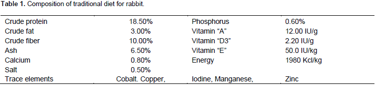
The rabbits handling and the experimental plan was set according to the Guidelines of the National Committee of Bio and Medical Ethics, King Abdul-Aziz City for Science and Technology (KACST), Saudi Arabia. Thirty white male rabbits were randomly divided into three groups as follows: the control (-), HFD (+) and HFD with camel milk, with the control group fed 100 g/day of NOR diet rabbit chow (USA) for three months, whereas the HFD group will be fed rabbit diet supplemented with 1.0% cholesterol plus 1.0% olive oil. The chemical composition of rabbit diet is shown in Table 1.
Biochemical analyses
For the same period as the control, blood samples were collected three months after the commencement of the treatment for the analysis of Malondiaaldehdye (MDA), Catalse (CAT), and glutathione peroxidase (GSH-Px). The total plasma cholesterol, triglyceride and high-density lipoprotein were measured using albacam kits, USA. The results were expressed as mean±SD. To assess the significant differences between the control group and the fat-fed group of rabbits for lipid profile, reduced glutathione (GSH) content in liver homogenate was determined colorimetrically by the method modified by Bulaj et al. (1998). Activities of antioxidant enzymes glutathione peroxidase (Paglia and Valentaine, 1979), superoxide dismutase (Spitz and Oberley, 1989) and catalase (Sinha, 1972) were colorimetrically determined using commercial assay kits.
Statistical analysis
Results were statistically analyzed using computer program Statistical package for Social Sciences (
SPSS). One-way analysis of variance (ANOVA), least significant differences (LSD) and Duncan test were used. The difference was considered significant at
P-value < 0.05 according to
Zar (1984).
The main objective is to assess the possible hypolipidemic effects of camel milk. The central idea for the current study is the relation between fat intake and the risk of cardiovascular disease and atherosclerosis. Recent investigations suggest that oxidative stress markers are useful in the evaluation of some types of abdominal pathology. The chemical composition of experimental diet for rabbits is outlined in Table 1. It is well-defined between hyperlipidemia and the development of oxidative stress, and the introduction of medicinal plants with higher antioxidant content proved effective (Tunsophon and Chootip, 2016). Previous studies indicated that a variety of plants extracts (Natal et al., 2016; Vivarelli et al., 2016; Sripradha et al., 2016), natural product (Zhu et al., 2016) and whey protein isolates (Betik et al., 2016) were used to ameliorate hyperlipidemia’s laboratory animal models. Hyperlipidemia significantly reduced the degree of lipid peroxidation (increase in MDA), and a decrease in individual antioxidant (Table 2). Owing to its high antioxidant composition, we hypothesized that camel milk could be effective in laboratory animal model subjected to high fat diet. In this regard, camel milk showed hypolipidemic effects in rats. The exact mechanism behind these effects may be well-related to the antioxidant status of camel milk.

The high fat diet supplemented rabbits showed elevated concentrations of TC (P < 0.001), TG (p < 0.001) and lower HDL-C (p < 0.001), compared to the control group. Dietary camel milk counteracted HFD-induced hyperlipidemia (Table 3). The exact mechanism may well relate to its chemical composition, in particular antioxidant profiles such as high vitamin C (Mohamed et al., 2005), and unique immunoglobulin (Jia et al., 2017). The camel milk showed hypolipidemic effects in diabetic rat model (Abdel Moneim et al., 2016). There is a well-documented relation between hyperlipidemia and the increased production of oxidants. The application of efficient nutraceuticals reducing the risk of hyperlipidemia attracts the attention of researcher in recent years. The higher intake of phytochemical-rich foods help in lowering the waist circumference and adiposity (Carnauba et al., 2017). The study indicated a clear significant difference between the control group and the other two groups. The supplementation of camel milk normalized TAC. These effects may be related to possible hypolipidemic effects of camel milk. In conclusion, the exact cause of these effects, in particular genomic studies on milk fat should be further investigated.

The author has not declared any conflict of interests.
REFERENCES
|
Abdel MA, Helmy H, Abdel-Reheim ES, Addaleel WW (2016). Camel milk ameliorates hyperglycemia-mediated hyperlipidemia and oxidative stress on streptozotocin-induced diabetic rats. Int. J. Diabetes Res. 5(4):63-69.
|
|
|
|
Ahamad SR, Raiash M, Ahmad A, Shakeel F (2017). Potential health benefis and metabolomics of camel milk by GC-MS and ICP-MS. Biol. Trace Elem. Res. 175(2):322-330.
Crossref
|
|
|
|
|
Betik AC, Aguila J, McConell GK, McAinch AJ, Mathai ML (2016). Tocotrienols and whey protein isolates substantially increase excericse endurance capacity in diet-induced obese male Sprague-dawley rats. PLoS One, 11(4):e0152562.
Crossref
|
|
|
|
|
Bulaj G, Kortemme T, Goldenberg D (1998). Ionization-reactivity relationship for cysteine thiols in polypeptides. Biochemistry 37:8965- 8972.
Crossref
|
|
|
|
|
Carnauba RA, Chaves DF, Baptistella AB, Paschoal V, Naves A, Buehler AM (2017): Assocition between high comsumption of phytochemical-rich foods and anthropometric measures: a systematic review. Int. J. Food. Sci. Nutr. 68(2):158-166.
Crossref
|
|
|
|
|
Chen B, Long P, Sun Y, Meng Q, Liu X, Cui HQ, Zhang L (2017). The chemical profiling of loqut leaf extract by hPLC-DAD-ESI-MS and its effects on hyperlipidemia and hyperglycemia in rats induced by a high-fat and fructose diet. Food Funct. In Press.
|
|
|
|
|
Jia S, Zhang W, Tan X, He W, Wang W (2017). The distribution of SIgA and IgG antibody-secreting cells in the palatine tonsils of Bactrian camels (Camelus bactrianus) of different ages. Histol. Histopathol. 32(5):511-521.
|
|
|
|
|
Meena S, Rajpur YS, Pandey AK, Sharma R, Singh R (2016). Camel milk ameliorates hyperglycemia and oxidative damage in type-1 diabetic experimental rats. J. Dairy Res. 83(3):412-419.
Crossref
|
|
|
|
|
Mohamed HE, Mousa HM, Beynen AC (2005). Ascorbic acid concentrations in milk from Sudanese camels. J. Anim. Physiol. Anim. Nutr. 89(1):35-37.
Crossref
|
|
|
|
|
Natal GD, Moreira CM, Miliao SM, Benjamin DA, Dantas S, Ribeiro MR, Martino SD (2016). Uba mango juices intake decreases adipocity and inflammation in high-fat diet-induced obese wistar rats. Nutrition, 32(9):1011-1018.
Crossref
|
|
|
|
|
Paglia DF, Valentaine WN (1979). Studies on glutathione and glutathione characterization of erythrocytes glutathione peroxidase. J. Lab. Clin. Med. 70:158-169.
|
|
|
|
|
Sripradha R, Sridhar MG, Maithilikiarpagaselvi N (2016). Antihyperlipdemic and antioxidant activities of the ethanolic extract of Garciniacambogia on high fat diet-fed rats. J. ComplemenIntegr. Med. 13(1):9-16.
|
|
|
|
|
Sinha KA (1972). Colorimetric assay of catalase enzyme. Anal. Biochem. 47:328-330.
Crossref
|
|
|
|
|
Tunsophon S, Chootip K (2016). Comparative effects of piperine and simvastatin in fat accumulation and antioxidative status in high fat-induced hyperlipidemic rats. Can. J. Physiol. Pharmacol. 94(12):1344-1348.
Crossref
|
|
|
|
|
Vivarelli F, Canistro D, Sapone A, De Nicola GR, BabotMarquillas C, Iori R, Antonazzo IC, Gentilini F, Paolini M (2016). Raphanus sativus cv. Sango Sprout Juice decreases diet-induced obesity in Sprague Dawley rats ameliorates related disorders. PLoS One, 11(3):e0150913
Crossref
|
|
|
|
|
Woodie N, Blythe S (2017). The differential effects of high-fat and high-fructose diets on physiology and behavior in male rats. Nutr. Neurosci. Pp.1-9.
Crossref
|
|
|
|
|
Yang W, Shi H, Zhang J, Shen Z, Zhou G, Hu M (2017). Effects of the duration of hyperlipidemia on cerebral lipids, vessels and neurons in rats. Lipids Health Dis. 16(1):26.
Crossref
|
|
|
|
|
Zhu H, Wang Y, Liu Z, Wang J, Wan D, Feng S, Yang X, Wang T (2016). Antidiabetic and antioxidant effects of catalpol extracted from Rehmannia glutinosa (di Huang) on rat diabetes induced by streptozotocin and high-fat, high-sugar feed. Chin. Med. 11(1):25.
Crossref
|
|
|
|
|
Zar JH (1984). Biostatistical Analysis. The 2nd Edition, Prentice-Hall, Englewood Cliffs, NJ, USA.
|
|