Full Length Research Paper

ABSTRACT
The study investigated the antioxidant potential as well as the inhibitory potential of the seed extracts of Picralima nitida on α-amylase and α-glucosidase enzymes. The methanolic, aqueous and coconut water extracts were obtained using 70% methanol, distilled water and coconut water, respectively. Antioxidant properties were studied in vitro using DPPH (1, 1-diphenyl-2-pricrylhydrazyl) radical scavenging, Ferric Reducing Antioxidant Power (FRAP), Total Antioxidant Capacity (TAC), Hydroxyl Radical Averting Capacity (HORAC), Total Phenol Content (TPC), and Total Flavonoid Content (TFC) assays. Different concentrations (0.1 - 0.5 mg/ml) of the extracts were subjected to α-amylase and α-glucosidase inhibitory assays using acarbose as standard. Absorbance was measured at 540 (for α-amylase) and 405 nm (α-glucosidase). The percentage of α-amylase and α-glucosidase inhibitory activity of the extracts and their IC50 values were determined. The seed extracts of P. nitida showed significant antioxidant potential. The inhibitory activity of the extracts on α-amylase and α-glucosidas compared favourably with acarbose with the methanolic extract possessing the highest inhibitory activity. The methanolic extract also possessed the highest antioxidant capacity with the lowest IC50 value among the extracts. The results indicate that the seed extracts of P. nitida possess significant antioxidant properties and are effective inhibitors of α-amylase and α-glucosidase enzymes.
Key words: Antioxidants, α-amylase, α-glucosidase, diabetes, phenolics, flavonoids, Picralima nitida.
INTRODUCTION
Cellular damage caused by free radicals is believed to play a central role in the aging and in disease progression (Sies et al., 1992). Antioxidants are the first line of defence against free radical damage, and are therefore important for maintaining optimum health and wellbeing. Imbalance between free radicals and antioxidants leads to oxidative stress which in turn results in the development of pathological conditions, one of which is diabetes. Studies have revealed the inference of oxidative stress in diabetes pathogenesis by lipid peroxidation, decreased vitamin C levels, impaired glutathione metabolism and alteration in enzymatic systems (Ullah et al., 2016).
Diabetes mellitus is a major cause of morbidity and mortality in the world. About 451 million people (aged 18-99 years) across the globe are reportedly suffering from diabetes (Cho et al., 2018). In 2015, diabetes was one of the leading causes of non-communicable diseases (NCD) death, contributing 1.5 million deaths globally (Wang et al., 2015) and 321,100 deaths in the African region (IDF, 2015). More than half of diabetic patients in Africa live in the most populous countries in the region - Nigeria, South Africa, Ethiopia and the Democratic Republic of Congo (IDF, 2015). Consumption of energy-rich diet, obesity and increase in sedentary lifestyle has been attributed to the rise in the number of diabetic cases (Sheeehan, 2003).
Diabetes mellitus is classified into two namely type 1 and type 2. Type 1, commonly referred to as insulin dependent diabetes mellitus (IDDM) is caused by immunological destruction of pancreatic cells which results in insulin deficiency (Hudson et al., 2002) while type 2, also known as non-insulin dependent diabetes mellitus (NIDDM), results from insulin resistance, a condition which is caused by reduced sensitivity of target tissues to the metabolic effect of insulin (Neuser et al., 2005). Although various conventional therapies abound, over 80% of rural dwellers in developing countries still depend on medicinal herbs (Brownlee, 2001). The side effects associated with the use of insulin and oral hypoglycemic agents have also led to an increase in the demand for alternative approaches to treat diabetes (Kahn et al., 2001). Hence, in modern days, huge attention has been directed towards recognition of plants with antidiabetic ability that may be used effectively for human consumption (Grover et al., 2002). A lot of plants have been screened for antidiabetic activity with promising results. Compounds responsible for antidiabetic activity in these plants include complex carbohydrates, alkaloids, glycopeptides, terpenoids, peptides, steroids, flavonoids, lipids, coumarines, sulphur compounds, and inorganic ions (Tchinda et al., 2008).
Picralima nitida is a species of the genus Picralima. It belongs to the hunterieae tribe of Apocynaceae family and is commonly called Osi-Igwe in Ibo and Abere in Yoruba (Duwiejua et al., 2002). In other parts of West Africa, the plant is called Gbe-Fondagne in Benin Republic, Adangme in Ghana, Abureebissi in Cote d’Ivoire and Susubalunyi in Sierra Leon (Kpodar et al., 2015). Picralima nitida is extensively distributed across West-Central Africa. The tree of P. nitida is under storey, reaching up to up to 4-35 m in height. Its trunk is about 5-60 m in diameter; cylindrical in shape and the wood is a pale yellow and hard (Okonta and Aguwa, 2007). The flowers are white (about 3 cm long) and they have ovoid fruits which become yellowish when mature. The leaves are broad (3-10 cm) and oblong (6-20 cm long) with tough tiny lateral nerves of about 14 to 24 pairs (Duwiejua et al., 2002).
P. nitida has diverse applications in West African traditional medicine. Various parts of the plant such as the leaves, seeds, stem, bark and roots are used by herbalists for the treatment of fever, hypertension, jaundice, gastro-intestinal disorders and malaria (Falodun et al., 2006). Preparations from different parts of the plant are employed as crude drug or crude herbal extract as remedy for various kinds of human diseases (Duwiejua et al., 2002). The seeds are widely used in West Africa especially in Nigeria, Cote d’Ivoire and Ghana as antipyretic, aphrodisiac agents, and for the treatment of malaria, pneumonia and other chest-conditions (Falodun et al., 2006). Herbalists have also made claims for the efficacy of the coconut water extract of P. nitida seeds in the treatment of many diseases, including diabetes (Adegoke and Oloyede, 2013). The presence of saponins, alkaloids, glycosides, steroids and tannins in the seeds of P. nitida has been reported (Sunmonu et al., 2014). It has been shown that seeds of the plant are rich in amino acids, vitamins A and E, as well as in mineral elements such as zinc, iron and manganese (Nwaogu, 2016). Investigations into the biological activity of the seed extracts also revealed their antimicrobial, larvicidal and hyperproteinaemic potential (Nwabor et al., 2014; Adegoke and Oloyede, 2013). This study is therefore aimed at evaluating the antioxidant properties of the seed extracts of P. nitida and their in-vitro anti-diabetic potential through the inhibition of alpha-amylase and alpha-glucosidase.
MATERIALS AND METHODS
Sample collection
Dried seeds of P. nitida were purchased from Ibode Market, Molete, Ibadan. The plant was identified and authenticated at the Herbarium, Department of Botany, Obafemi Awolowo University, Ile-Ife, Nigeria and deposited with the voucher number FPI – 2112.
Preparation of extracts
The seeds of P. nitida were air-dried and ground into fine powder. The methanolic, aqueous and coconut water extracts were separately prepared by mixing 20 g of the powder with 500 ml each of 70% methanol, distilled water and coconut water respectively. The various mixtures were stored in appropriately labeled conical flasks at room temperature for 48 h. The mixtures were sieved using cheese cloth to obtain the supernatants. The supernatants were then concentrated to dryness using a rotary evaporator to obtain the crude extracts: methanolic extract (ME), aqueous extract (AE) and coconut water extract (CE).
Determination of 1, 1-Diphenyl-2-Picraylhdrazyl hydrate (DPPH) radical scavenging activity
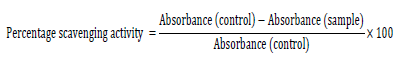
The 1, 1-diphenyl-2-picraylhdrazyl hydrate (DPPH) radical scavenging activity was measured according to the method of Blois (1958) . To 1 ml of different concentrations of the extract and the standard (ascorbic acid), 1 ml of 0.3 mM DPPH in methanol was added and allowed to react. The mixture was vortexed and kept in a dark chamber at room temperature for 30 min to allow for reaction. The absorbance was then measured at 517 nm against a DPPH control containing only 1 ml of methanol instead of the extract. The antioxidant activity was then calculated using this formula:
The 50% inhibitory concentration (IC50) was obtained from a linear regression plot of percentage inhibition against concentration of the extract.
Ferric reducing antioxidant power (FRAP) assay
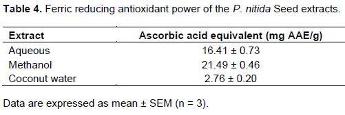
FRAP assay was carried out as described by Benzie and Strain (1999). FRAP reagent was obtained by mixing a 300 mmol/L acetate buffer (pH 3.6) with 2, 4, 6-tri-(2-pyridyl)-1, 3, 5-triazine and 20 mmol/L FeCl3.6H2O. An aliquot (50 μl) of the extract at 5 mg/ml and 50 μl of standard solutions of ascorbic acid (20, 40, 60, 80, and 100 μg/ml) were added to 1 ml of FRAP reagent and thoroughly mixed. Absorbance was measured at 593 nm wavelength after 10 min against the blank which contained 50 μl of distilled water. All measurements were taken at room temperature with samples protected from direct sunlight. The reducing power was expressed as ascorbic acid equivalent (mg AAE/g extract) which is defined as the concentration of antioxidant that gives a ferric reducing ability equivalent to that of the ascorbic acid standard.
Evaluation of total antioxidant capacity
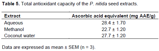
The total antioxidant assay was carried out based on the reduction of Mo (VI) to Mo (V) and the subsequent formation of a green phosphate/Mo (V) complex at acidic pH (Prieto et al., 1999) . The extract (0.3 ml) was mixed with 3 ml of reagent solution (0.6 M sulphuric acid, 28 mM sodium phosphate and 4 mM ammonium molybdate). The tubes containing the reaction solutions were incubated at 95°C for 90 min. The solution was cooled to room temperature and absorbance was then measured at 695 nm against the blank containing 0.3 ml of methanol. The antioxidant activity was expressed as ascorbic acid equivalent (mg AAE/g extract) which served as positive control.
Hydroxyl radical averting capacity (HORAC)
The ability of the seed extracts of P. nitida to scavenge the hydroxyl radical generated by the Fenton reaction was measured according to the modified method of Chung et al. (1997) . The reaction mixture containing 200 μl of 10 mM FeSO4, H2O, 200 μl of 10 mM EDTA and 200 μl of 10 mM 2-deoxyribose was added to 1.2 ml of 0.1 M phosphate buffer (pH 7.4) containing 200 μl of the extract. Thereafter, 200 μl of 10 mM H2O2 was added to the mixture and incubated for 4 h at 37°C. After incubation, 1 ml of 28% Trichloroacetic acid (TCA) and 1 ml of 1% Thiobarbituric acid (TBA) were added and placed in a boiling water bath for 10 min. The resultant mixture was then allowed to cool to room temperature and absorbance was measured at 532 nm in a UV-VIS spectrophotometer.
Determination of total phenol content
The total phenol content of the extracts was determined using the Folin-Ciocalteu’s method of Singleton and Rossi (1995) as described by Gulcin et al. (2003). To 0.1 ml of 5 mg/ml of extract was added 0.9 ml of distilled water. 0.2 ml of 10 % Folin reagent was then added. The resulting mixture was vortexed. After 5 min, 1 ml of 7% Na2CO3 solution was then added to the mixture. The solution was diluted to 2.5 ml with distilled water and then incubated for 90 min at room temperature. The absorbance at 750 nm was then read against the blank. The total phenol content of the extract was then calculated as shown in the equation below and expressed as mg gallic acid equivalent (GAE)/g fresh weight. Analysis was done in triplicates.

where C = total content of phenol compound in gallic acid equivalent (GAE); c = concentration of gallic established from the calibration curve (mg\ml); V = volume of extract/fractions (ml) and m = weight of the crude extract obtained.
Determination of total flavonoid content
The estimation of the total flavonoid content of the plant extracts was based on the aluminium chloride colorimetric method according to the method of Zhilen et al. (1999) and as described by Miliauskas et al. (2004). To 0.2 ml of extract was added 0.4 ml of distilled water. This was followed by the addition of 0.1 ml of 5% (w/v) sodium nitrite. After 5 min, 0.1 ml of 10% (w/v) aluminium chloride and 0.2 ml of 4% sodium hydroxide solution were added and the volume made up to 2.5 ml with distilled water. The absorbance was measured against the blank at 500 nm. The total flavonoid content of the plant extract was calculated with the equation below and expressed as mg rutin equivalents per gram of the extract. Analysis was done in triplicates.


where X = total content of flavonoid compound in rutin equivalent; q = concentration of rutin established from the standard curve; V = volume of extract (ml) and w = weight of the crude extract obtained.
In vitro alpha-amylase inhibitory activity
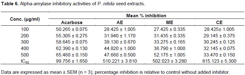
Alpha-amylase inhibitory activity of the different seed extracts (methanolic, aqueous and coconut water) of P. nitida was carried out according to the method of Miller (1959) with slight modifications. In a 96-well plate, reaction mixture containing 50 µl phosphate buffer (100 mM, pH = 6.8), 10 µl α–amylase (2 U/ml), and 20 µl of varying concentrations of the extracts (0.1, 0.2, 0.3, 0.4, and 0.5 mg/ml) was pre-incubated at 37°C for 20 min. Then, 20 µl of 1% soluble starch (100 mM phosphate buffer pH 6.8) was added as the substrate and incubated further at 37°C for 30 min; 100 µl of the DNS color reagent was then added and boiled for 10 min. The absorbance of the resulting mixture was measured at 540 nm using Multiplate Reader (Multiska thermo scientific, version 1.00.40). Acarbose at various concentrations (0.1-0.5 mg/ml) was used as the positive control while blank was used as the negative control. Each experiment was performed in triplicates. The results were expressed as percentage inhibition, which was calculated using the formula,
Inhibitory activity (%) = (1 − As/Ac) ×100
where As is the absorbance in the presence of test substance and Ac is the absorbance of control.
In vitro alpha-glucosidase inhibitory activity
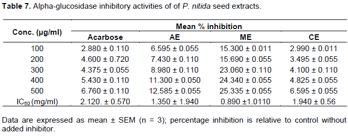
Alpha-glucosidase inhibitory activity of the different seed extracts of P. nitida was carried out according to the method reported by Ranila et al. (2010) with slight modifications. In a 96-well plate, reaction mixture containing 50 µl phosphate buffer (100 mM, pH = 6. 8), 10 µl alpha-glucosidase (1 U/ml), and 20 µl of varying concentrations of extract (0.1, 0.2, 0.3, 0.4, and 0.5 mg/ml) was preincubated at 37°C for 15 min. Then, 20 µl P-NPG (5 mM) was added as a substrate and incubated further at 37°C for 20 min. The reaction was stopped by adding 50 µl NaCO3 (0.1 M). The absorbance of the released p-nitrophenol was measured at 405 nm using Multiplate Reader. Acarbose at various concentrations (0.1-0.5 mg/ml) was included as the positive control while blank served as the negative control. Each experiment was performed in triplicates. The results were expressed as percentage inhibition, which was calculated using the formula,
Inhibitory activity (%) = (1 − As/Ac) ×100
where As is the absorbance in the presence of test substance and Ac is the absorbance of control.
Statistical analysis
All the measurements were done in triplicate and results are expressed in terms of mean ± standard error of mean. IC50 values were calculated using GraphPad Prism 5 version 5.01 (Graph pad software, Inc., La Jolla, CA, USA.) statistical software.
RESULTS AND DISCUSSION
Percentage yield of extracts
The percentage yields of the methanolic, aqueous and coconut water extracts were 14.90, 16.55 and 33.75% of the starting material respectively.
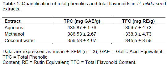
Total phenol and total flavonoid content
The results of total phenolic contents in P. nitida extracts are shown in Table 1. The aqueous extract contains the highest amount of phenols while coconut water extract has the lowest phenolic content. Coconut water extract was shown to have the highest flavonoid content while the aqueous extract contained the least amount of flavonoids.
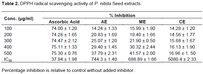
DPPH radical scavenging activity of P. nitida seed extracts
The extracts were found to possess concentration dependent inhibitory activity against DPPH radical as shown in Table 2. The order of decreasing activity was methanol extract > aqueous extract > coconut water extract. DPPH free radical scavenging potential of all the extracts was found to be lower than that of the reference compound ascorbic acid.
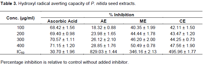
Hydroxyl radical averting capacity
The order of decreasing activity was methanol > coconut water > aqueous extract as seen in Table 3. Hydroxyl free radical scavenging potential of all the extracts was found to be lower than that of the reference compound ascorbic acid. The extracts possess concentration-dependent inhibitory activity against hydroxyl radical.
Ferric reducing antioxidant power
Table 4 shows the results of the FRAP assay. The methanolic extract had the highest FRAP value while the coconut water extract has the least FRAP value.
Total antioxidant capacity
The order of total antioxidant capacity as shown in Table 5 is aqueous extract > coconut water extract > methanol extract.
Alpha-anylase inhibitory activity
Table 6 shows the results alpha-amylase inhibitory activity of P. nitida seed extracts. The methanolic, aqueous and coconut water extracts of the seeds of P. nitida showed reasonable inhibitory activity against α-amylase when compared with acarbose.
Alpha-glucosidase inhibitory activity
The methanolic, aqueous and coconut water extracts of the seeds of P. nitida showed reasonable inhibitory activity against α-glucosidase when compared with acarbose. The methanolic extract exhibited the highest activity as shown in Table 7. Free radicals are harmful by-products produced during cellular metabolism, which could lead to oxidative damage in the body (Abidi and Ali, 1999). Antioxidants play a significant role in the body’s defence system against damage by free radicals. Several studies have described antioxidant compounds with radical scavenging activity present in herbs, fruits, vegetables and cereals extracts (Gray et al., 2002; Nuutila et al., 2003; Hou et al., 2005).
Phenols are important plant constituents due to their free radical scavenging ability by virtue of their hydroxyl groups. Flavonoids are one of the most diverse and widespread group of natural phenolics (Shimoi et al., 1996). The results of the present study indicate that the seed extracts of P. nitida are rich in phenols and flavonoids which may be attributable to their high antioxidant activity. The leaves and root bark of the methanolic extract of P. nitida have also been shown to contain significant amounts of phenols and flavonoids (Saravanan and Parimelazhagun, 2014; Teguwa et al., 2013). The aqueous extract contained the highest amount of phenols while the methanolic extract contained the highest amount of flavonoids according to the results in Table 1. Several studies have reported a higher phenol and flavonoid content in methanolic extracts relative to extracts of other solvents (Anokwuru et al., 2011; Babbar et al., 2014; Kopjar et al., 2015). However, Stanojevic et al. (2016) and Reis et al. (2016) reported a higher phenolic and flavonoid content of ethanol extracts when compared to other solvents that were investigated.
DPPH scavenging activities involve hydrogen atoms transfer and electrons transfer (Saravanan and Parimelazhagun, 2014). The results of the present study indicate that the seed extracts of P. nitida may contain some hydrogen donor molecules which may help in the reduction of free radical production. However, all seed extracts exhibited lower scavenging properties than the reference compound ascorbic acid as seen in their IC50 values in Table 2. The methanolic extract showed the highest DPPH radical scavenging activity among the seed extracts of P. nitida. Similar findings of the DPPH scavenging activity of the extracts of the root bark and leaf extracts of P. nitida have been reported (Erharuyi et al., 2014; Teguwa et al., 2013).
The hydroxyl radicals are extremely reactive oxygen species that can react with every possible molecule in living organisms, especially with proteins, DNA, and lipids (Mohammed et al., 2009). Hydroxyl radicals can rapidly initiate lipid peroxidation process by extracting hydrogen atoms from unsaturated fatty acids (Aruoma, 1998). They are able to do this because of the aromatic hydroxylation at the ortho-position of their phenolic rings (Lipinski, 2011). The electron or proton donation capacities of P. nitida seed extracts were further confirmed by the Fenton reaction system in a concentration-dependent manner similar to their DPPH radical scavenging activity. Nwankwo et al. (2017) reported a significant free radical scavenging activity of the ethanolic extract of P. nitida seeds on malaria-induced albino mice. This may be attributable to their high phenol and flavonoid content.
FRAP assay measures the reducing potential of an antioxidant reacting with a ferric tripyridyltriazine (Fe3+-TPTZ) complex to produce a coloured ferrous tripyridyltriazine (Fe2+-TPTZ) (Gordon, 1990. Antioxidants are strong reducing agents and this is primarily based on the redox potential of their hydroxyl groups (Oboh et al., 2012). The results indicate that there is a possible correlation between total phenol and total flavonoid content and FRAP assay which agrees with the findings of Zheng et al. (2001) and Rajurkar and Hande, (2011) who reported strong correlations between total phenol content and FRAP assay.
Total antioxidant activity of P. nitida seed extracts was evaluated by the phosphomolybdenum method (Prieto et al., 1999). This involves the transformation of Mo (VI) to Mo (V) by the antioxidant compound phosphor-molybdenum complex with a maximal absorption at 695 nm. The results obtained from phosphomolybdic acid assay are shown in Table 5 with the aqueous extract having the highest total antioxidant capacity. Polyphenolic compounds contribute significantly to the total antioxidant capacity of plants. A major goal in the treatment of diabetes mellitus is to maintain near normal blood glucose levels in both the fasting and postprandial state (Bailey, 2000). One therapeutic method used to decrease postprandial hyperglycemia is to suppress glucose production and/or absorption from the gastrointestinal tract through inhibition of either α-amylase or α-glucosidase enzymes (Cheng and Funtus, 2005; Kim et al., 2006; Matsui et al., 2007; Bhandari et al., 2008). Alpha amylase catalyzes polysaccharides (starch) into various oligosaccharides and disaccharides. Disaccharides produced by α-amylase are hydrolyzed further by α-glucosidase to yield glucose and other monosaccharides, which are readily absorbed in the small intestines (Shaw et al., 2010). Both animal studies (Okoli et al., 2010; Singh et al., 2001, Matsui et al., 2007) and clinical studies (Chiasson et al., 2002; Rhabasa-Lhoret and Chiason, 2004) have shown that inhibitors of both α-amylase and α-glucosidase can suppress glucose production and absorption from the small intestine. Currently, some inhibitors of α–amylase and α-glucosidase such as acarbose, phaseolamin and vogiblose are being used to suppress postprandial glucose levels in diabetic patients (Kim et al., 2006).
The hypoglycemic effects of the coconut extract of the seeds of P. nitida in alloxan-induced diabetic rats have been reported (Ajao et al., 2009). Adegoke and Oloyede (2013) also reported that the coconut water extract of the leaves of the same plant significantly lowered blood glucose and protein levels in alloxan-induced diabetic rats. The results obtained from the present study indicate reasonable α-amylase inhibitory activity of the methanolic, aqueous and coconut water extracts of the seeds of P. nitida, comparing favourably with acarbose with the methanolic extract exhibiting the inhibitory highest activity among the other extracts (Table 5). Similar in vitro studies have attributed the α-amylase inhibitory activity of some plant extracts to the presence of tannins (Bhandari et al., 2008), flavonoids, polyphenols and their glycoside derivatives (Campos et al., 2003). The methanolic, aqueous and coconut water extracts of the seeds of P. nitida also showed reasonable α-glucosidase inhibitory activity in comparison with acarbose. Again, the methanolic extract was observed to be the most potent inhibitor of α-glucosidase among the seed extracts of P. nitida. The α-glucosidase inhibitory activity of some plant extracts to the presence of flavonoids, polyphenols as well as their glycoside derivatives (Casirola and Ferraris, 2006; Andrade-Cetto et al., 2008). The order of activity of the extracts (methanolic > aqueous > coconut) was observed to be the same for both α-amylase and α-glucosidase. The difference in activity can be explained by the different polarities of the solvents, which selectively extracted targetable bioactive compounds relating to the inhibition of carbohydrate metabolism from seeds of P. nitida. Extracts of leaves of P. nitida have been reported to inhibit alpha-amylase and alpha-glucosidase (Kazeem et al., 2013).
CONCLUSION
The results of these investigations indicate that the seeds of P. nitida possess significant antioxidant properties. The results also support the traditional of the use of P. nitida seed extracts in the treatment of diabetes (Aguwa et al., 2001).
CONFLICT OF INTERESTS
The authors have not declared any conflict of interests.
REFERENCES
|
Abidi S, Ali A (1999). Role of ROS modified human DNA in the pathogenesis and etiology of cancer. Cancer Letters 142:1-9. |
|
|
Adegoke BM, Oloyede BO (2013). Antihyperglycemic and antihyperproteinaemic activity of extracts of Picralima nitida seed and Tapinanthus bangwensis leaf on alloxan-induced diabetic rabbits. International Journal of Innovation and Applied Studies 3(4):1125-1131. |
|
|
Aguwa CN, Ukwe CV, Inya-Agha SI, Okonta JM (2001). Antidiabetic effect of Picralima nitida aqueous seed extract in experimental rabbit model. Journal of Natural Remedies 1:135–139. |
|
|
Ajao SM, Olayaki LA, Oshiba OJ, Jimoh, RO, Jimoh SA, Olawepo A, Abioye AIR (2009). Comparative study of the hypoglycemic effects of coconut water extract of Picralima nitida seeds (Apocynaceae) and Daonil in alloxan-induced diabetic albino rats. African Journal of Biotechnology 8(4):574-576. |
|
|
Andrade-Cetto A, Becerra-Jiménez J, Cárdenas-Vázquez R (2008). Alpha-glucosidase inhibiting activity of some Mexican plants used in the treatment of type 2 diabetes. Journal of Ethnopharmacology 116(1):27-32. |
|
|
Anokwuru CP, Esiaba I, Ajibaye O, Adesuyi AO (2011). Polyphenolic content and antioxidant activity of Hibiscus sabdariffa calyx. Research Journal of Medicinal Plant 5(5):557-566. |
|
|
Aruoma OI (1998). Free radicals, oxidative stress, and antioxidants in human health and disease. Journal of the American Oil Chemists' Society 75(2):199-212. |
|
|
Babbar N, Oberoi HS, Sandhu SK, Bhargav VK (2014). Influence of different solvents in extraction of phenolic compounds from vegetable residues and their evaluation as natural sources of antioxidants. Journal of Food Science and Technology 51(10):2568–2575. |
|
|
Bailey CJ (2000). Potential new treatments for type 2 diabetes. Trends in Pharmacological Sciences 21(7):259-265. |
|
|
Benzie IF, Strain YT (1999). Total antioxidant capacity of teas by the ferric redcuing antioxidant power assay. Journal of Agricultural and Food Chemistry 47(2):633-636 |
|
|
Bhandari MR, Jong-Anurakkun N, Hong G, Kwabata J (2008). Alpha glucosidase and alpha amylase inhibitory activities of Nepalese medicinal herb Pakhanbhed (Bergenia ciliate, Haw). Food Chemistry 106:247-252. |
|
|
Blois MS (1958). Antioxidant determinations by the use of a stable free radical. Nature 26:1199-1200. |
|
|
Brownlee M (2001). Biochemistry and molecular biology of diabetic complications. Nature 414(6865):813. |
|
|
Campos KE, Diniz YS, Cataneo AC, Faine LA, Alves MJ, Novelli ELB (2003). Hypoglycaemic and antioxidant effects of onion, Allium cepa: dietary onion addition, antioxidant activity and hypoglycaemic effects on diabetic rats. International Journal of Food Sciences and Nutrition 54:241-246. |
|
|
Casirola DM, Ferraris RP (2006). α-Glucosidase inhibitors prevent diet-induced increases in intestinal sugar transport in diabetic mice. Metabolism 55(6):832-841. |
|
|
Cheng AY, Funtus IG (2005). Oral antihyperglycemic therapy for type 2 diabetes mellitus. Canadian Medical Association Journal 172(2):213-226. |
|
|
Chiasson JL, Josse RG, Gomis R, Hanefeld M, Karasik A, Laakso M (2002). Acarbose for prevention of type 2 diabetes mellitus: the STOP-NIDDM randomised trial. The Lancet 359(9323): 2072-77. |
|
|
Cho NH, Shaw JE, Karuranga S, Huang Y, da Rocha Fernandes JD, Ohlrogge AW, Malanda B (2018). IDF diabetes atlas: Global estimates of diabetes prevalence for 2017 and projections for 2045. Diabetes Research and Clinical Practice 138:271-281. |
|
|
Chung SK, Osawa T, Kawakishi S (1997). Hydroxyl radical scavenging effects of spices and scavengers from Brown Mustard (Brassica nigra). Bioscience, Biotechnology, and Biochemistry 61(1):118-123. |
|
|
Duwiejua M, Woode E, Obiri DD (2002). Pseudo-akuammigine, an alkaloid from Picralima nitida seeds, has anti-inflammatory and analgesic actions in rats. Journal Ethnopharmacology 81(1):73-79. |
|
|
Erharuyi O, Falodun A, Langer P (2014). Medicinal uses, phytochemistry and pharmacology of Picralima nitida (Apocynaceae) in tropical diseases: A review. Asian Pacific Journal of Tropical Medicine 7(1):1-8. |
|
|
Falodun A, Nworgu ZA, Ikponmwonsa MO (2006). Phytochemical components of Picralima nitida Stapf and its effect on isolated non-pregnant rat uterus in oestrus. Pakistani Journal of Pharmaceutical Sciences 19(3):256–258. |
|
|
Gordon MH (1990). The mechanism of antioxidant action in vitro. In Food antioxidants. Springer, Dordrecht pp. 1-18. |
|
|
Gray DA, Clarke MJ, Baux C, Bunting JP, Salter AM (2002). Antioxidant activity of oat extracts added to human LDL particles and in free radical trapping assays. Journal of Cereal Science 36(2): 209-218. |
|
|
Grover JK, Yadav S, Vats V (2002). Medicinal plants of india with antidiabetic potential. Journal of Ethnopharmacology 81(1):81-100. |
|
|
Gulcin I, Buyukokuroglu ME, Oktay M, Kufrevioglu OI (2003). Antioxidant and analgesic activities of turpentine of Pinus nigra Arn. Subsp. Pallsiana (Lamb.) Holmboe. Journal of Ethnopharmacology 86(1):51-58. |
|
|
Hou WC, Lin RD, Lee TH, Huang YH, Hsu FL, Lee MH (2005). The phenolic constituents and free radical scavenging activities of Gynura formosana Kiamnra. Journal of the Science of Food and Agriculture 85(4):615-621. |
|
|
Hudson BI, Hofmann MA, Bucciarelli W, Thoralf M, Bernhard L, Yan Q, Wu S, David MD, Vivette YSF, Grant PJ, Schmidt AM (2002). Glycation and diabetes: The RAGE connection. Current Science 1515-1521. |
|
|
International Diabetes Federation (IDF) (2015). IDF Atlas. 7th edition, Brussels, Belgium. Diabetesatlas.org 12-week prospective trial. |
|
|
Jia W, Gao WY, Tang PG (2003). Antidiabetic drugs of plant origin used in China: Compositions, pharmacology, and hypoglycemic mechanisms. China Journal of Chinese Materia Medica 28(2):108-113.Not Cited |
|
|
Kahn SE, Montgomery B, Howell W (2001). Importance of early phase insulin secretion to intravenous glucose tolerance in subjects with type 2 diabetes mellitus. Journal of Clinical Endocrinology Metabolism 86:5824-5829. |
|
|
Kazeem MI, Adamson JO, Ogunwade IA (2013). Modes of inhibition of α-amylase and α-glucosidase by aqueous extract of Morinda lucida Benth leaf. BioMed Research International. |
|
|
Kim YM, Jeong YK, Wang WY, Lee MH, Rhee HI (2006). Anti-diabetic activity of SMK001, a poly herbal formula in streptozocin-induced diabetic rats therapeutic study. Biological and Pharmaceutical Bulletin 29(3):477-482. |
|
|
Kopjar M, Tadić M, Piližota V (2015). Phenol Content and Antioxidant Activity of Green, Yellow and Black Tea Leaves. Chemical and Biological Technologies in Agriculture 2(1):1-6. |
|
|
Kpodar S, Lawson-Evi P, Bakoma B (2015). Ethnopharmacological survey of plants used in the treatment of diabetes mellitus in south of Togo (Maritime Region). Journal of Herbal Medicine 5(3):147-152. |
|
|
Lipinski B (2011). Hydroxyl radical and its scavengers in health and disease. Oxidative Medicine and Cellular Longevity 809696:1-9. |
|
|
Matsui T, Tanaka T, Tamura S, Toshima A, Miyata Y, Tanaka K (2007). Alpha-glucosidase inhibitory profiles of catechins and theaflavins. Journal of Agricultural and Food Chemistry 55:99-105. |
|
|
Miliaukas G, Venskutonis PR, Van BT (2004). Screening of radical scavenging activity of some medicinal and aromatic plant extracts. Food Chemistry 85(2):231-237. |
|
|
Miller GL (1959). Use of dinitrosalicylic reagent for determination of reducing sugar. Analytical Chemistry 31:426-428 |
|
|
Mohammed H, Ons M, Yosra ET, Rayda S, Neji G, Moncef N (2009). Chemical composition and antioxidant and radical-scavenging activities of Periploca laevigata root bark extracts. Journal of the Science of Food and Agriculture 89(5):897-905. |
|
|
Neuser D, Benson A, Brückner A, Goldberg RB, Hoogwerf BJ, Petzinna D (2005). Safety and tolerability of Acarbose in the treatment of type 1 and 2 diabetes mellitus. Clinical Drug Investigation 25(9):579-587. |
|
|
Nuutila AM, Puupponen-Pimia R, Aarni M, Oksman-Caldentey KM (2003). Comparison of antioxidant activity. Food Chemistry 81(4):485-493. |
|
|
Nwabor OF, Dibua UM, Ngwu GI, Onyenma NC, Odiachi O, Nnamonu EI, Okoro, JO, Eze TR, Okeke IS (2014). Evaluation of the antimicrobial and larvicidal potentials of seed extracts of Picralima nitida. International Research Journal of Natural Sciences 2(2):23-30. |
|
|
Nwankwo NE, Nwodo FOC, Joshua PE (2017). Free radical scavenging property of Picralima nitida Seed Extract on Malaria-Induced Albino Mice. American Journal of Life Sciences 5(5):125-133. |
|
|
Nwaogu LA (2016). Chemical profile of Picralima nitida seeds used in ethnomedicine used in West Africa. Futo Journal Series 2(2):110-122. |
|
|
Oboh G, Akinyemi AJ, Ademiluyi AO (2012). Antioxidant properties and inhibitory effect of ethanolic extract of Struchium sparganophora (Ewuro Odo) leaf on α-amylase and α-glucosidase activities. African Journal of Traditional, Complementary and Alternative Medicines 9(3):342-349. |
|
|
Okoli CO, Ibiam AF, Ezike AC, Akah PA, Okoye TC (2010). Evaluation of antidiabetic potentials of Phyllanthus niruri in alloxan diabetic rats. African Journal of Biotechnology 9(2):248-259. |
|
|
Okonta JM, Aguwa CN (2007). Evaluation of hypoglycaemic activities of glycosides and alkaloids extracts of Picralima nitida Stapf. (Apocynaceae) seed. International Journal of Pharmacology, 3(6):505-509. |
|
|
Prieto P, Pineda M, Aguilar M (1999). Spectrophotometric quantitation of antioxidant capacity through the formation of a phosphomolybdenum complex: specific application to the determination of vitamin E. Analytical Biochemistry 269(2):337-341. |
|
|
Rajurkar NS, Hande SM (2011). Estimation of phytochemical content and antioxidant activity of some selected traditional Indian medicinal plants. Indian Journal of Pharmaceutical Sciences 73(2):146-151. |
|
|
Ranila LG, Kwon YI, Apostoldis E, Shetty K (2010). Phenolic compounds, antioxidant activity and in vitro inhibitory potential against key enzymes relevant for hyperglycemia and hypertension of commonly used medicine plants, herbs and spices in Latin America. Bioresource Technology 101(12):4676-4689. |
|
|
Reis PM, Dariva C, Vieira GBA, Heinse H (2016). Exraction and evauation of antioxidant potential of the extracts obtained from tamarind seeds (Tamarindicus indica), sweet variety. Journal of Food Engineering 173:116-123. |
|
|
Rhabasa-Lhoret R, Chiasson JL (2004). α-Glucosidase Inhibitors. In; International Textbook of Diabetes Mellitus (3rd Edition). London, UK: Wiley and Sons pp. 901-914. |
|
|
Saravanan S, Parimelazhagan T (2014). In vitro antioxidant, antimicrobial and anti-diabetic properties of polyphenols of Passiflora ligularis Juss. fruit pulp. Food Science and Human Wellness 3(2):56-64. |
|
|
Shaw JE, Sicree RA, Zimmet PZ (2010). Global estimates of the prevalence of diabetes for 2010 and 2030 (Review), pharmacological basis of therapeutics. Diabetes Research and Clinical Practice 87(1):4-14. |
|
|
Shimoi K, Masuda S, Shen B, Furugori B, Kinae N (1996). Radio-protective effect of antioxidative plant flavonoids in mice. Mutatation Research 350(1):153-161. |
|
|
Sies H (1992). Antioxidant functions of vitamins: Vitamins E and C, Betaâ€Carotene, and other carotenoids a. Annals of the New York Academy of Sciences 669(1):7-20. |
|
|
Singh SN, Vats P, Suri S, Shyam R, Kumria ML, Ranganathan S, Sridharan K (2001). Effect of an antidiabetic extract of Catharanthus roseus on enzymic activities in streptozotocin induced diabetic rats. Journal of Ethnopharmacology 76(3):269-277. |
|
|
Tchinda AT, Tchuendem I, Khan SN, Omar I, Ngandeu F, Nkeng PA, Choudhary IM (2008). Antioxidant activity of the crude extract of the fruits of Pycnanthus angolensis and α-glucosidase inhibitory activity of its constituents. International Journal of Pharmacology 1:422-431. |
|
|
Teguwa CM, Mejiato PC, Zofou D, Tchinda BT, Boyom FF (2013). Antioxidant and antidiabetic properties of two African medicinal plants: Picralima nitida (Apocynaceae) and Sonchus oleraceaus (Asteraceae). BMC Contemporary and Alternative Medicine 13(1):1755-183. |
|
|
Ullah A, Khan A, Khan I (2016). Diabetes mellitus and oxidative stress - A concise review. Saudi Pharmaceutical Journal 24(5):547-553. |
|
|
Wang L, Song R, Chen S, Wang J, Ling F (2015). Prevalence of depressive symptoms and factors associated with it in type 2 diabetic patients: A cross-sectional study in China. BMC Public Health 15:188. |
|
|
Zheng W, Wang SY (2001). Effect of plant growth temperature on antioxidant capacity in strawberry. Journal of Agricultural and Food Chemistry 49:4977-82. |
|
Copyright © 2024 Author(s) retain the copyright of this article.
This article is published under the terms of the Creative Commons Attribution License 4.0


