Full Length Research Paper

ABSTRACT
Salix humboldtiana Willd. (Salicaceae) colonizes Brazilian riverine forests and, besides other congeneric species, is traditionally used to treat fever. In folk medicine, it is even used as a Quinine substitute to treat malaria fever. Although its occurrence worldwide was largely reduced between 2000-2015, in Brazil, the case numbers induced by Plasmodium falciparum have increased. Thus, new tools and strategies are necessary against malaria according to the World-Malaria-Report. In the present study the characterization of flavonoids, phenolic acids, phenolic glycosides, and salicylates detected in the ethanol extract of willow leaves, and the antiplasmodial activity of extract of S. humboldtiana and its fractions are firstly reported. Chromatography, UV-Visible spectroscopy, and mass spectrometry were used to characterize substances, and the antiplasmodial activity was accessed by cellular DNA fluorescence measurement with ethidium bromide on a flow cytometer. Kaempferol, apigenin, salicylic acid, and salicylates were found in the extract among other phenolics. The bioassays revealed that lyophilized extract of S. humboldtiana Willd showed antiplasmodial activity. Hexane, AcOEt, and Methanol fractions were active against P. falciparum at IC50 of 0.655, 0.929 and 0.688 μg/mL, respectively. Quinine, used as a reference, showed an IC50 of 0.147 μg/mL. The polyphenols - salicylates and flavonoids, especially kaempferol and catechin, detected in LESh, exhibit biochemical interference on the plasmodial metabolism, as already reported, and they could also promote the antiplasmodial activity detected. This alleged medicinal use has been already reported for some Salix species. Moreover, this work presents scientific evidence for the antiplasmodial activity of S. humboldtiana, which can be helpful in the development of antiplasmodial phytomedicine.
Key words: Salix humboldtiana Willd, extracts, phenolic compounds, antiplasmodial activity.
INTRODUCTION
The genus Salix is a species-rich taxon comprising five subgenres, and includes about 400 species of deciduous trees and shrubs (Freischmidt et al., 2012). Salix humboldtiana Willd. is the single willow species native to South America, belonging to the subgenus Protitea according to molecular data (Argus, 2011) and settles in riparian floodplains as a pioneer tree species in a wide distribution area, ranging from southern North America to Patagonia, in southern South America. The willow species is adapted to tropical and subtropical climates (Hernandez-Leal et al., 2019).
In Europe, “the success of the willow bark in the cure of the ague” was already highlighted by Stone (1763) and he provided the first convincing demonstration of a potent antipyretic effect of the willow containing salicylates. Moreover, in Europe at that time, the most problematic ‘plague’ (intermittent fever) to be effectively cured, with white willow (Salix alba L.) bark, was probably, caused by malaria (Wood, 2015). In folk medicine, leaves and bark of some Salix species are used to treat fever, including malaria (Frei et al., 2009; Deharo et al., 2001), rheumatism (Leporatti and Ivancheva, 2003), and constipation (Scarpa, 2004). In South American folk medicine, Salix is even used as a Quinine substitute to treat fever associated with malaria indicating that S. humboldtiana could be included in the research for new natural active pharmaceutical ingredients to treat malaria as reported by Milliken (1997) and Felix-Cuenca et al., (2022).
According to the recent World-Malaria-Report (WHO, 2019) new malaria-facing agents and strategies will be critical to accelerate the pace of malaria-eliminating progress. This is necessary since, despite the malaria burden reduction in the period of 2000-2015, the rate of malaria eliminating progress has slowed in recent years and, e.g., in Brazil the casuistics of Plasmodium falciparum malaria even increased. Willows are known for their medicinal properties for millennia, and from them, in 1828, salicylic acid was isolated, becoming the precursor of the anti-thermic and anti-inflammatory drug, aspirin (FAO, 2014).
Generally, secondary metabolites, like phenolics, are biosynthesized, aiming mainly to defend plants against infections and predation. Thus, they can be employed as anti-infectious agents and insecticides (Zaynab et al., 2018). In this way, phenolics of willow seem to exert a protective function, since high concentrations of them, in the leaves, present a negative effect on mammalian herbivory (Stolter et al., 2013). Particularly, phenolic glycosides can be very abundant in willows compared to other secondary metabolites. Concentrations of up to 30% of plant dry weight have been reported (Donaldson et al., 2006) and extracts containing phenolic glycosides can be analyzed by GC, TLC, and HPLC, which is the most frequently reported analytical method using gradient elution and UV detection (Boeckler et al., 2011).
The sixth edition of the Brazilian Pharmacopoeia (2019) presents a monograph of S. alba and the second edition of the Brazilian Pharmacopoeia Formulary of Phytomedicines (2021) describes formulations obtained from S. alba and other Salix species. Also, the World Health Organization (WHO, 2004) and European Medicines Agency (EMA, 2020) report monographs on Salix species and preparations, respectively.
Considering the information found in the relevant literature and presented in this paper, the central aim of this work is to detect and characterize the main phenolic substances found in S. humboldtiana by chromatographic and spectrometric techniques, and secondarily to assess the antiplasmodial activity of its ethanolic extract.
MATERIALS AND METHODS
Plant material
The plant material was collected from willow formations on the sandy levees of the Aquidauana River, Anastácio, Mato Grosso do Sul, Brazil, at the site 20º28’42.55” S 55º48’9.46”0 with original coordinates Lat: -20.478486 Long: -55.812778 WGS84 Alt: 146 m on August 23rd, 2015. Ten of these willow samples were identified as S. humboldtiana Willd. by both botanists Geraldo Damasceno Junior and Heike Markus-Michalczyk. A voucher specimen is deposited in herbarium at the Federal University of Mato Grosso do Sul under number GGMS 52608. Willow branches with leaves were collected from these ten specimens.
The plant material was stored in plastic boxes to keep them fresh until further analyses. This material was cleaned, dried, and ground to produce the herbal derivative as a semi-coarse powder.
Analytical methods
All the reported phytochemical analyses of the present study were performed in the Laboratory of Chromatography and Mass Spectrometry (LCMS) of the Pharmacy Faculty at the Para Federal University on a freeze-dried extract of dried leaves of S. humboldtiana Willd. (LESh). For detecting different classes of secondary metabolites, common chemical reactions were performed, in triplicate, on the herbal extract. Among these classes of secondary metabolites are saponins, organic acids, reducing sugars, polysaccharides, proteins and amino acids, phenols and tannins, flavonoids, cardiac glycosides, catechins, sesquiterpenes and other lactones, alkaloids, purines, steroids, and triterpenoids, azulenes, depsides and dapsones, coumarin derivatives and anthraquinones, according to Barbosa et al. (2020).
Extraction and fractionation
The extract of S. humboldtiana Willd. was prepared by macerating about 200 g of ground leaves in 1000 mL 96ºGL Ethanol (RDD 1:5). After 7 days, maceration was filtered and concentrated on a rotary evaporator under reduced pressure until the complete removal of ethanol. The aqueous residue was then frozen and lyophilized, furnishing a lyophilized extract of S. humboldtiana Willd. (LESh). The dry weight of the extract was 23.6 g (yield 11.8%).
An aliquot of 5 g LESh was then treated with solvents of increasing polarity: Hexane, Dichloromethane, Ethyl Acetate, and Methanol. After evaporation of the solvents, the following fractions were obtained: HFSh, from Hexane; DFSh, from Dichloromethane; AFSh, from Ethyl Acetate; MFSh, from Methanol.
Thin layer chromatography (TLC)
The qualitative analysis of the extract (10 mg/mL), polar fractions (10 mg/mL), and standard catechin (1 mg/mL), were performed by TLC using silica gel chromatoplates of 0.20 mm thickness. The qualitative analysis of the extract (10 mg/mL), polar fractions (10 mg/mL), and standard catechin (1 mg/mL), were also performed as the mobile phase ethyl acetate, toluene, and formic acid (85:10:5) with observation under ultraviolet radiation (254 nm and 365 nm) and after sprayed with vanillin sulfuric (ethanolic vanillin 1% followed by ethanolic sulfuric acid 10% with heating at 110ºC for 5 min) (Wagner and Blat, 2001) and pulverization with a methanolic solution of 2, 2-diphenyl-1-picrylhydrazyl radical (DPPH) 0.05% (Wang et al., 2012).
Thin layer chromatography chromatoplates were sized 20 cm x 20 cm, covered with a 0.2 mm thick 60G - F254 silica gel layer, containing fluorescence indicator (MERCK®). Catechin, Rutin, Chlorogenic acid, and DPPH solution, and all other chemicals, including solvents HPLC grade, used in this work were purchased from Sigma-Aldrich (Darmstadt, Germany).
High-pressure liquid chromatography (HPLC)
Preparation of samples: Standard substances, extract, and fractions
High-pressure liquid chromatography was conducted as LC-DAD analysis and as LC-MS analysis. These are analytical techniques for the separation of phenolic compounds in high-performance reverse-phase liquid chromatography, with diode array detection (DAD) and mass spectrometry (MS) detection, respectively. This is a combination of techniques commonly used for separation, identification, and quantification of phenolic compounds (Kähkönen et al., 2001).
Aliquots of LESh were dissolved in MeOH-HPLC at 20 mg/mL and 1 mg/mL for LC-DAD and LC-MS analyses, respectively. All samples were centrifuged for 10 min at 14,000 rpm (Centrifuge 5418, Eppendorf) before each injection. The substances used as standard in the LC-DAD analyses were dissolved at 1 mg/mL (m:v), also in MeOH-HPLC.
LC-DAD analyses
LC–DAD analyses were performed using an Agilent 1260 model G1361A Infinity series instrument coupled to a Diode Array Detector G1315C, managed by Open Lab ChemStation software version A.01.05 (Agilent Technologies).
The sample injection volume was 10 μL and separation occurred on a C18 column (Zorbax Eclipse XDB-C18) with particle size 5 μm, 4.n6 x 150 mm maintained at 35ºC. Two solvents form the mobile phase in a gradient elution system using H2O at pH 3.2 by formic acid (0.03% v/v) (A) and acetonitrile (B) at a flow of 0.8 mL/min. The gradient proportions were: 0 min: 90% A; 10 min: 75% A; 20 min: 10% A; 30 min: 0% A, 32 min: 90% A. The UV–vis spectra were recorded between 200 and 600 nm and the chromatograms were registered at 280 and 330 nm, respectively.
LC-MS analyses
The LC-MS system used was an Agilent 1260 model 6460 Triple Quad LC/MS managed by Agilent MassHunter Qualitative Analysis Software Version B.07.00. The same conditions as sample injection volume, column, analysis time, and flow rate were used for the UPLC–MS analysis. For the ESI (negative mode) source, the following inlet conditions were employed: scan spectra from m/z 100 to 1500, the gas temperature used was 350ºC; the Nitrogen flow rate, 6 L/min; nebulizer pressure, 20 psi and capillary voltage, 3500 V. The applied fragmentation energy values were: 60, 80, 120, and 150 V, where the 120 V generated the best profiles.
The sample injection volume was 4 μL and the separation occurred on a C8 column (Zorbax Eclipse XDB-C8) with particle size 5 μm, 4.6 x 150 mm maintained at 27ºC. Two solvents form the mobile phase in a gradient elution system using H2O at pH 3.2 by Formic Acid (0.03% v/v) (A) and Acetonitrile (B) at a flow of 0.4 mL/min. The gradient proportions were: 4 min: 80% A; 6 min: 60% A; 9-20 min: 5% A. The UV–vis spectra were recorded between 200 and 600 nm, respectively and the chromatograms were registered at 254, 280, 315, 330, and 350 nm, respectively.
The relevant peaks of the samples were identified by comparison of retention time of authentic reference substance (catechin and chlorogenic acid) and UV-Vis spectrum with the same experimental data. Some peaks of the sample were also identified based on of data reported in the literature, including the results obtained by mass spectrometry.
Evaluation of antiplasmodial activity
The FRC3 P. falciparum strains were provided by Leônidas and Maria Deane Institute – FIOCRUZ-Amazônia. The parasites were cultivated according to Trager and Jensen (2005) and the tests carried out based on Rabelo et al. (2014) and Bolson et al. (2019). The samples were dissolved in Dimethylsulfoxide (DMSO), in a serial dilution (1:2), providing concentrations from 0.39 to 50 μg/mL, and added to the RPMI 1640 culture medium (Gibco, Waltham, USA). Correspondingly, the final solvent concentration in the wells was ≤ 0.5%, a concentration that did not interfere with the parasite's viability.
Blood samples containing P. falciparum at 1% parasitemia and 2% hematocrit were equally distributed in the test wells, which were completed with culture medium up to 100 μL / well. The plates were incubated for 72 h at 37°C in a low concentration of oxygen and carbon dioxide, following the traditional microaerophilic technique of candle burning in a desiccator. After this time, the plates were centrifuged at 800 rpm for 5 min. Subsequently, each well was treated with 50 μL of an ethidium bromide solution (1:50) for 30 min. Thereafter, the plates were washed twice with 200 μL of PBS buffer 1X. After that, the samples were resuspended in 200 μL of PBS buffer. Finally, the samples were analyzed on a BD FACSCanto II flow cytometer (BD Biosciences, San Jose, USA), on channel FL-1 using software Getting Started with BD FACSDiva™ and FlowJo™ version 10. The parasitic growth in 0.5% DMSO was used as a positive control and non-parasitized erythrocytes as a negative control. Quinine was used as a reference substance, in the same concentrations as phytochemical samples (Dose/Concentration).
The percentage of parasite growth inhibition was determined using the formula by Lopes et al. (2009):

The concentration responsible for 50% inhibition of total parasitemia (IC50) was calculated using the GraphPad Prism 8 software based on a logarithmic graph of dose versus inhibition (expressed as a percentage in relation to the control) by non-linear regression analysis.
RESULTS AND DISCUSSION
Previous phytochemical tests on the lyophilized extract of S. humboldtiana - LESh – implemented to characterize the presence of secondary metabolites, following an analytic protocol described by Barbosa et al. (2020) showed the presence of phenols and tannins, catechins, steroids, triterpenoids, and coumarin derivatives. These metabolic classes were described by Evans et al. (1995) and Jeon et al. (2008), besides flavonoids, which have been connected to different biological activities, including antiplasmodial (Miliken, 1997; Felix-Cuenca et al., 2022).
Thin layer chromatography (TLC)
The presence of a phenolic substance, like catechin, in LESh was confirmed by TLC, since both shows remarkably close Rf values and the same red color. When the chromatogram was treated with vanillin, according to Wagner and Blat (2001), and Stahl and Schild (1981), flavonoids were also detected. Moreover, these spots reacted with DPPH reagent, indicating their antioxidant capacity. The same reagent indicated the presence of salicylates by discoloration (Wagner and Bladt, 2001).
The results agree with the study on phenolics and flavonoids in extracts of S. aegyptiaca of Enayat and Banerjee (2009), which were described as significant amounts of catechin and rutin in the aqueous extract of the leaves and in the ethanolic extract of bark, respectively.
High-pressure liquid chromatography (HPLC-DAD)
The results of the conducted HPLC-DAD on LESh are shown in Figures 1 and 2. Peak 1, shown in Figure 2, at 7.22 min, corresponds to a UV-Visible spectrum of catechin showing λmax = 280 nm. By overlying the chromatograms registered under the same conditions, both for the catechin standard and the sample, the retention times are remarkably close. In addition, the absorption maxima at 280 nm observed in both UV–Visible spectra are superimposed, depicting the structural similarity (Figure 2A). "In addition, the LC-MS analyses show a signal at m/z 288.9 that corroborate with data reported by Pohjamo at al (2003)."
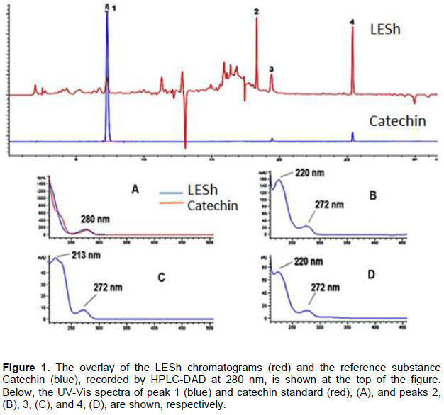
Peaks 2, 3, 4 shown in Figure 2 at 18.30 min; 19.40 min; 25.38 min, respectively, correspond to UV-Vis spectra, and present band 1 at 272 nm, and band 2 at 220 nm, Figure B; band 1 at 272 nm, and band 2 at 213 nm, Figure C; and band 1 at 272 nm, and band 2 at 220 nm, Figure D (Figure 2B, C, D). These spectra are similar to those described by Abreu et al. (2011) for salicylates, as phenolic glycoside type, present spectra with band 1, between 215 nm and 220 nm and a weaker band 2 around 270 nm.
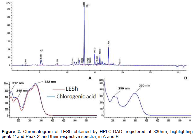
Figure 3 shows a chromatogram of LESh registered at 330 nm by HPLC-DAD. Peak 1 shown in Figure 3 at 5.24 min, corresponding to UV-Vis spectrum presents absorption maxima at 217, 245, and 322 nm, respectively.
This is similar to that obtained from chlorogenic acid, a standard substance (Figure 3-A), a derivative of hydroxycinnamic acid, and an isomer of caffeoylquinic acid. These data combined with that of the LC-MS analyses (Figure 3), a peak in m/z = 352.9 [M-H]- (Table 1), allow inferring that chlorogenic acid is present in this sample, although slight differences in retention times and in the resolution of the UV-Vis spectrum, at low wavelength, may be due to the different purity grade of the acid as isolated standard and in the mixture of a complex matrix.
Peak 2 shown in Figure 3 at 13.02 min corresponds to a UV-Vis spectrum (Figure 3-B) with the band I at 350 nm and band II around 250 nm, characteristic of flavonoids. As described by Mabry et al. (1970) and Merker and Beecher (2000), flavone and flavanols spectra exhibit two absorption maxima, band I (300-380 nm) and band II (240-280 nm).
Investigation on the polyphenol content of six different Salix species pointed out that luteolin and apigenin, along with their derivatives, are the main flavones present in their leaves, while the main flavonols are myricetin and quercetin, and their derivatives, along with with isorhamnetin-3-glucoside (Nyman and Julkunen-Tiitto, 2005).
Furthermore, according to El-Wakil et al. (2015), the methanolic extract of Salix tetrasperma and its fractions show antioxidant activity and high content of phenolic compounds, especially in the ethyl acetate fraction.
Liquid chromatography electrospray ionization mass spectroscopy (LC-ESI-MS)
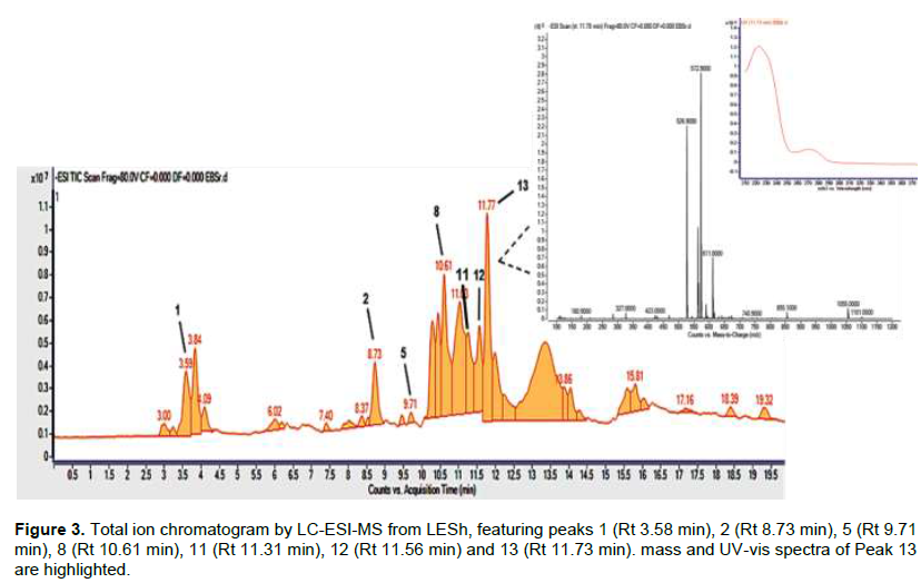
Results of the analyses on LESh using Electrospray Ionization (ESI) Mass Spectroscopy (ESI-MS) are shown in Figures 3 and 4.
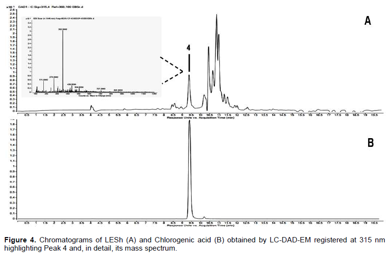
Figure 3 shows the Total Ion Chromatogram - TIC -, using ESI-MS, with peaks of phenolic compounds, like phenolic acids, phenolic glycosides, and polyphenols. The electrospray source (ESI) in negative mode [M-H]- generated ions that produced peaks 1, 2, 5, 8, 11, 12 and 13 corresponding to the following ions m/z: 178.9; 330.9; 352.9; 288.9; 468.9; 285.9; 572.9, respectively.
Caffeic acid (peak 1) is a constituent of the extract, derived from caffeoylquinic acid, identified by ESI-MS in the crude extract based on m/z= 170.9 [M-H]-. This acid and its derivatives have been widely reported in the leaf and bark of Salix species (Enayat and Banerjee, 2009; Poblocka-Olech et al., 2010; El-Wakil et al., 2015).
In this research, catechin was characterized by CCD and by HPLC-DAD in LESh. This substance (peak 5) is suggested as a constituent of the extract, identified by ESI-MS based on m/z= 288.9 [M-H]-. Catechin is a polyphenol, precisely a flavan-3-ol, and is known for its antioxidant (Karakaya et al. 2001; Kim et al., 2003), and antiplasmodial activities (Tasdemir et al., 2006). Catechin was identified in bark extracts of other Salix species (Tawfeek et al., 2021). Other flavonoids identified by ESI-MS in negative mode are kaempferol, peak 12, (Plazonic et al., 2009; Abu-Reidah et al., 2015), also an antiplasmodial substance (Tasdemir et al., 2006), and an apigenin derivative, peak 6, (Plazonic et al., 2009).
Figure 2 shows the chromatogram of LESh (A) and chlorogenic acid (B) obtained by LC-DAD-EM registeredat 315 nm. Peak 4 (Rt= 9.46 min) was recorded at 315 nm. In comparison with the retention times of this peak and the chlorogenic acid pattern obtained under the same conditions, it is inferred that both show high equivalence. In addition, the corresponding mass spectrum, with a base peak if m/z= 352.9 [M-H]- characteristic of chlorogenic acid, confirms the identity of the substance. This acid was also identified by El-Wakil et al. (2015) in the leaves of S. tetrasperma.
Other salicylates were identified by ESI-MS in LESh (Figure 3). These are: salicin 2 (m/z 330.9 [M-H + HCCOH]-), Salicortin 8 (m/z 468.9 [M -H + HCCOH]-), Tremuloidine 11 (m/z 434.9 [M-H + HCCOH]-) and Tremulacin 13 (m/z 572.9 [M-H + HCCOH]-). Finally, salicylic acid 14 and hydroxybenzoic acid were identified by their mass in the subfraction AFSh6 (Figure 4).
Salicin, the main phenolic glycoside present in bioactive extracts of Salix species, is considered the pharmacologically active substance due to its aspirin-like structure. In animal models, extracts of leaves of S. aegyptiaca and male flowers showed anti-inflammatory effects in Carrageenan-induced paw edema and hot plaque tests (Karawya et al., 2010; Rabbani et al., 2010).
Antiplasmodial activity
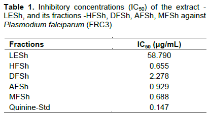
The bioassay model used in this work revealed that fractions of a lyophilized extract of S. humboldtiana leave - LESh - treated with solvents of increasing polarity (Hexane - HFSh; Dichloromethane - DFSh; Ethyl acetate. - AFSh; Methanol - MFSh) showed antiplasmodial activity after evaporating the solvents. HFSh presented the best result regarding the antiplasmodial activity against P falciparum showing an IC50 of 0.655 μg/mL, better than MFSh and AFSh, which showed IC50 of 0.688 μg/mL and IC50 of 0.929 μg/mL, respectively (Table 1).
The antiplasmodial activity of HFSh and MFSh, fractions of LESh, against P. falciparum is encouraging even when compared to the standard substance used (quinine), considering that the samples are complex matrices containing many substances, among which are flavonoids and phenolics. Ogunlanaa et al. (2015) assayed the antiplasmodial activity of an ethanolic extract and of an Ethyl Acetate derivative and found IC50 values higher than those here reported: (>92.0±0.04) and (16.0±0.01), respectively. These samples contain flavonoids derivatives, which were isolated and tested. The phytochemical investigation of AFSh (ethyl acetate fraction), which is here reported in detail, provides a remarkably interesting phenolic profile, which results from the detection and characterization of salicylates and flavonoids, like apigenin, catechin, and kaempferol. Soré et al. (2018) reported the antiplasmodial activity of such substances derivatives. Additionally, five phenolic glycosides detected in a Salix species present antiplasmodial activity as described by Kim et al. (2014), and their IC50 ranged from 6.6 to 20.5 μM.
CONCLUSION
The hydroethanolic extract of S. humboldtiana leaves presents antiplasmodial activity, which according to published data can be connected to the flavonoids kaempferol, and catechin, whose presence in the analyzed samples was evidenced by TLC and LCMS. This attribute justifies upcoming efforts aiming at innovative Salix formulations to treat malaria, as urged by WHO, and to accelerate the pace of malaria-fighting. Also, six salicylates were characterized in the samples, which can be considered quimiomarker of the genus Salix, useful to standardize extracts and control the quality of herbal ingredients. Further investigations aiming to identify the antiplasmodial activity detected for HFSh and MFSh and the substances of this species that allow the standardization of the active pharmaceutical substance are recommended. This may support the development of a phytomedicine based on S. humboldtiana Willd., and the elucidation of the action mechanism of the active pharmaceutical substance.
CONFLICT OF INTERESTS
The authors have not declared any conflict of interests.
ACKNOWLEDGMENTS
The authors thank their respective Institutions for the research infrastructure and materials, and the workforce mobilized to carry out this research at the undergraduate, masters, and doctoral levels.
REFERENCES
|
Abreu IN, Ahnlund M, Moritz T, Albrectsen BR (2011). UHPLC-ESI/TOFMS Determination of salicylate-like phenolic glycosides in Populus tremula leaves. Journal of Chemical Ecology 37(8):857-870. |
|
|
Abu-reidah IM, Ali-shtayeh MS, Jamous RM, Arráez-román D, Segura-carretero A (2015). HPLC-DAD-ESI-MS/MS screening of bioactive components from Rhus coriaria L. (Sumac) fruits. Food Chemistry 166:166-179. |
|
|
Argus GW (2011). Guide to Salix (Willow) in the Canadian Maritime Provinces (New Brunswick, Nova Scotia, and Prince Edward Island). Canadian Museum of Nature, Ottawa, Ontario, Canada. |
|
|
Barbosa WLR, Santa Brígida A, Remigio MSN (2020). Alfafito - Alfarrábios de Fitoquímica. CRV Publ, Curitiba, Paraná, Brazil. |
|
|
Boeckler GA, Gershenzon J, Unsicker SB (2011). Phenolic glycosides of the Salicaceae and their role as anti-herbivore defenses. Phytochemistry 72(13):1497-1509. |
|
|
Bolson GCM, Barros LB, Ribeiro CV, Lima JA, França TCC, Santos I, Orlandi PP, Veiga Junior VF (2019). Chemical composition and biological activities of Metania and Drulia (Metaniidae) freshwater sponges from Amazonia. Chemistry and Biodiversity 16(8):e1900318. |
|
|
Brazilian Pharmacopoeia (2021). Brazilian Pharmacopoeia Formulary of Phytomedicines Salix spp., ANVISA, Brasília pp. 173-175. |
|
|
Brazilian Pharmacopoeia (2019).Brazilian Pharmacopoeia, 6th Ed (2019). Salicis cortex, PM079-00, ANVISA, Brasilia. |
|
|
Deharo E, Bourdy G, Quenevo C, Muñoz V, Ruiz G, Sauvain M (2001). A search for natural bioactive compounds in Bolivia through a multidisciplinary approach. Part V. Evaluation of the antimalarial activity of plants used by the Tacana Indians. Journal of Ethnopharmacology, Limerick 77(1):91-98. |
|
|
Donaldson JR, Stevens MT, Barnhill HR, Lindroth RL (2006). Age-related shifts in leaf chemistry of clonal aspen (Populus tremuloides). Journal of Chemical Ecology 32(7):1415-1429. |
|
|
El-Wakil EA, Abdel-Hameed ESS, El-Sayed MM, Abdel-Lateef EE (2015). Identification of the chemical composition of the methanolic extract of Salix tetrasperma Roxb. using LC-ESI-MS and evaluating its potential as antioxidant agent. Der Pharma Chemica 7(2):168-177. |
|
|
Enayat S, Banerjee S (2009). Comparative antioxidant activity of extracts from leaves, bark, and catkins of Salix aegyptiaca sp. Food Chemistry 116(1):23-28. |
|
|
European Medicine Agency (EMA) (2017). European Union herbal monograph on Salix [various species including S. purpurea L., S. daphnoides Vill., S. fragilis L.], cortex. |
|
|
Evans TP, Clausen TP, Reichardt PB (1995). Structurally Intriguing Glucosides from Alaskan Littletree Willow (Salix arbusculoides). Journal of Natural Products 58(12):1897-1900. |
|
|
FAO (Food and Agricultural Organization of the United Nations) (2014). Poplars and willows of the world, with emphasis on Silvicultural important species. In: Isebrands, JG, Richardson, J (eds.), Poplars and Willows: Trees for Society and the Environment. CABI Publishing, Oxon 699 p. |
|
|
Felix-Cuencas L, Delis-Hechavarria E, Jarro A, Parola-Contreras I, Escamilla-García A, Torres-Pacheco I, García-Trejo JF, Soto-Zarazúa GM, Guevara-González RG (2022). Bioactivity characterization of herbal molecules. In Herbal Biomolecules in Healthcare Applications, pp. 145-183). Academic Press, Amsterdam. |
|
|
Frei B, Baltisberger M, Sticher O, Heinrich, M (2009). Medical ethnobotany of APG III. An update of the Angiosperm Phylogeny Group classification for the orders and families of flowering plants. Botanical Journal of the Linnean Society, Oxford 161:105-121. |
|
|
Freischmidt A, Ju?rgenliemk G, Kraus B, Okpanyi SN, Mu?ller J, Kelber O, Weiser D, Heilmanna J (2012). Contribution of flavonoids and catechol to the reduction of ICAM-1 expression in endothelial cells by a standardized Willow bark extract. Phytomedicine 19(3-4):245-252. |
|
|
Hernandez?leal MS, Suarez?atilano M, Pinero D, Gonzalez-?rodriguez A (2019). Regional patterns of genetic structure and environmental differentiation in willow populations (Salix humboldtiana Willd.) from Central Mexico. Ecology and Evolution 9(17):9564-9579. |
|
|
Jensen TW, Human JB (1976). Malaria parasites in continuous culture. Science 193(4254):673-675. |
|
|
Jeon SH, Chun W, Choi YJ, Kwon YS (2008). Cytotoxic constituents from the bark of Salix hulteni. Archives of Pharmacal Research 31(8):978-982. |
|
|
Kähkönen MP, Hopia AI, Heinonen M (2001). Berry phenolics and their antioxidant activity. Journal of Agricultural and Food Chemistry 49(8):4076-4082. |
|
|
Karakaya S, El SN, Tas AA (2001). Antioxidant activity of some foods containing phenolic compounds. International Journal of Food Sciences and Nutrition 52(6):501-508. |
|
|
Karawya MS, Ammar MN, Hifnawy MS, Al-okbi SY, Mohamed DA, Elanssary AA (2010). Phytochemical study and evaluation of the anti-inflammatory activity of some medicinal plants growing in Egypt. Medical Journal Islamic World Academy Sciences 18(4):139-150. |
|
|
Kim CS, Kwon OW, Kim SY, Choi SU, Kim JY, Han JY, Choi SI, Choi JG, Kim KH, Lee KR (2014). Phenolic Glycosides from the Twigs of Salix glandulosa. Journal of Natural Products 77(8):1955-1961. |
|
|
Kim D, Jeong SW, Lee CY (2003). Antioxidant capacity of phenolic phytochemicals from various cultivars of plums. Food chemistry 81(3):321-326. |
|
|
Leporatti ML, Ivancheva S (2003). Preliminary comparative analysis of medicinal plants used in the traditional medicine of Bulgaria and Italy. Journal of Ethnopharmacology, Limerick 87(2-3):123-142. |
|
|
Lopes SCP, Blanco YC, Justo GZ, Nogueira PA, Rodrigues FLS, Goelnitz U, Wunderlich G, Facchini G, Brocchi M, Duran N, Costa FTM (2009). Violacein extracted from Chromobacterium violaceum inhibits Plasmodium growth in vitro and in vivo. Antimicrobial Agents and Chemotherapy 53(5):2149-2152. |
|
|
Mabry TJ, Markham KR, Thomas MB (1970). The Ultraviolet Spectra of Flavones and Flavonols. In: The Systematic Identification of Flavonoids. Springer, Berlin, Heidelberg. |
|
|
Merker HM, Beecher GR (2000). Measurement of food flavonoids by high-performance liquid chromatography: A review. Journal of Agricultural and Food Chemistry 48:577-579. |
|
|
Milliken W (1997). Plants for Malaria. Plants for Fever, The Royal Botanic Gardens, Richmond. |
|
|
Nyman T, Julkunen-tiitto R (2005). Chemical variation within and among six northern willow species. Phytochemistry 66 (24):2836-2843. |
|
|
Ogunlanaa OO, Kimb H-S, Watayab Y, Olagunjuc JO, Akindahunsid AA, Tane NH (2015). Antiplasmodial flavonoid from young twigs and leaves of Caesalpinia bonduc (Linn) Roxb. Journal of Chemical and Pharmaceutical Research 7(1):931-937. |
|
|
Plazonic A, Bucar F, Males Z, Mrnar A, Nigovic B, Kujundzic N (2009). Identification and Quantification of Flavonoids and Phenolic Acids in Burr Parsley (Caucalis platycarpos L.), Using High-Performance Liquid Chromatography with Diode Array Detection and Electrospray Ionization Mass Spectrometry. Molecules 14(7):2466-2409. |
|
|
Poblocka-olech L, Krauze?Baranowska M, G?ód D, Kawiak A, ?ojkowska E (2010). Chromatographic analysis of simple phenols in some species from the genus Salix. Phytochemical Analysis 21(5):463-469. |
|
|
Pohjamo SP, Hemming JE, Willför SM, Reunanen MH, Holmbom BR (2003). Phenolic extractives in Salix caprea wood and knots. Phytochemistry 63(2):165-169. |
|
|
Rabbani M, Sajjadi Se, Rahimi F (2010). Anxiolytic effect of flowers of Salix aegyptiaca L. in mouse model of anxiety. Journal of Complementary and Integrative Medicine 7(1):18-22. |
|
|
Rabelo DM, Pinheiro MLB, Barison A, Salomé KS, Costa EV, Silva FMA, Chaves YO, Bastos IS (2014). Isoquinoline alkaloids and investigation of the antibacterial and antiplasmodial activities of Guatteria citriodora (Annonaceae). Química Nova 37(9):1453-1458. |
|
|
Scarpa GF (2004). Medicinal plants used by the Criollos of Northwestern Argentine Chaco. Journal of Ethnopharmacology, Limerick 91:115-135. |
|
|
Soré H, Sanon S, Hilou A (2018). Antiplasmodial properties of plants isolated flavonoids and their derivatives. International Journal of Herbal Medicine 6(5):43-56. |
|
|
Stahl E, Schild W (1981). Pharmazeutische Biologie. Drogenanalyse II: Inhaltsstoffe und Isolierungen. Gustav Fischer Verlag. |
|
|
Stolter C, John P, Ball R, Julkunen-tiitto (2013). Seasonal differences in the relative importance of specific phenolics and twig morphology result in contrasting patterns of foraging by a generalist herbivore. Canadian Journal of Zoology 91:338-347. |
|
|
Stone E (1763). An account of the success of the bark of the willow in the cure of agues. Philosophical transactions of the Royal Society of London (53):195-200. |
|
|
Tasdemir D, Lack G, Brun R, Ruedi P, Scapozza L, Perozzo R (2006). Inhibition of Plasmodium falciparum Fatty Acid Biosynthesis: Evaluation of FabG, FabZ and FabI as Drug Targets for Flavonoids. Journal of Medicinal Chemistry 49(11):3345-3353. |
|
|
Tawfeek N, Mahmoud MF, Hamdan DI, Sobeh M, Farrag N, Wink M, El-Shazly AM (2021). Phytochemistry, Pharmacology and Medicinal Use of Plants of the Genus Salix: An Updated Review. Frontiers in Pharmacology 12:593856. |
|
|
Trager W, and Jensen JB (2005). Human malaria parasites in continuous culture. Journal of Parasitology 91(3):484-486. |
|
|
Wagner H, Blat S (2001). Plant Drug Analysis: A thin layer chromatography atlas. 2. Ed. Library of Congress Cataloging-in-Publication Data. |
|
|
Wang J, Yue Y-d, Tang F, Sun J (2012). TLC Screening for antioxidant activity of extracts from fifteen Bamboo Species and identification of antioxidant flavone glycosides from leaves of Bambusa textilis McClure. Molecules 17:12297-12311. |
|
|
World Health Organization (WHO) (2004). Monographs on selected medicinal plants, 4, WHO, Salerno, Italy. |
|
|
World Health Organization (WHO) (2019). World-Malaria-Report. |
|
|
Wood JN (2015). From plant extract to molecular panacea: a commentary on Stone (1763) 'An account of the success of the bark of the willow in the cure of the agues'. Philosophical Transactions of the Royal Society B: Biological 370:20140317. |
|
|
Zaynab M, Fatima M, Abbas S, Sharif Y, Umair M, Zafar MH, Bahadar K (2018). Role of secondary metabolites in plant defense against pathogens, Microbial Pathogenesis 124:198-202. |
|
Copyright © 2024 Author(s) retain the copyright of this article.
This article is published under the terms of the Creative Commons Attribution License 4.0


