Full Length Research Paper

ABSTRACT
Canthium multiflorum (Thonn.) Hiern (Rubiaceae) is a popular herb used by traditional healers in western Burkina Faso. C. multiflorum leaves are widely used in decoction to treat malaria. The present study aims to evaluate its in vivo potential against malaria parasites in mice. The antimalarial activity of the organic and aqueous extracts of C. multiflorum leaves was evaluated on Plasmodium berghei Anka in NMRI mice using the Peters 4-day suppressive test. The fractions of the extracts were also tested. The acute toxicity study was performed according to Lorke method and sub-acute toxicity by Seewaboon method. Phytochemical analysis of extracts was carried out according to Ciulei method. The results showed that ethanolic and decoctions were the best inhibitors of parasites’ growth. The ethanolic extract exhibited an inhibition of 22.5, 30.8 and 81.9% at 100, 250 and 500 mg/kg body weight, respectively. While the decoction with water, an inhibition of 10.1, 25.9 and 74.2% at the same doses. Fractions’ extracts showed moderate activities at dose of 250 mg/kg bw. In addition, no mortality was recorded with the ethanolic extract. No signs of toxicity were observed in animals in the sub-acute toxicity study. The phytochemical constituents of the extracts were mainly steroids and/or triterpenes, flavonoids, emodols, carotenoids, coumarins, tannins, saponins, anthocyanosides and reducing compounds. Ethanol and decoctions of C. multiflorum leaves have been shown to have significant antimalarial activity in infected mice, with no toxicity. Phytochemical analysis confirmed the previously chemical groups found in the roots of the plant.
Key words: Canthium multiflorum, malaria, toxicity, Plasmodium berghei, in vivo, phytochemical.
INTRODUCTION
Malaria remains a public health problem in Burkina Faso, especially during the peak transmission season (June- September), where 60% of cases occur in children (Tiono et al., 2014). Currently, WHO-recommended effective treatments are based on artemisinin-based combination therapies (ACTs), the costs of which are still prohibitively expensive for populations in endemic countries (Beiersmann et al., 2007). Plasmodium falciparum resistance to antimalarial drugs is also an obstacle to access effective treatment of the disease. In addition,
mosquito resistance to insecticide is increasing, limiting the effectiveness of vector control programs. Since the dawn of time, malaria has been treated with medicinal plants mainly recommended by traditional healers. Numerous antimalarial recipes are available worldwide and some have been scientifically validated from the most historic, Cinchona officinalis (Rubiaceae) to the most recent, Artemisia annua, Cryptolepis sanguinolata (Periplocaceae) and many others (Willcox et al., 2004). Malaria remains a challenge for researchers and finding new therapeutic agents from plants can be a solution. Efforts to integrate traditional medicine into national health systems are under way in Burkina Faso, where some thirty herbal medicines are recognized and marketed (OMS, 2011).
In the western region of Burkina Faso, the decoction of leaves of Canthium multiflorum (Schumach. &Thonn.) Hiern (Rubiaceae) is widely used to treat malaria (orally and bath). The water decoction extract of plant leaves is also used to reduce high blood pressure and fever (Ouédréogo-Nacoulma, 1996).
Ethyl acetate and dichloromethane extracts from the roots of C. multiflorum, in previous studies, had shown interesting antiplasmodial activity in vitro against the parasite Plasmodium falciparum 3D7 (IC50= 7 μg/ml). Of the active extracts, 19a-hydroxy-3-oxo-ursa-1,12-dien- 28-oic acid was isolated, with a moderate antiplasmodial effect on parasites (IC50 = 26 μg/ml) without inducing a change in the shape of the erythrocyte membranes (Traore, 2008). So far, no in vivo antimalarial study has been performed on the plant. The objective of this study was to evaluate antimalarial activity and toxicity of C. multiflorum leaves in mice.
MATERIALS AND METHODS
Collection of plant
The leaves of C. multiflorum were collected from “Tin” surroundings, in the Western region of Burkina Faso in September 2017. C. multiflorum was identified by Dipama Pascal at IRSS (Institut de recherche en sciences de la santé), and voucher specimen CmL2017 was deposited in the unit of pharmacognosy at IRSS Bobo Dioulasso. The leaves were thoroughly rinsed with water, reduced to small pieces, air-dried at room temperature about two week, and powdered with a mechanical grinder.
Chemicals
Hydrochloric acid, ethyl acetate, ethanol and hexane were purchased from SIGMA (SIGMA-ALDICH, Germany). Dragendorff’s reagent (SIGMA, USA), ethanoic anhydride, ferric chloride (Labosi, France), methanol, n-butanol, dichloromethane, Fehling solution, and sulfuric acid (CARLO ERBA) were also used for this study. All reagents were of analytical grade.
Extraction
Two organic extracts (dichloromethane and ethanol) and two water extracts were prepared for the test. About 100 g of dried powdered leaves were macerated with 500 mL of water, dichloromethane or ethanol at room temperature for about 24 h with stirring. The water decoction was prepared with 100 g of powdered leaves in 500 mL of boiling water. Sequential solid-liquid extractions (called fractions) were also carried out using solvents of increasing polarity (hexane/ethyl acetate, ethyl acetate, n-butanol, ethanol and methanol).
Extracts (decoction and maceration) of leaves were filtered using Whatman N°3 and were evaporated to dryness with an R-200 rotary evaporator (BUCHI Labortechnik AG, United Kingdom) at constant temperature (60°C) and at a pressure below 1 bar, while the water extracts were freeze-dried and stored in dried containers until use.
Phytochemical analysis
One gram of each extract (extract macerated with water, extract decocted with water, extract, ethanol and dichloromethane extracts) were dissolved in 30 mL of distilled water, acid-hydrolyzed (HCl) for 30 min, then liquid-liquid extracted with dichloromethane. From each extract, 3 sub-extracts were thus obtained: dichloromethane fraction, hydrolysed aqueous and ethanol fractions and unhydrolyzed aqueous and ethanolic fractions, all subjected to the phytochemical analysis. Tests were also made with unhydrolyzed extracts.
Qualitative phytochemical analysis was performed in tubes according the method of Ciulei (1982). The phytochemical constituents of the extracts were characterized for their main metabolites, namely alkaloids, steroids and/or triterpenes, tannins, flavonoids, carotenoids, coumarins, emodols, anthocyanosides and reducing compounds using known characteristic tests summarized in the following.
Steroids and/or triterpenes
Solution (2 mL) of ethanoic anhydride and 1 mL of sulfuric acid (H2SO4) were added to 2 mL of dichloromethane extract; the appearance of a blue-green color indicated the presence of steroids and/or triterpenes.
Carotenoids
Extracts (10 ml) were evaporated to dryness. The residue was dissolved in 2 mL of 50% ethanol while heating. This was followed by the addition to the residue of a few drops of antimony trichloride (SbCl3) saturated in CHCl3. A blue-green color that could change to red on time indicates the presence of carotenoids.
Flavonoids
Approximately 0.3 g of the extract was diluted with 2 mL of 50% methanol. The pink-red color after adding 2 to 3 fragments of magnesium ribbon and 1 mL of concentrated hydrochloric acid to the extract indicates the presence of flavonoids.
Emodols by Borntrager’s reaction
Extracts (3 ml) were mixed with 1 mL of 25% NH3. The mixture is shaken. The development of a red color indicates the presence of emodols.
Anthracenosides
To 1 mL of dichloromethane fraction in the test tube, 1 ml of 25% NH3 was added while stirring. A cherry red color indicates the presence of anthracenosides.
Coumarins
Hydrolysed aqueous and ethanolic fractions (2 mL) were evaporated to dryness. The residue was dissolved in 2 mL of water by heating and then divided into equal volume in two tubes. 0.5 mL of 10% NH3 was added to the tubes, which were observed under ultraviolet light. The blue-green fluorescence indicates the presence of coumarins.
Saponins
Extracts (unhydrolysed aqueous and ethanolic fractions, 2 mL) were diluted with 2 mL of distilled water and put in a graduated cylinder. The solution was shaken vigorously for 15 min and the presence of 1 cm foam layer persistence for more than 30 min indicates the presence of saponins.
Tannins
Water (2 mL) and 2-3 drops of dilute 2% ferric chloride solution was added to 2 mL of the extract. A dark green solution indicates the presence of tannins.
Alkaloids
Unhydrolyzed aqueous and ethanolic fractions (10 mL) were evaporated to dryness. The residue was dissolved in 2 ml of 2% HCl. The solution was divided into two equal volumes. One was used as a reference. A few drops of Dragendorff’s reagent were added to the test tube. An orange red precipitate indicates the presence of alkaloids.
Anthocyanosides
Hydrolysed aqueous and ethanolic fractions (2 mL) dissolved in distillated water, 2 mL of HCl and 2 ml of NH3 are added; the pink-red color turning to violet blue indicates the presence of anthocyanosides.
Reducing compounds
Water (1 mL) and Fehling solution (1 mL) mixture were heated in the water bath. Brick-red precipitate indicates the presence of reducing compounds.
Parasites and experimental animals
Female NMRI albino mice aged 8 to 12 weeks and weighing 25±2 g from the International Centre for Research and Development on Livestock in Sub-humid Areas (CIRDES) in Bobo-Dioulasso were used. The animals were housed in the same environmental conditions (temperature 25°C, 12 h photoperiod) and fed with standard food provided by “Service Régional d’Elevage de Bobo-Dioulasso”, Burkina-Faso. Experiments were conducted in accordance with the international guidelines for animal care. P. berghei strain originally obtained from Malaria Research Reagent Resource Center (MR4) was maintained by serial passage of blood from infected to uninfected mice.
Ethical considerations
This study protocol has been approved by the national ethic review board of “Comité d’Ethique pour la recherche en santé” (ref:2015-5-056/CERS). The animals were used in accordance to the Directive 86/609/EEC and in compliance with animal welfare assurance guidelines for foreign institutions (OLAW A5926-01). All animals have been provided by the lab of animal production in Centre International de Recherche-Développement sur l’Elevage en zone Subhumide (CIRDES), reférence center in West and Central Africa.
In vivo antiplasmodial activity test
Three doses (100, 250 and 500 mg/kg bw) of each extract were used to treat the animals. The dichloromethane extracts were dissolved in distilled water mixed with 5% (v/v) of Tween 80. The ethanol and aqueous extracts were dissolved in distilled water. The experiments were performed in mice according to Peters’ 4-day suppressive test (Peters and Robinson , 1992). Mice were randomly divided into 6 mice per group. On day 0, the mice were inoculated with 107 parasitized red blood cells by intraperitoneal route. Two hours after infection, the animals received orally (PO), 200 µl of each dose of extract once a day from day 0 to day 4. The control group received only the solution used to dissolve the extracts. At day 5 of the test, thin blood smears were made from blood drawn from the tail of mice, fixed with methanol and stained with Giemsa 10%. The slides were read with an optical microscope, under a 100X oil immersion objective. Each experiment was repeated three times. The mean parasitaemia of each group of mice was then calculated.
Acute toxicity test
The acute toxicity (LD50) of the active ethanol extract was evaluated using the Lorke method (Lorke et al., 1983). NMRI mice, aged 8-12
weeks weighing 25±2 g were used. The animals were divided into five groups of three mice (five groups of females and five groups of males) and received the following extract doses: 3797, 5695, 8543
and 12814 mg/kg/bw. The extracts previously dissolved were administered orally as a single dose. The control group received the solvent used to dissolve the extract; and animals were monitored for 14 days. At the end of the observation period, the animals were weighed; mortality as well as behavioral and physical changes was recorded first at the beginning and on days 7 and 14. Each experiment was also repeated three times.
Sub-acute toxicity test
The sub-acute toxicity test with the active ethanol extract was evaluated using the seewaboon method (Seewaboon et al., 2013). Wistar rats, 8-12 weeks old and weighing 200±10 g were used. The animals were divided into three groups of three mice (three groups of males and three groups of females) and received orally 125 and 250 mg extract/kg body weight once a day for 14 days; the control group received the solvent used to dissolve the extract, under the same experimental conditions. The rats were followed for 28 days; body weight, signs of toxicity and mortality were recorded. The day after the test, all rats were fasted and anesthetised. Blood was collected for evaluation of hematological parameters. Serum was tested for biochemical parameters such as alanine amino-transferase (ALT), aspartate aminotransferase (AST), cholesterol (TC), triglycerides (Trig), glucose (Glu), urea and creatinine (Crea) using an automated analyser (ARCHITECT). Animals were then sacrificed under diethyl ether anaesthesia; major organs (heart, lungs, spleen, liver and kidneys) were collected for visual detection of macroscopic lesions in both treatment and control groups.
Statistical analysis
Microsoft Excel 2013 were used for data analysis. The percent inhibition of parasitaemia was calculated according to the formula described by Fidock et al. (2004).
Values are presented as mean ± standard deviation. Statistical significance was determined at p=0.05 level and performed by One-way analysis of variance (ANOVA) followed by Dunnett’s post-test.
RESULTS
Phytochemical analysis
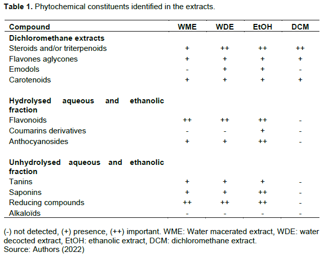
Phytochemical analysis revealed the presence (noted +) of steroids and/or triterpenes, flavonoids, carotenoids, emodols, saponins, tannins, anthocyanosides and reducing compounds in the ethanol and the water decocted extracts (WDE) (Table 1). The presence (+) of steroids and/or triterpenoids, flavones aglycones, emodols, and carotenoids compounds was noted in the dichloromethane extract (DCM). The content in phytochemical constituents varied within polar and non-polar extracts. The importance of a constituent in an extract is noted by (++). It was found that the ethanol extract contained qualitatively and quantitatively more compounds, followed by decocted and macerated extracts. The dichlomethane extract was found to contain fewer compounds than the two other extracts (Table 1).
Acute toxicity
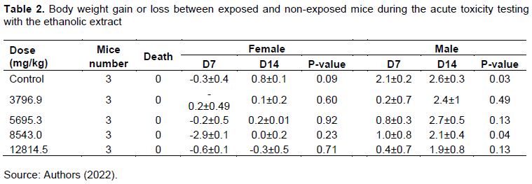
No deaths among animals were recorded at the highest dose tested (12814.45 mg/kg/bw). Physical signs observed in mice included a slight 30-min depression at the highest dose and a change in body weight (-0.6 ± 0.1 to -0.2 ± 0.5 g at D7) after drug administration (Tables 2 and 3).
Subacute toxicity
Body and organ weight
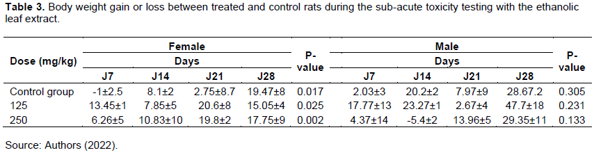
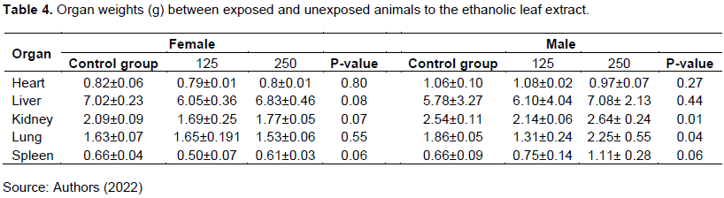
No evidence of toxicity was observed in rats receiving the ethanolic extract of C. multiflorum at 125 and 250 mg/kg bw. Only, weight loss in the first two weeks was noted (-5.4 ± 2 g at D14 male group dose 250 mg/kg bw), but after that, weight in all groups increased (Table 3). A slight difference in organ weight was observed between treated rats (kidney 1.77 ± 0.05 g) and control (kidney 2.09 ± 0.09 g) rats at 250 mg/kg bw (Table 4).
Biochemical parameters
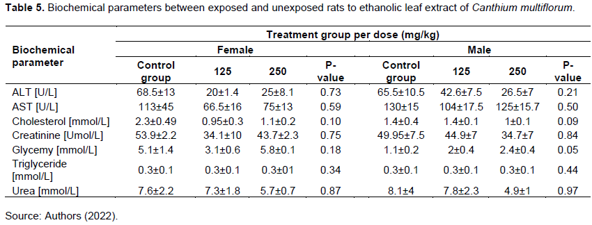
ALT and AST levels were lower between the treated group and control groups at 125 and 250 mg/kg bw but the difference was not significant (P = 0.73 and P = 0.59). A decreased in glucose level but not significant (P = 0.26) was noted in the treated female animals (3.1 ± 0.6 mmol/L) compared to the control (5.1 ± 1.4 mmol/L) at 125 mg/kg/bw. But there was a slight increase (5.8 ± 0.1 mmol/L) at 250 mg/kg/bw (Table 5).
Hematological measurements
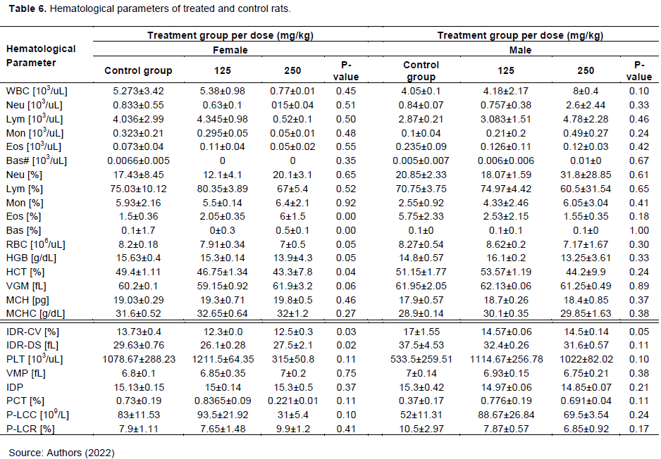
A slight change not significant increase (P = 0.45) in the number of white blood cells was observed between exposed females rats and controls (Table 6).
Antiplasmodial activity
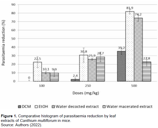
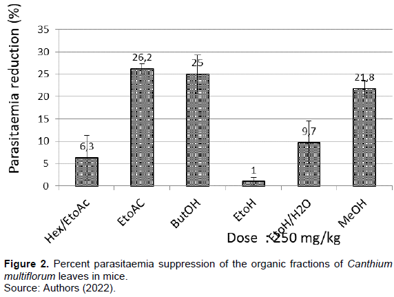
The overall findings showed a dose-dependent effect of the extracts of C. multiflorum on the parasites a part from to water extract macerated. The crude ethanol extract exhibited 22.5, 30.8 and 81.9% inhibition, respectively at 100, 250, and 500 mg/kg/bw. Similar inhibition was observed with the decocted extract, which gave 10.1, 25.9, and 74.2% inhibition at 100, 250, and 500 mg/kg bw. The extract macerated with water showed an inhibition of 9.9, 28.7 and 22.8% at the same doses. An inhibition of 2.4 and 35.2%, respectively at 250 and 500 mg/kg/bw was obtained with the dichloromethane extract. No inhibition with this extract was observed at 100 mg/kg/bw (Figure 1). Fractions or sub extracts, hexane/ethyl acetate (1:1), ethyl acetate, n-butanol, ethanol, methanol and aqueous extracts gave 6.3, 26.2, 25, 1%, 21.8, and 9.7% parasite reduction at 250 mg/kg bw, respectively (Figure 2).
DISCUSSION
C. multiflorum (Schumach. & Thonn.) Hiern used as an antimalarial by traditional healers in western Burkina Faso has been investigated for its antimalarial activity in vivo. The plant had already been studied in vitro against Plasmodium falciparum and had given promising results (Traoré et al., 2008; Akomo et al., 2009). Our results showed that the ethanolic and decocted extracts significantly reduced the parasitaemia of infected mice, confirming their antimalarial potential; both extracts had a higher activity than the others (p≤0.04) and the difference in activity between the two extracts was not significant (p=0.90). The dichloromethane extract had a weak activity while the water macerated extract had a moderate activity (Muñoz et al., 2000; Rasoanaivo et al., 2004). These results confirm the good antiplasmodial activity of C. multiflorum previously demonstrated by Traoré et al. (2008) and Akomo et al. (2009). Canthium henriquesianum, another species, showed an activity on P. falciparum (Ilboudo et al., 2013). Fractions or sub-extracts were found to be less active than the crude extracts (ethanol, dichloromethane and water). The stability of the compound in light, the heat, the nature of the extraction solvent and compounds, the polarity and number of compounds are known to influence the activity of secondary metabolites in plants. This is the first time that C. multiflorum is tested in vivo in mice, an animal model of human disease. The same model was used for the toxicity tests, the results of which show an ethanolic extract of the leaves of the plant, relatively safe. Indeed, no significant body weight loss and no physical changes in mice were observed. Slight changes may be more related to stress from infection than to extract (Table 2). Decreased AST and ALT levels in exposed rats suggest hepato-protective activity of the extract (Ozer et al., 2010; Mehta et al., 2009). In addition, increased white blood cell counts and decreased in glucose levels in treated rats suggest that C. multiflorum may have immune and hypoglycaemic effects.
The phytochemical screening showed that C. multiflorum leafextracts contained terpenoids, carotenoids, coumarins, tannins and flavonoids (; 1).
All of the compounds identified in our extracts could be responsible for the antimalarial activity in vivo. Some of these chemical groups have already been demonstrated to have antiplasmodial activity against P. falciparum (Khalid et al., 1986; Cubukcu et al., 1990; Traoré et al., 2009). In addition, polyphenols and flavonoids, known as antioxidant compounds may also play an important role in the antiplasmodial activity of the plant (Traoré et al., 2009; Akomo et al., 2009). Indeed, antioxidant compounds inhibit the polymerization of heme, fatal for the intra-erythrocyte parasite (Taramelli et al., 1999).
These results complement the in vitro antiplasmodial study of C. multiflorum, where crude extracts were found to be more active than fractions and molecules (Traoré et al., 2008b). The plant appears to be able to kill parasites in vivo and in vitro, but its efficacy may depend on the concentration of chemical compounds in the extracts, the doses administered and the type of chemical compounds in the extracts.
CONCLUSION
The results of this study showed that C. multiflorum leaves have a real in vivo antimalarial potential against P. berghei infection in mice. The leaf ethanol extract, the most active extract in mice is relatively safe when administered orally. The decocted extract, the form used by traditional healers had also a promising activity against the parasites. These finding support the use of C. multiflorum in the treatment of malaria in the west of the country. The results of the sub-acute toxicity tests revealed more therapeutic potential of the plant.
CONFLICT OF INTERESTS
The authors have not declared any conflict of interests.
REFERENCES
|
Akomo EF, Zongo C, Karou SD, Obame LC, Savadogo A, Atteke C, Traore AS (2009). In vitro antiplasmodial and antibacterial activities of Canthium multiflorum Schum and Thonn (Rubiacea) extracts. Pakistan Journal of Biological Sciences: PJBS 12(12):919-923.. |
|
|
Beiersmann C, Sanou A, Wladarsch E, De Allegri M, Kouyaté B, Müller O (2007). Malaria in rural Burkina Faso: local illness concepts, patterns of traditional treatment and influence on health-seeking behaviour. Malaria Journal 6(1):1-9. |
|
|
Ciulei I (1982). Methodology for analysis of vegetable drugs. Practical Manual on the industrial Utilisation of Medicinal and Aromatic Plants Center Building, Romania pp. 67-81. |
|
|
Cubukcu B, Bray DH, Warhurst DC, Mericli AH, Ozhatay N, Sariyar G (1990). In vitro antimalarial activity of crude extracts and compounds from Artemisia abrotanum L. Phytotherapy Research 4(5):203-204. |
|
|
Fidock DA, Rosenthal PJ, Croft SL, Brun R, Nwaka S (2004). Antimalarial drug discovery: efficacy models for compound screening. Nature reviews Drug Discovery 3(6):509-520. |
|
|
Ilboudo DP, Basilico N, Parapini S, Corbett Y, D'Alessandro S, Dell'Agli M, Taramelli D (2013). Antiplasmodial and anti-inflammatory activities of Canthium henriquesianum (K. Schum), a plant used in traditional medicine in Burkina Faso. Journal of Ethnopharmacology 148(3):763-769. |
|
|
Khalid SA, Farouk A, Geary TG, Jensen JB (1986). Potential antimalarial candidates from African plants: an in vitro approach using Plasmodium falciparum. Journal of Ethnopharmacology 15(2) :201-209. |
|
|
Lorke D (1983). A new approach to practical acute toxicity testing. Archives of Toxicology 54(4):275-287. |
|
|
Mehta AK, Arora N, Gaur SN, Singh BP (2009). Acute toxicity assessment of choline by inhalation, intraperitoneal and oral routes in Balb/c mice. Regulatory Toxicology and Pharmacology 54(3):282-286. |
|
|
Muñoz V, Sauvain M, Bourdy G, Callapa J, Bergeron S, Rojas I, Deharo E (2000). A search for natural bioactive compounds in Bolivia through a multidisciplinary approach: Part I. Evaluation of the antimalarial activity of plants used by the Chacobo Indians. Journal of Ethnopharmacology 69(2):127-137. |
|
|
Ouédréogo-Nacoulma OG (1996). Plantes medicinales et pratiques medicales traditionnelles au Burkina Faso: cas du plateau central. Faculté des Sciences et Techniques (FAST), Université de Ouagadougou. Thèse de Doctorat d'Etat, Tome 1:1-320,.. |
|
|
OMS (2011). Ministère de la Santé du Burkina Faso en collaboration avec l'OMS. Profil pharmaceutique du pays. 14, 2011.OMS, Profil pharmaceutique du Burkina Faso. 14. |
|
|
Ozer JS, Chetty R, Kenna G, Palandra J, Zhang Y, Lanevschi A, Ramaiah SK (2010). Enhancing the utility of alanine aminotransferase as a reference standard biomarker for drug-induced liver injury. Regulatory Toxicology and Pharmacology 56(3):237-246. |
|
|
Peters W, Robinson BL (1992). The chemotherapy of rodent malaria. XLVII. Studies on pyronaridine and other Mannich base antimalarials. Annals of Tropical Medicine and Parasitology 86(5):455-465. |
|
|
Philippe R, Eric D, Suzanne R, François FRP, Ramanitrahasimbola D, Rafatro H (2004). "Guidelines for the nonclinical evaluation of the efficacy of traditional antimalarials." Chapitre 16 in Traditional Medicinal Plants and Malaria Edited by Merlin Wilcox, Philippe Rasoanaivo and Gerard Bodeker, CRC Press. |
|
|
Seewaboon S, Urarat N, Supaporn V, Natthakarn C, Supachai S, Pennapa S, Pornthip T, Parunkul T, Anchalee C, and Kanjana J (2013). Acute and subchronic toxicity study of tud-rak-ka-sai-puu recipe in rats. African Journal of Traditional, Complementary and Alternative Medicines 10(1):142-148. |
|
|
Taramelli D, Monti D, Basilico N, Parapini S, Omodeo-Sale F, Olliaro P (1999). A fine balance between oxidised and reduced haem controls the survival of intraerythrocytic plasmodia. Parassitologia 41(1-3):205-208. |
|
|
Tiono AB, Kangoye DT, Rehman AM, Kargougou DG, Kaboré Y, Diarra A, Ouedraogo E, Nébié I, Ouédraogo A, Okech B, Milligan P (2014). Malaria incidence in children in South-West Burkina Faso: comparison of active and passive case detection methods. PloS ONE 9(1):e86936. |
|
|
Traore M (2008a). Phytochemical studies of plants used as antimalarials in Burkina Faso, PhD thesis, Faculty of Pharmaceutical Sciences, University of Copenhagen. PhD:144:2008a. |
|
|
Traoré M, Jaroszewski JW, Olsen CE, Ouédraogo JB, Pierre GI, Nacoulma OG, Christensen SB (2008b). A new oxygenated ursane derivative from Canthium multiflorum. Planta Medica 74(05):560-562. |
|
|
Traoré ÃM, Ziegler H L, Olsen CE, Millogo H, Ouà JB, Guissou IP Christensen SB (2009). Antiplasmodial Triterpenoids Isolated From Canthium Multiflorum. African Journal of Traditional, Complementary and Alternative Medicines 23(12):1108-1111. |
|
|
Willcox ML, Bodeker G (2004). Traditional herbal medicines for malaria. Bmj 329(7475):1156-1159. |
|
Copyright © 2024 Author(s) retain the copyright of this article.
This article is published under the terms of the Creative Commons Attribution License 4.0


