Journal of
Medicinal Plants Research

  • Abbreviation: J. Med. Plants Res.
  • Language: English
  • ISSN: 1996-0875
  • DOI: 10.5897/JMPR
  • Start Year: 2007
  • Published Articles: 3835

Full Length Research Paper

Leaves of Schinus polygamous (Cav.) Cabrera (Anacardiaceae) are a potential source of hepatoprotective and antioxidant phytochemicals

Abeer Mohamed El Sayed
  • Abeer Mohamed El Sayed
  • Pharmacognosy Department, College of Pharmacy, Cairo University, Kasr El-Einy Street, 11562, Cairo, Egypt.
  • Google Scholar


  •  Received: 26 January 2016
  •  Accepted: 20 April 2016
  •  Published: 03 May 2016

 ABSTRACT

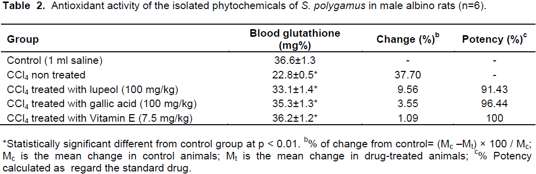
Phytochemical investigation of the crude ethanol extract of the leaves of Schinus polygamus (Cav.) Cabrera (Anacardiaceae) results in isolation of eight compounds identified as triterpenoids, 3-O-acetyllupeol (I), βeta-sitosterol (II), lupeol (III). In addition to the polyphenols, gallic acid (IV), methyl gallate (V), quercetin-3-α-O-rhamnoside (VI), kaempferol (VII) and quercetin (VIII). In Egypt, degenerative diseases in general and toxic hepatitis in particular remain a serious public health problem. The hepatoprotective, curative and anti-oxidant properties of the major phytochemicals, compounds III and IV isolated from leaves of S. polygamus were investigated. Liver damage was induced by CCl4 (1 mlkg-1); a well-known toxicant and exposure to this chemical is known to induce oxidative stress and causes tissue damage by the formation of free radicals. Silymarin (25 mgkg-1) and vitamin E (7.5 mgkg-1) were used as standard drugs to compare hepatoprotective and antioxidant effects of the phytochemicals, respectively. Oral administration of 50 to 100 mgkg-1 body weight of compounds III and IV were significantly protected from CCl4 induced elevation in aspartate aminotransferase (AST), alanine aminotransferase (ALT) and alkaline phosphatase (ALP) in adult male albino rats. The antioxidant effect in the liver was estimated by measuring the activity of antioxidant enzyme reduced glutathione. Detection of gallic acid and lupeol in S. polygamus as a member of family Anacardiaceae support the claim that both compounds could be considered as chemotaxonomic markers for plants belong to family Anacardiaceae. Results of the present study strongly reveal that both compounds III and IV have potent antioxidant and hepatoprotective effects against CCl4-induced hepatic intoxication.

Key words: Schinus polygamus (Cav.) Cabrera, Anacardiaceae, triterpenes, phenolics, hepatoprotective, antioxidant effect.


 INTRODUCTION

Metabolic processes of the body produce significant amounts of reactive oxygen species (ROS). Liver is a major organ attacked by ROS. Damaging effects brought by these ROS are being counteracted by the cellular antioxidant defense
 
system of the body (Hopps et al., 2010; Santiago and Mayor, 2014). However, at certain conditions, oxidative stress is triggered due to the imbalance between the production of ROS and the antioxidant systems of the body (Pepe et al., 2009). High risk health conditions such as neurodegenerative diseases, hepatitis, diabetes, cancer and inflammation are usually related to oxidative stress (Fearon and Faux, 2009; Fang et al., 2002). Based on growing interest in free radical biology and lack of effective therapies for most chronic diseases, the usefulness of antioxidants in protection against these diseases is warranted (Huang et al., 2010). In this regard, phytochemicals from various natural products had become the subject of most drug development researches (Santiago and Mayor, 2014). Plants are the most commonly known sources of natural antioxidants which includes polyphenolic and terpenoids compounds (Grassmann, 2005; Dimitrios, 2006).
 
Family Anacardiaceae (Bailey, 1953) has long reputation in folk medicine for its nutritional value of edible fruits and seeds, and for variable ailments such as treatment of bowel complain, chronic wounds, jaundice, hepatitis and relieving inflammatory conditions (Abbasia et al., 2010). There is an extensive body of literature addressing the escalated distribution of hepatic diseases among the people in Egypt (Shabana et al., 2011; Lehman, 2008; Strickland et al., 2002). Hepatoprotective herbal drugs can offer help by blocking absorption of toxins into liver cells and the formation of inflammatory substances that contribute to liver degeneration (Shabana etal., 2011).
 
As part of on-going study of medicinal plants belongs to family Anacardiaceae, the closely related genera, Harpephyllum caffrum Bernh, Rhus coriaria L and Schinus polygamus Cav. were subjected to biological testing in order to confirm the claimed herbal benefits of these drugs by different communities (Van Wyk et al., 2009; Rayne and Mazza, 2007; Shabana et al., 2008, 2011, 2013).
 
Schinus polygamus (Cav.) Cabrera, family Anacardiaceae is a tree cultivated for ornamental purpose. Traditionally the leaves have been used for wounds cleansing, while the bark produces a balsamic resin, is used to treat arthritic and chronic bronchitis (Munoz et al., 1981). The presence of well-known flavonoids was reported in S. polygamus, namely, kaempferol, quercetin and quercetin-3-O-galactoside (Mandich et al., 1984). The n-hexane extract of S. polygamus were reported to contain β-sitosterol to which analgesic, anti-inflammatory and antipyretic activities of the Argentine and the Chilean species were attributed (Erazo et al., 2006; Gonzalez et al., 2004). The essential oil obtained from S. polygamus leaves exhibited antimicrobial activities (Gonzalez et al., 2004).
 
Nowadays, the current research on plant based medications focus on isolation of biologically active substances from potent plants (Kingston, 2011). However, nothing could be traced on phytochemicals of Egyptian S. polygamus and its biological potentiality. Considering the health benefits one can derive from S. polygamus (Mandich et al., 1984; Munoz et al., 1981), its phytochemical, biochemical and pharmacological studies were scarce. Moreover, chemical profiling of the antioxidative components present in the plant is not yet been fully elucidated. Therefore this study was undertaken to back up its ethnobotanical uses while intensively evaluating the antioxidative components of S. polygamus as well as assessing its potential role as a powerful antioxidant and hepatoprotective


 MATERIALS AND METHODS

Plant
 
The leaves of S. polygamus (Cav.) Cabrera were obtained from trees growing in El Orman botanical garden, Giza in April 2013. The plant was kindly authenticated by Dr. Mohamed El-Gebaly, Botany Specialist. Voucher specimen 1842013 is kept in the herbarium of the Pharmacognosy Department, Faculty of Pharmacy, Cairo University.
 
Preparation of the extracts
 
Two kilograms of air-dried powdered leaves of S. polygamus were extracted by cold percolation with 90% ethanol (5 × 1 L). The ethanol extract was evaporated under reduced pressure to give 350 g green semi-solid residue. 250 g of the crude extract were successively partitioned between n-hexane, chloroform, ethyl acetate and n-butanol saturated with water. The solvent in each case was completely evaporated under reduced pressure to yield 30, 10, 150 and 3 g, respectively.
 
Material for phytochemical study
 
For chromatographic studies pre-coated silica plates 60 GF 254, cellulose plates (20×20 cm) from Fluka (Sigma-Aldrich
 
chemicals-Germany) for thin layer chromatography (TLC), silica gel 60 for normal phase column chromatography (CC), silica gel H for vacuum liquid chromatography (VLC) (E-Merck Darmstadt, Germany) polyamide (E-Merck Darmstadt, Germany), sephadex LH 20 and silica gel RP-18 (70 to 230 mesh) for reversed phase column chromatography were obtained from Fluka (Sigma-Aldrich chemicals-Germany). For developing the chromatograms, the following solvent systems were used: S1=n-hexane: chloroform (9:1 v/v), S2=n-hexane:chloroform (7:3 v/v), S3=n-hexane:ethylacetate (6:4 v/v), S4=n-butanol:acetic acid: water (4:1:5 v/v/v), S5=benzene: ethyl acetate: formic acid (5.5:4.5:1 v/v/v) and S6=ethyl acetate: methanol: formic acid: water (8:2:0.5:1 v/v/v/v). Spots were visualized by spraying with the following spray reagents: (I) p-anisaldehyde-sulphuric acid for triterpenoids, (II) 1% aluminium chloride spray reagent for flavonoids, (III) ferric chloride spray reagent for phenolic compounds (Mabry et al., 1996).
 
Apparatus for phytochemical study
 
Ultraviolet lamp (λmax=254 and 330 nm), Shimadzu, a product of Hanovia lamps for localization of spots  on  chromatograms.  EI-MS were recorded with a Varian Mat 711, Finnigan mass SSQ 7000 Mass spectrometer, 70 eV. IR spectra were observed as KBr discs on Shimadzu IR-435, PU-9712 infrared spectrophotometer. 1HNMR (300 MHz) and 13CNMR (75 MHz) spectra were recorded on Jeol EX-300MHz and Bruker AC-300 spectrometer operating at 300 (1H) and 75 (13C) MHz in CDOD3 and CDCl3 as a solvent and chemical shifts were given in δ (ppm).
 
Phytochemical investigation of the n-hexane soluble fraction
 
Thirty grams of the n-hexane soluble fraction were chromatographed on a VLC column, 210 g silica gel (12.5 × 7 cm) using n-hexane, n-hexane-chloroform and chloroform-ethyl acetate mixtures as elutes. Fractions, 200 ml each, were collected and monitored by TLC. Similar fractions were pooled together to obtain three major fractions (A-C).
 
Fraction A (4.0 g), eluted with n-hexane, was rechromatographed on silica gel sub-column using n-hexane and1% chloroform in n-hexane as solvent systems to obtain compound I (0.02 g).
 
Fraction B (2.5 g), eluted with 15% chloroform in n-hexane, was purified on silica gel column using n-hexane-chloroform mixtures (10% chloroform in n-hexane) as an eluent to obtain compound II (0.03 g).
 
Fraction C (2.0 g), eluted with 35% chloroform in n-hexane, was purified on silica gel column using n-hexane-chloroform mixtures (80:20 and 75:25), respectively as eluents revealed two spots. Further rechromatography on successive silica gel columns using n-hexane-chloroform mixtures yielded compounds III (0.25 g).
 
Phytochemical investigation of the ethyl acetate soluble fraction
 
Ethyl acetate soluble fraction (150 g) was purified on column chromatography (300 g polyamide, 50 × 3 cm) using 100% water and water-methanol mixtures in order of decreasing polarity. Fractions, 100 ml each, were collected and monitored by TLC. Similar fractions were pooled together to obtain two major fractions (a,b).
 
Fraction a (2 g), eluted with 15 and 25% methanol in water revealed the presence of three major spots. Further re-chromatography using sephadex LH 20 and water-methanol in decreasing polarity as eluent led to the isolation of two phenolic compounds IV and V (0.5 and 0.08 g), respectively.
 
Fraction b (5.37 g), eluted with 30 to 50% methanol in water, was further subjected to rechromatography on RP silica column using water- methanol as elute and resulted in separation of three compounds VI (0.03 g), VII (0.05 g) and VIII (0.03 g).
 
Material for biological study
 
The tested solutions were prepared by dissolving them in distilled water containing few drops of Tween 80 to yield a concentration of 5% w/v.
 
Animals
 
Adult Sprague-Dawely rats weighing 160 ± 10 g were provided from the animal-breeding unit of National Research Center, El-Dokki, Giza, Egypt. They were kept under standard conditions with temperature at 23 ± 2°C and a 12/12 h light/dark cycle and allowed free access to food and water throughout the experiment. This study was conducted in accordance with the standard guidelines used in handling of the experimental animals and approved by the Institutional Animal Care and Use Committee (IACUC)  (No. 9-031), College of Pharmacy, Cairo University.
 
Drugs and chemicals
 
Carbon tetrachloride (Analar, El-Gomhoreya Co., Cairo, Egypt, for induction of liver damage; 5 ml kg-1 of 25% carbon tetrachloride in liquid paraffin, IP) (Paget and Barnes, 1964), Silymarin (Sedico Pharmaceutical Co., 6 October City, Egypt, standard hepato-protective drug; 25 mgkg-1 b.wt.), Biodiagnostic kits for assessment of serum alanine aminotransferase (ALT), aspartate amino-transferase (AST) and alkaline phosphatase enzymes (ALP) estimated kits were purchased from Bio-Merieux Co. (France). Vitamin E (7.5 mgkg-1 b.wt.) (dlα-tocopheryl acetate): Pharco Pharmaceutical Co. Biochemical kits: Biodiagnostic glutathione kit.
 
Experimental design
 
For testing the hepatoprotectiveeffect on the liver, 36 adult male albino rats were used in the experiment and divided into six groups (each of 6). Group served as control, received saline (1 ml kg-1/day) and received CCl4 1 mlkg-1(1:1 CCl4 in liquid paraffin orally) once daily for 7 days. Group received CCl4 1 mlkg-1 and silymarin 25 mgkg-1 orally for 7 days as a reference drug. Four groups were administered the tested compounds at a daily dose of 50 and 100 mgkg-1 b.wt. (Paget and Barnes, 1964). The compounds, as well as, the reference drug, were administered to the rats for another seven days after liver damage.
 
For testing the antioxidant effect on liver, five groups of animals (6 animals each) were divided. One group was kept as a control, while for the other groups, group with liver injury was kept non-treated, another group received daily the reference drug (Vitamin E) and the other groups received the tested isolates of S. polygamus daily in the given doses. Blood samples were taken after 7 days.
 
Estimation of hepatoprotective activity
 
Measurement of AST, ALT and ALP serum levels
 
The animals were scarified and blood sample was collected from the retro-orbital sinus plexus under mild ether anesthesia. Collected blood was allowed to clot and serum was separated at 3500 rpm for 15 min for carrying out biochemical investigation serum levels of aspartate aminotransferase (AST) (Thewfeld, 1974), alanine aminotransferase (ALT) (Thewfeld, 1974), and alkaline phosphatase (ALP) (Kind and King, 1954; Klassan and Plaa, 1969) enzymes were measured in each group at zero time, after 7 days of receiving the tested drug, 72 h after induction of liver damage and after 7 days of treatment with the tested samples.
 
Estimation of antioxidant activity
 
The antioxidant activity was calculated by the determination of glutathione in blood of CCl4-induced liver injury rats adopting the methods of (Beutler, 1975; Beutler et al., 1963) using vitamin E as a reference drug. Blood samples were taken after 7 days for the determination of glutathione.
 
Statistical analysis
 
All data were expressed as mean ± standard error (SE). The statistical comparison of difference between the control group and the treated groups were carried out using two-way analysis of variance (ANOVA) followed by Duncan's multiple range test.


 RESULTS

Phytochemical investigation      
 
Column chromatographic fractionation of the n-hexane soluble fraction of the crude ethanol extract of leaves of S. polygamus led to the isolation of three compounds (I to III), whereas, the ethyl acetate soluble fraction afforded five compounds (IV to VIII) which were characterized through their physicochemical and spectral data. 
 
Compound I
 
Lupeol acetate (0.04 g, crystallize from methanol as white needle crystals, m.p. (218°C), was suggested to be a triterpenoid since it responded positively to libermann-Burchard’s test. It gives characteristic violet color with ρ-anisaldehyde/H2SO4 spray reagent. The IR√MaxKBR Spectrum, incorporated absorption bands at 1735 cm-1 which is characteristic for ester carbonyl group and exomethylene group at 1638 and 877 cm-1 (C=CH2), 2924 and 2854 cm-1 which is characteristic for (C-H) stretching, 1459 cm-1(C=C), 1246 cm-1 (C-O) and bending at 812. The EI/Mass (70 eV) m/z[M+] at 468 m/z calculated for the molecular formula C32H52O2. In addition to, the characteristic fragmentation peaks at 453 [M+-CH3] and 408 [M+-CH3COO] suggesting the presence of acetyl group in the structure.
 
The presence of m/z 249 ion indicates the presence of CH3CO at C-3 position. Skeletal mass peaks are represented by m/z 218 and 203 and the base peak at 189 all in accordance with lupene skeleton of triterpenoid. The 1HNMR spectrum (300 MHz in CDCl3) exhibited four tertiary methyl signals of the lupene skeleton at 0.80, 0.86, 0.95, and 1.05 as singlets. The C-30 vinylic methyl at δ1.75 ppm (s) acetyl methyl resonate at δ1.97 ppm (s), where α-proton at C-3 appear at δ4.22 ppm dd (J=3, 6 Hz). The C-29 olefinic protons Ha, Hb appeared at δ5.71 and 5.60 ppm as broad singlets. 13CNMR (75 MHz, in CDCl3 chemical shift assignments for compound I is revealed the presence of oxycarbon signal at δ83.0 assigned to (C-3) with downfield shift about (+4.0 ppm) compared with that of lupeol, which confirm the substitution with acetyl group (Chatterjee et al., 2000). Two signals assigned for olefinic carbons (C-20 and C-29) at δ=145.09 and 109.3 ppm, respectively and showed the presence of a carbonyl at δ172.8. All the aforementioned resonances are in accordance with compound I proposed structure and comparable to lupeol acetate (Jamal et al., 2008). As far as the available literature is concerned, this is the first report on isolation of lupeol acetate (C32H52O2) from S. polygamus.
 
Compound II
 
Beta-sitosterol white powder (0.03 g); m.p.:134 to 135°C; MS (m/z): exhibited molecular ion 414 (M+), and other fragmentation pattern as 396, 339, 325, 310, 298, 257, 140, 139, 125, 97, 71, and 57. Its mass spectral data suggested the molecular formula as C29H50O. Compound II also showed positive Liebermann-Burchard reaction indicated its sterol nature. The 1HNMR spectra (CDCl3, 300 MHz) showed the presence of six methyl signals that appeared as two methyl singlet at δ0.68 and 1.01; three methyl doublets that appeared as δ0.81, 0.83, and 0.93; and a methyl triplet at δ0.84. Olefinic proton was presented at δ5.37 suggested the presence of tri substituted double bond at C-5/C-6 in its structure. The 1HNMR spectra showed a proton corresponding to the proton connected to the C-3 hydroxy group which appeared as a triplet of doublet of doublets at δ3.53. The 13CNMR showed twenty nine carbon signal including six methyls, eleven methylenes,ten methane together with three quaternary carbons. Thus, the structure of II was assigned as β-sitosterol that was consistent to the reported literature values (Jamal et al., 2008).
 
Compound III
 
Lupeol (0.25 g, crystallize from methanol as white needle crystals, m.p. (210 to 212), positive test for triterpenoid skeleton Rf values, 0.76 in n-hexane: ethylacetate (6:4 v/v) and 0.35 n-hexane: chloroform (7:3 v/v); violet color with p-anisaldehyde/H2SO4 spray reagent. IR√MaxKBR Spectrum: incorporated absorption bands at 3415 cm-1 (OH), 2948, 2867 cm-1 (CH) and 1640, 885 cm-1   for (C=CH2). EI Mass (70 eV) m/z showed a molecular ion peak (M+) at 426.69 calculated for C30H50O with characteristic fragment ions at 411 (M+-Me), 393 (M+-Me-H2O), 365, 299, 297, 245,  fragment ions at m/z 220 and 207 (allocate the hydroxyl group at C3 position), m/z 218, 205 and 189  all in accordance with lupene skeleton. 1HNMR (300 Hz CDCl3): revealed signals for seven tertiary methyl groups at 0.81 (3H,s,C28-CH3), 0.86 (3H,s,C23-CH3), 0.87 (3H,s,C24-CH3), 0.88 (3H,s,C25-CH3), 0.96 (3H,s,C27-CH3), 1.05 (3H,s,C26-CH3) and 1.70 (3H,s,vinylic methyl, C30-CH3), 3.14 (1H,dd,J=6 Hz, 10.5 Hz indicative of C3-H is α- oriented) and 4.67 (1H,s,C29-Hb), 4.55 (1H,s,C29-Ha). 
 
Compound IV
 
Gallic acid (0.50 g), brownish white powder crystallized from water as colorless needles m.p. (248 to 250°C). Rf values (0.80 in n-butanol: acetic acid: water (4: 1: 5) v/v/v). In visible light deep brown color with NH3.  Violet fluorescence in UV and deep blue color with FeCl3. UV λmax 274 nm in MeOH. IR√MaxKBR Spectrum: shows absorption peaks at 3238 cm-1 (OH), 1683 cm-1(C=O), and 1613 cm-1   for C=C aromatic, and at 719, 771, 869 cm-1 (-C-H) aromatic. EI Mass (70 eV) m/z; [M+] 170, 153 ( M+-H2O), 125 [M+-H-CO2],79. 1HNMR (300 Hz  CD3OD): showed signals at 6.98 (2H, s, H-2, H-6).
 
Compound V
 
Methylgallate (0.08 g grayish white powder crystallized from absolute methanol give rise to  colorless needles   m.p. (199 to 203°C). Rf values (0.78 in n-butanol: acetic acid: water (4: 1: 5 v/v/v) shiny violet fluorescence in UV, deep brown color with NH3 in visible light, and dark blue color with FeCl3 reagent.IR √Max  KBR Spectrum: shows  absorption peaks at 3521, 3367 cm-1 (OH), 1700 cm-1 for ester carbonyl group (C=O),1617 cm-1 and at 867, 758 cm-1 (-C-H) aromatic. UVλmax 220 and 275 nm in MeOH. EI Mass (70 eV) m/z; [M+]=m/z 184, 153 (M+-OCH3), [M+-CO2Me] =125 1HNMR (270 Hz CD3OD): showed signals at 6.95 ( 2H,s,H-2,H-6) and 3.58 (3H,s,OMe).
 
Compound VI
 
Quercitrin (0.03 g, yellow powder needle crystallize from ethanol: water m.p. (182-184°C). 0.71 in S6=Ethylacetate: methanol: formic acid: water (80: 20: 5: 1 v/v/v/v), Visible Yellow. UV: dark purple. UV/NH3:Yellow. AlCl3; Yellow. UVλmax nm: MeOH:257,297sh,352 (flavonol) NaOMe: 270, 355, 405n nm (free OH on ring A&B). AlCl3:268, 333, 430 (free OH on ring A&B) AlCl3/HCl: 272, 353, 405 (free OH at C-5). NaOAc: 267, 322 sh, 372 (free OH at C-5&ring B) NaOAc/H3BO3: 260,300sh, 367 (ortho groups at ring B. 1HNMR spectrum showed, Aglycone: 7.34 (1H,d, J=2.1 Hz, H-2`), 7.32 (1H,dd, J=2.1, 8.4 Hz, H-6`), 6.92 (1H,d, J=8.4 Hz, H-5`), 6.37 (1H,d, J=2.1 Hz, H-8), 6.20 (1H, d, J=2.1 Hz, H-6). Sugar: 5.35 (1H,d, J=1.2 Hz, H-1``), 4.2 (1H,dd, J=1.5, 3.4 Hz, H-2``), 3.71 (1H,dd, J=3.4, 8.9 Hz, H-3``), 3.47-3.32 (m, H-4``,5``) and 0.95 (3H,d, J= 6 Hz, CH3-rhamnose, 13CNMR spectrum showed: 177.43 (C-4), 166.1 (C-7), 160.08 (C-5), 158.38 (C-9), 158.18 (C-2), 149.8 (C-4`), 143.85 (C-3`), 136.0 (C-3), 123.13 (C-1`), 123.5 (C-6`), 116.25 (C-5`), 117.26 (C-2`), 104.3 (C-10), 102.54 (C-1``), 96.1 (C-6), 94.7 (C-8), 72.16 (C-5``), 72.04 (C-3``), 71.94 (C-2``), 73.28 (C-4``), 17.65 (C-6``).
 
Compound VII
 
Kaempferol (0.03 g, yellow powder soluble in methanol m.p. (276 to 278°C). Rf value 0.88 in S6: Ethylacetate: methanol: formic acid: water (80:20:5:1 v/v/v/v) color of spot in visible pale yellow. UV: dark yellow, NH3/UV: dark yellow. AlCl3/visible: bright yellow. AlCl3/UV: fluorescent green. UVλmax nm MeOH: 268, 329sh, 369 (flavonol). NaOMe: 278, 326sh, 416 (free OH on ring A&B). AlCl3: 268, 304sh, 345, 424 (free OH on ring A&B). AlCl3/HCl: 269, 303sh, 350, 424 (free OH at C-5&no ortho OH at ring B). NaOAc: 275, 300 sh, 387 (free OH at C-7). NaOAc/H3BO3:267, 295sh, 373 (no ortho   OH  groups  at ring B). 1HNMR (300 MHz CD3OD), 8.29 (2H,d, J=9Hz, H-2`,H-6`), 7.11 (2H,d, J=8.7 Hz, H-3`,H-5`), 6.59 (1H,d, J=1.8 Hz, H-8), 6.38 (1H,d, J= 1.8 Hz, H-6).
 
Compound VIII
 
Quercetin (0.05 g, yellow powder needle crystallize from ethanol: waterm.p. (182-184°C). Rf value 7.0 in S6 = Ethyl acetate: methanol: formic acid: water (80:20:5:1 v/v/v/v) color of spot in Visible: Yellow. UV: dark purple. UV/NH3: Yellow. AlCl3/visible: bright yellow. AlCl3/UV: Yellow. UVλmaxnm MeOH: 255, 301sh, 368 (flavonol). NaOMe: 246, 406 (free OH on rings A&B). AlCl3: 269, 457 (free OH on rings A&B). AlCl3/HCl: 266, 303sh, 353, 430 (free OH at C-3, C-5 & ortho OH at ring B). NaOAc: 271, 329 sh, 390 (free OH at C-7 & ring B). NaOAc/H3BO3: 260, 384 (ortho OH groups at ring B). 1HNMR (300 MHz CD3OD), 7.91 (2H,d, J= 2.1 Hz, H-2`), 7.82 (1H,dd, J=2.1, 9 Hz, H-6`) 7.07 (1H,d, J= 8.4 Hz, H-5`), 6.57 (1H,d,J= 2.1 Hz, H-8), 6.37 (1H,d, J= 1.8 Hz, H-6).
 
The isolated compounds I to VIII were identified by comparison of MS, IR, 1H-NMR and 13C-NMR data to previously reported ones and were identified as lupeol acetate (I) (Ogunkoya, 1981) β-sitosterol (II) (Saha et al., 2011; Goad and Akihisa, 1997) and lupeol (III) (Dantanrayana et al., 1982), gallic acid (IV), methyl gallate (V), quercetin (VI), kaempferol (VII, quercetin-3-α-O-rhamnoside (VIII) (Hussein et al., 2003; Gutzeit et al., 2007; Marzouk 2008). It is noteworthy to mention that this is the first report of compounds I (lupeol acetate), III (lupeol) and VIII (quercetin-3-α-O-rhamnoside) from the leaves of S. polygamus growing in Egypt. Compounds III (lupeol) and IV (gallic acid) were detected as major compounds from the leaves of the plant so the biological activity, hepatoprotection and antioxidant effects against toxicity induced by CCL4 was carried out on them. Shabana et al. (2013) reported that the crude ethanol extract of leaves at dose 100 mgkg-1exhibited a significant hepatoprotective activity.  Therefore, this study was undertaken to investigate the phytochemicals to which these activity could be attributed.
 
In vivo hepatoprotective activity
 
The control group (received saline) served as a baseline for all the biochemical parameters. A significant increase in the activity of the serum enzymes AST, ALT and ALP (P<0.05) was observed upon treatment with CCl4. Positive control (Silymarin group) showed highly significant decrease (P<0.05) in AST, ALT and ALP as compared to negative control group. The treatment of intoxicated rats with compounds III and IV at a dose of (50 and 100 mg kg-1) produced a significant hepatoprotective effects. Hepatic protection was evidenced by the ability of gallic acid to normalize the high enzyme parameters in a dose-dependent manner (50 and 100 mgkg-1) by 47.9 and 102.9% for AST comparable to silymarin. Gallic acid (100 mgkg-1) exhibited a highly significant reduction (p<0.05) in AST, ALT and ALP better than lupeol. Results presented in Table 1 showed that low dose, 50 mgkg-1 of lupeol exhibited normalization by 93%, for AST comparable to silymarin.
 
 
In vivo antioxidant activity
 
Concerning the antioxidant activity, the phytochemicals of the leaves of S. polygamus exhibited significance antioxidant activity. Results (Table 2) revealed that lupeol and gallic acid in dose 100 mgkg-1 exerted a remarkable antioxidant activity (91.4 and 96.4%), respectively. The reduced level of glutathione in intoxicated rats was greatly restored by lupeol and gallicacid relative to vitamin E (potency 100%). These results clearly elucidated the significant in vivo antioxidant activity provided by both lupeol and gallic acid, so they could be considered as powerful antioxidant agents. Gallic acid showed   highly   significant  rise  (p<0.05)  in  glutathione better than lupeol.


 DISCUSSION

Free radicals and reactive oxygen species (ROS) play a role in liver diseases pathology and progression, dietary antioxidants have been proposed as therapeutic agents to counteract liver damage (Higuchi and Gores, 2003). Natural antioxidant may act as protectors but, more importantly, may exert modulator effects in cells through actions in antioxidant, drug metabolizing and repairing enzymes as well as working as signaling molecules in important cascades for cell survival (Singh et al., 2008).
 
Terpenoids are useful phytochemicals isolated from plants (Firn, 2010). They can regulate the activity of nuclear hormone receptors, which are involved in maintain the energy homeostasis in the body and manage obesity induced metabolic disorders including type 2 diabetes, hyperlipidemia, insulin resistance and cardiovascular disease (Goto et al., 2010).
 
In the present study, the in vivo antioxidant and hepatoprotective activities of major isolated compounds (lupeol, gallic acid) from the  leaves  of  S. polygamus  on experimental rats were evaluated. The hepatoprotection showed that the tendency of these molecules to preserve liver enzymes was particularly pronounced with gallic acid. The results of the present study are in accordance with the previously reported reviews, which demonstrated that lupeol ameliorates aflatoxin B1-induced peroxidative hepatic damage in rats (Preetha et al., 2006). Hepatoprotective effect of lupeol on tissue defense system in cadmium-induced hepatotoxicity in rats was known (Sunitha et al., 2001). In relation to that, it was observed that lupeol can substantially normalized degenerative alterations in the hepatocytes with granular cytoplasm (Akçam et al., 2013). Additionally, lupeol induced growth inhibition and apoptosis in hepatocellular carcinoma SMMC7721 cells by down-regulation of the death receptor expression (Zhang et al., 2009). Moreover, have some effect on lipid peroxidation and antioxidant status in rat kidney after chronic cadmium exposure (Nagaraj et al., 2000).
 
Henceforth, Lupeol has several biological activities, anti-inflammatory, and anti-tumor activities wherein according to some review (Wal et al., 2011; Gallo and Sarachine, 2009; Ragasa et al., 2009), was previously identified in family Anacardiaceae (Shabana et al., 2011). Lupeol and its ester derivative, lupeollinoleate were investigated for their possible hepatoprotective effect against cadmium-induced toxicity in rats (Sunitha et al., 2001). The mechanism of cadmium-mediated acute hepatotoxicity involves two pathways, one for the initial injury produced by direct effects of cadmium and the other for the subsequent injury produced by inflammation. Primary injury appears to be caused by the binding of Cd2+ to sulfhydryl groups on critical molecules in mitochondria. Thiol group inactivation causes oxidative stress, the mitochondrial permeability transition, and mitochondrial dysfunction (Rikans and Yamano, 2000). The fact that antioxidant agents inhibit carbon tetrachloride-induced liver damage (Wu et al., 2009) prompt to study the antioxidant effect of the isolated compounds.
 
Phenolics have been known to possess a capacity to scavenge free radicals (Valenzuela et al., 2003).Gallic acid, phenolic acid possesses antioxidant and anticancer activities (You et al., 2010; Ji et al., 2006; Chanwitheesuk et al., 2007). Gallic acid with the lowest hydrophobicity than methylgallate and alpha-tocopherol was found to be the most active antiradical agent (Asnaashari et al., 2014). The low hydrophobicity of gallic acid can be treated by using gallic acid–lecithin complex (Liu et al., 2014). The hepatoprotective and antioxidant effects of gallic acid were evaluated against paracetamol-induced hepatotoxicity in mice (Rasool et al., 2010). Gallic acid showed significant protection against paracetamol induced liver injury in rats by improving the antioxidant defense condition, lessening lipid peroxidation and conserving the pathological changes of the liver. Hepatotoxicity, results not from paracetamol itself, but from one of its metabolites N-acetyl-P-benzoquinoneimine (NAPQI), which depletes the liver's natural  antioxidant glutathione and directly damages cells in the liver, leading to liver failure (Rasool et al., 2010).
 
Carbon tetrachloride is a well-known hepatotoxic agent and the preventive action of drugs on liver damage by CCl4 has been widely used as an indicator of their liver protective activity. Changes associated with CCl4-induced liver damage are similar to that of acute viral hepatitis (Huang et al., 2010). Carbontertachloride treatment initiated lipid peroxidation, caused leakage of enzymes like alanine tranaminase and lactate dehydrogenase (Singh et al., 2008; Krithika et al., 2009) levels. CCl4-induced lipid peroxidation as well as covalent binding of CCl4 metabolites to cell components, and also restored lipoprotein metabolism (Boll et al., 2001). The anti-oxidant vitamin E (alpha-tocopherol) blocked lipid peroxidation, but not covalent binding, and secretion of lipoproteins remained inhibited. The covalent binding of the CCl3* radical to cell components initiates the inhibition of lipoprotein secretion and thus steatosis, whereas reaction with oxygen, to form CCl3-OO*, initiates lipid peroxidation. The former process may result in adduct formation and, ultimately, cancer initiation, whereas the latter results in loss of calcium homeostasis and, ultimately, apoptosis and cell death (Boll et al., 2001).
 
In CCl4-induced liver injury model, oxidative stress could be provoked, which prompts lipid peroxidation that injure hepatocellular membrane,  followed  by  substantial release of pro-inflammatory chemokines and cytokines, which in consequence of liver damage (Feng et al., 2011). A large amount of plants, especially medicinal plants, has been investigated to eliminate the hepatic damage stimulated by CCl4. For example, Coptidis rhizome, a traditional Chinese medicinal plant used to clear heat and scavenge toxins, belongs to liver meridian in Chinese medicinal practice (Feng et al., 2010, 2012).
 
Anti-oxidative therapy, mainly using natural antioxidants, represents a reasonable therapeutic approach for the prevention and/or treatment of liver diseases due to the role of oxidative stress in initiation and progression of hepatic damage (Li et al., 2015). However, although concept of anti-oxidative therapy has been improved nowadays, there is a long way to go for the application of antioxidants in liver disease. Therefore, translational research is of great importance for anti-oxidative therapy. Considering ROS and oxidative stress act positively in certain circumstances and the difference between animals and humans, the effective dose and safe dose, duration of treatment, absorption and bio-availability of antioxidants require thorough investigation. Furthermore, in the future, large-scale samples and appropriate duration of anti-oxidative treatment for liver diseases should be performed (Li et al., 2015).


 CONCLUSION

The current study provides a scientific basis for the claims that S. polygamus is effective against certain liver–related diseases (Shabana et al., 2013). Taken together, our findings provide evidence that the major isolated compounds (lupeol and gallic acid) from S. polygamus exhibit hepatoprotective and antioxidant activities. These compounds might be useful for the prevention of toxic-induced and free radical-mediated liver diseases since it has been suggested that antioxidant compounds may be used as prophylactic agent.
 
Moreover, this study demonstrates that S. polygamus have potential hepatoprotective activity which is mainly attributed to the antioxidant potential, which might occur by reduction of lipid peroxidation and cellular damage. Previous reports were prevailed the isolation of lupeol and gallic acid from different genera belongs to Family Anacardiaceae (Shabana et al., 2008, 2011). As a part of the ongoing investigation of plants belongs to family Anacardiaecae (Shabana et al., 2008, 2011, 2013), it can be concluded that gallic acid and lupeol could be consider as potent hepatoprotective agents and chemotaxonomic markers for plants which belongs to family Anacardiaecae.  


 CONFLICT OF INTEREST

The authors have not declared any conflict of interests. 


 ACKNOWLEDGEMENT

The authors sincerely thank Prof. Dr. Mohamed El-Gebaly, Botany Specialist for plants authentication and Prof. Dr. Amany Sleem, Department of pharmacology, National Research Center, El-Dokki, Giza, Egypt for carring out the biological experiments.


 REFERENCES

Abbasia AM, Khana MA, Ahmada M, Zafara M, Jahanb S, Sultanaa S (2010). Ethnopharmacological application of medicinal plants to cure skin diseases and in folk cosmetics among the tribal communities of North-West Frontier Province, Pakistan. J. Ethnopharmacol. 128(2):322-35.
Crossref


Akçam M, Artan R, Yilmaz A, Ozdem S, Gelen T, NazıroÄŸlu M (2013). Caffeic acid phenethyl ester modulates aflatoxin B1-induced hepatotoxicity in rats. Cell Biochem. Funct. 31(8):692-697.
Crossref


Asnaashari M, Farhoosh R, Sharif A (2014). Antioxidant activity of gallic acid and methyl gallate in triacylglycerols of Kilka fish oil and its oil in-water emulsion. Food Chem.159:439-444.
Crossref


Bailey LH (1953). The Standard Cyclopedia of Horticulture III, New York. The Macmillan Company. pp. 2952-2954.


Beutler E (1975). Glutathione. In: Red Cell Metabolism, a Manual of Biochemical Methods. Grune and Stratton; Beutler E (eds). New York, USA. pp. 112-114.


Beutler E, Duron O, Kelly B (1963). Improved method for the determination of blood glutathione. J. Lab. Clin. Med. 61:882-888.


Boll M, Weber LW, Becker E, Stampfl A (2001). Mechanism of carbon tetrachloride-induced hepatotoxicity. Hepatocellular damage by reactive carbon tetrachloride metabolites. Z Naturforsch C. 56(7-8):649-59.
Crossref


Chanwitheesuk A, Teerawutgulrg A, Kilibum JD, Rakariythm N (2007).Antimicrobial gallic acid from Caeslpinia mimosoides Lamk. Food Chem. 100:1044-1048.
Crossref


Chatterjee PA, Kouzi S A,Pezzuto JM, Hamann M T (2000). Biotransformation of the antimelanoma agent betulinic acid by Bacillus megaterium ATCC 13368. Appl. Environ. Microbiol. 66(9):3850-3855.
Crossref


Dantanrayana AP, Savitri NK, Muthukuda PM, Wazeer MI (1982). A lupane derivative and the 13C NMR chemical shifts of some lupanols from Pleurostylia opposite. Phytochemistry 21(8):2065-2068
Crossref


Dimitrios B (2006). Sources of natural phenolics and antioxidants. Trends Food Sci. Technol. 17:506-512.
Crossref


Erazo S, Delporte C, Negrete R, Gracia R, Zaldivear M, Iturra G, Caballero E, Lopez JL, Backhouse N (2006). Constituents and biological activities of Schinus polygamous. J. Ethnopharmacol. 107(3):395-400.
Crossref


Fang YZ, Yang S, Wu G (2002). Free radicals, antioxidants and nutrition. Nutrition 18(10):872-879.
Crossref


Fearon IM, Faux SP (2009). Oxidative stress and cardiovascular disease: Novel tools give free radical insight. J. Mol. Cell Cardiol. 47(3):372-81.
Crossref


Feng YB, Wang N, Zhu MF, Zhang ZJ, Tong Y, Tsao S (2010). Interdisciplinary approaches in study of Chinese medicines: Case of coptis. Int. J. Mol. Med. 26:S21.


Feng Y, Wang N, Ye X, Li H, Feng Y, Cheung F, Nagamatsu T (2011). Hepatoprotective effect and its possible mechanism of Coptidisrhizoma aqueous extract on carbon tetrachloride-induced chronic liver hepatotoxicity in rats. J. Ethnopharmacol. 138:683-690.
Crossref


Feng Y, Wang N, Tong Y, Tsao S (2012). Berberine: An old drug but new use for liver diseases. Planta Med. 78:1091.
Crossref


Firn R (2010). Nature's Chemicals. The natural products that shaped our world. Oxford University Press, UK. pp 74-75. Available at: 

View


Gallo MBC, Sarachine MJ (2009). Biological activities of lupeol. Int. J. Biomed. Pharm. Sci. 3(1):46-66.


Goad J, Akihisa T (1997). Analysis of Sterols (1stedn)Blackie Academic and Professional Press, Champan and Hall, London.


Gonzalez S, Guerra P, Bottaro H, Molares S, Demo M, Oliva M, Zunino M, Zygadlo J (2004). Aromatic plants from Patagonia Part I. Antimicrobial activity and chemical composition of Schinus polygamus (Cav.) Cabrera essential oil. Flavour Fragr. J. 19:36-39.
Crossref


Goto T, Takahashi N, Hirai S, Kawada T (2010). Various terpenoids derived from herbal and dietary plants function as PPAR modulators and regulate carbohydrate and lipid metabolism. PPAR Res. 7:216-226.
Crossref


Grassmann J (2005). Terpenoids as plant antioxidants. VitamHorm. 72:505-535.
Crossref


Gutzeit D, Wray V, Winterhalter P, Jerz G (2007). Preparative isolation and purification of flavonoids and protocatechuic acid from sea Buckthorn juice concentrate (Hippophae rhamnoides L. ssp. rhamnoides) by high speed counter current chromatography. Chromatographia 65(1):1-7.


Higuchi H, Gores GJ (2003). Mechanisms of liver injury: an overview. Curr. Mol. Med. 3:483-490.
Crossref


Hopps E, Noto D, Caimi G, Averna MR (2010). A novel component of metabolic syndrome: the oxidative stress. Nutr. Metab. Cardiovasc. Dis. 20(1):72-77.
Crossref


Huang B, Ban X, He J, Tong J, Tian J, Wang Y (2010). Hepatoprotective and antioxidant activity of ethanolic extracts of edible lotus (Neumbomucifera Gaertn) leaves. Food Chem.120:873-878.
Crossref


Hussein SAM, Hashem ANM, Seliem MA, Lindequist U, Nawwer MAM (2003). Polyoxygenated flavonoids from Eugenia edulis. Phytochemistry 64:883-9.
Crossref


Jamal AK, Yaacob WA, Laily BD (2008). A chemical study on Phyllanthus reticulatuse. J. Phys. Sci. 19(2):45-50.


Ji HF, Zhng HY, Shen L (2006). Proton dissociation is important to understng structure-activity relationship of gallic acid antioxidants. Bloog. Med. Chem. Lett. 16:4095-4098.
Crossref


Kind PR, King EG (1954). A colorimetric method for the determination of serum alkaline phosphatase. J. Clin. Pathol. 7:322.
Crossref


Kingston DGI (2011). Modern natural products drug discovery and its relevance to biodiversity conservation. J. Nat. Prod. 74(3):496-511.
Crossref


Klassan CD, Plaa GL (1969). Comparison of the biochemical alteration elicited in liver of rats treated with CCl4 and CHCl3. Toxicol. Appl. Pharmacol.18:2019.


Krithika R, Mohankumar R, Verma RJ, Shrivastav PS, Mohamed IL, Gunasekaran P, Narasimhan S (2009). Isolation,characterization and antioxidative effect of phyllanthin against CCl4-induced toxicity in HepG2 cell line. Chem. Biol. Interact. 181:351-358.
Crossref


Lehman EM (2008). Dynamics of Liver Disease in Egypt: Shifting Paradigms of a Complex Etiology. Ph D (Epidemiological Science). USA, The University of Michigan. pp. 1-11.


Li S, Tan H-Y, Wang N, Zhang Z-J, Lao L, Wong C-W, Feng Y (2015). The role of oxidative stress and antioxidants in liver diseases. Int. J. Mol. Sci. 16(11):26087-26124.
Crossref


Liu C, Chen C, Mo H, Ma H, Yuan E, Li Q (2014). Characterization and DPPH Radical Scavenging activity of gallic acid–lecithin complex. Trop. J. Pharm. Res. 13(8):1333-1338.
Crossref


Mabry JT, Markham KR, Thomas MB (1996). The Systematic Identification of Flavonoids, 2nd edition. New York-Berlin-Heidelberg, Springer-Verlag.


Mandich L, Bittner M, Silva M, Barros C (1984). Phytochemical screening of medical plants. Studies of flavonoids. Rev. Latinoam. Quím. 15:80-82.


Marzouk MSA (2008). Acylated flavonol glycosides and hydrolysable tannins from Euphorbia cotinifolia L. leaves. Bull. Fac. Pharm. Cairo Univ. 46:181-91.


Munoz M, Barrera E, Meza I (1981). El uso medicinal y alimenticio de plantasnativas y naturalizadas de Chile, Edici’onocasional, 33. Museo Nacional de Historia Natural, Santiago, Chile. P 17.


Nagaraj MS, Suniyha S, Varalakshmi P (2000). Effect of lupeol, a pentacyclictriterpene on lipid peroxidation and antioxidant status in rat kidney after chronic cadmium exposure. J. Appl. Toxicol. 20:413-417.
Crossref


Ogunkoya L (1981). Application of mass spectrometry in structural problems in triterpenes. Phytochemistry 20:121-126.
Crossref


Paget GE, Barnes JM (1964). Evaluation of Drug Activities Sited in LaboratoryRats. New York, Academic Press Inc.


Pepe H, Balci SS, Revan S, Akalin PP, KurtoÄŸlu F (2009). Comparison of oxidative stress and antioxidant capacity before and after running exercises in both sexes. Gend. Med. 6(4):587-595.
Crossref


Preetha SP, Kanniappan M, Selvakumar E, Nagaraj M, Varalakshmi P (2006). Lupeol ameliorates aflatoxin B1-induced ive hepatic damage in rats. Comp. Biochem. Physiol. C. Toxicol. Pharmacol. 143:333-339.
Crossref


Ragasa CY, Tsai P, Shen CC (2009). Terpenoids and Sterols from the endemic and endangered Philippine Trees, Ficus pseudopalma and Ficus ulmifolia. Philipp. J. Sci. 138(2):205-209.


Rasool MK, Sabina EP, Ramya SR, Preety P, Patel S, Mandal N, Mishra PP, Samuel J (2010). Hepatoprotective and antioxidant effects of gallic acid in paracetamol-induced liver damage in mice. J. Pharm. Pharmacol. 62(5):638-43.
Crossref


Rayne S, Mazza G (2007). Biological activities of extracts from Sumac (Rhus spp.): A Review. Plant Foods Hum. Nutr. 62:165-175.
Crossref


Rikans LE, Yamano T (2000). Mechanisms of cadmium-mediated acute hepatotoxicity. J. Biochem. Mol. Toxicol. 14(2):110-7.
Crossref


Saha S, Subrahmanyam EVS, Kodangala C, Shastry SC (2011). Isolation and characterization of triterpenoids and fatty acid ester of triterpenoid from leaves of Bauhinia variegate. Der Pharm. Chem. 3(4):28-37.


Santiago LA, Mayor ABR (2014). Lupeol: An antioxidant triterpene in Ficuspseudopalma Blanco (Moraceae). Asian Pac. J. Trop. Biomed. 4(2):109-118.
Crossref


Shabana MM, El Sayed, Aly M, Yousif MF, El Sayed Abeer M, Sleem AA (2011). Bioactive constituents from Harpephyllum caffrum Bernh. and Rhus coriaria L. Pharmacogn. Mag. 7(28):298-306.
Crossref


Shabana MM, El Sayed, AM, Yousif MF, El Sayed AM, Sleem AA (2013). Phytochemical and biological study of Shinus polygamus growing in Egypt. Int. J. Tradit. Herbal Med. 1(5):136-146.


Shabana MM, El SayedA M, Yousif MF, El Sayed AM, Sleem AA (2008). Phytochemical and biological study of Sumac. Egypt J. Pharm. Sci. 49:83-101.


Sing N, Kamath V, Narasimhamurthy K, Rajini PS (2008). Protective effect of potato peel extracts against carbon tetrachloride-induced liver injury in rats. Environ. Toxicol. Pharmacol. 26:241-246.
Crossref


Strickland GT, Elhefni H, Salman T, Waked I, Abdel-Hamid M, Mikhail NN, Esmat G, Fix A (2002). Role of hepatitis C infection in chronic liver disease in Egypt. Am. J. Trop. Med. Hyg. 67:436-442.


Sunitha S, Nagaraj M, Varalakshmi PH (2001). Hepatoprotective effect of lupeol and lupeollinoleate on tissue antioxidant defence system in cadmium-induced hepatotoxicity in rats. Fitoterapia 72(5):516-23.
Crossref


Thewfeld W (1974). Enzymatic method for determination of serum AST and ALT. Dtsch Med. 99:343.


Valenzuela A, Sanhueza J, Nieto S (2013). Cholesterol oxidation health hazard and the role of antioxidants in prevention. Biol. Res. 36:291-302.


Van WykB-E, Van Oudtshoom B, Gericke (2009). Medicinal Plants of South Africa" 2 nded.146, Briza Publication, Pretoria.


Wal P, Wal A, Sharma G, Rai AK (2011). Biological activities of lupeol. Syst. Rev. Pharm. 2(2):96-103.
Crossref


Wu YX, Zhang XM, Hu MH, Wu XM, ZHoa Y (2009). Effect of Laggeraalata on hepatocyte damage induced by Carbon tetrachloride invitro and in vivo. J. Ethnopharmacol. 126:50-56.
Crossref


You BR, Kim S Z, Kim S H, Park WH (2010). Gallic acid inhibits the growth of Hela cervical cancer cells via apoptosis and/or necrosis. Food Chem.Toxicol. 48:1334-1340.
Crossref


Zhang L, Zhang Y, Zhang L, Yang X, Lv Z (2009). Lupeol, a dietary triterpene, inhibited growth and induced apoptosis through down-regulation of DR3 in SMMC7721 cells. Cancer Investig. 27:163-170.
Crossref


          */?>