Full Length Research Paper

ABSTRACT
The aim of this study was to quantify and evaluate the antibacterial activity of tannins extracted from leaves of Psidium guineense, obtained using two different isolation methods, against Staphylococcus aureus and Pseudomonas aeruginosa. The first extraction method was used to isolate condensed tannins (Method A) and the second to isolate mixtures rich in esters of gallic acid and glucose (Method B). Both extraction methods yielded high concentrations of tannins, with 312 and 257 mg/g of dry material obtained using Methods A and B, respectively. These compounds formed halos of growth inhibition in S. aureus and P. aeruginosa cultures. Tannins isolated by method B gave rise to larger inhibition halos than those isolated by method A. Analysis of the antibacterial activity of tannins isolated by method B revealed a minimum inhibitory and bactericidal concentration of 256 μg/ml for S. aureus and 512 μg/ml for P. aeruginosa. These results demonstrate that P. guineense is a promising source of pharmacologically active tannin molecules, and further studies are therefore necessary to determine the toxicity of the plant and ensure its safe use for animal and human health.
Key words: Psidium guineense, tannins, isolation, antibacterial activity, Staphylococcus aureus, Pseudomonas aeruginosa.
INTRODUCTION
Psidium guineense, commonly known as araçá, is a shrub of the family Myrtaceae with a height of 2 to 2.5 m and is native to and widely dispersed in tropical America (González et al., 2005). The roots of P. guineense have been traditionally used in Brazil as antidiarrheals and diuretics, and the bark is used in tanneries due to its high tannin content (Rodrigues and Carvalho, 2001). A study by González et al. (2005) demonstrated the antimicrobial potential of the pulp of P. guineense fruit and Fernandes et al. (2012) realized a growth inhibitory effect of P. guineense extract on antimicrobial agents against methicillin-resistant Staphylococcus aureus strains due for it combination with beta-lactams, carbapenems and fluoroquinolones in antibiotic, corroborating the hypothesis that P. guineense extract have synergistic effect in antimicrobial activity.
Some species of the family Myrtaceae are promising targets for exploration through chemical and pharmaco-logical studies. Studies of Psidium guajava and Eugenia uniflora have demonstrated that extracts from the leaves of these plants show diverse pharmacological activities, such as anti-inflammatory, antimicrobial, antimalarial, hypoglycemic, antispasmodic and antioxidant effects (Schapoval et al., 1994; Lee et al., 2000; Gutiérrez et al., 2008). Multiple classes of chemicals with important pharmacological activities have already been isolated from plants belonging to this family (Meckes et al., 1996; Begum et al., 2002; Salib and Michael, 2004).
The ability of tannins to form complexes with macro-molecules is the basis both for their ecological properties in controlling insects, fungi and bacteria and for their pharmacological activities (Santos and Mello, 1999). Plants rich in tannins are employed in traditional medicine to treat a number of diseases (Haslam, 1996; De Bruyne et al., 1999). Extensive analyses have revealed diverse biological activities of this class of substances (Matsuo et al., 1994; Haslam, 1996; Lee et al., 2000), including their ability to inhibit the growth of microorganisms (Scalbert, 1991; Akiyama et al., 2001; Ferreira et al., 2012).
Considering the inhibitory effect of tannins on micro-organisms, the notable presence of these compounds in the leaves of P. guineense and the paucity of studies concerning this species, this study aimed to quantify and evaluate the antibacterial activity of tannins extracted from leaves of P. guineense.
MATERIALS AND METHODS
Collection of the botanical material and preparation of the extract
Leaves of P. guineense were collected from 10 randomly chosen wild plants in (Savannah Brazilian area) northern area of the state of Minas Gerais, Brazil (16°51'00''S, 43°41'49''W). A sample of the botanical material was deposited in the herbarium of the State University of Montes Claros (n° 606). The extraction method was adapted from Salminen et al. (1999). Briefly, 20 g of dry and pulverized leaves were suspended in 100 ml of 70% acetone with 0.1% ascorbic acid and agitated for one hour. After centrifugation at 10,000 g for 10 min, the pellet was re-extracted four times with the same solvent. The acetone from the combined extracts was then evaporated in a rotary evaporator under a vacuum at 30°C, and the extract was subsequently filtrated using filter paper Whatman’s No. 1 and concentrated in a forced air convection oven at 30°C until the volume was reduced as much as possible. A sample of the resulting raw extract was used for evaluation of the minimum inhibitory con-centration. The remaining portion was diluted with an equal volume of 95% ethanol and centrifuged at 10,000 g in order to remove the pellet.
Isolation of tannins
The crude extract was then subjected to one of two methods of tannin isolation, designated here as A and B. Method A was proposed by Asquith and Butler (1985) for the isolation of fractions rich in tannins. The hydrophilic fraction resulting from the extraction process, which was dissolved in 95% ethanol, was applied to a Sephadex LH-20 column (3 × 30 cm) equilibrated with 95% ethanol. The column was washed with 95% ethanol at a flow rate of 1 ml/min. Fractions of 10 ml were collected and read with a spectro-photometer at a wavelength of 280 nm until their absorbances reached approximately zero. The ethanol used in the washes was discarded after calculating the yield of the combined fractions. The column was then eluted with 50% aqueous acetone at a flow rate of 1 ml/min until the Sephadex returned to its characteristic color (white) and the elute became less intense in color. Fractions of 4 ml were collected, and their absorbances at 435 nm were determined. The fractions were analyzed for the presence of tannins using the gelatin precipitation test (Strumeyer and Malin, 1975). Tannin-positive fractions were combined, and the acetone was completely removed from the combined fractions by evaporation under reduced pressure at 30°C. The aqueous sample was extracted three times with an equal volume of ethyl acetate, and the organic phase (upper) was discarded. After the third extraction, traces of ethyl acetate remaining in the aqueous fractions were removed in a forced air convection oven at 30°C. The aqueous sample was then lyophilized and subjected to antibacterial activity testing, and the radial diffusion in agarose gel method was used for total tannin quantification.
Method B was proposed by Hagerman and Klucher (1986) to isolate tannins from mixtures rich in esters of gallic acid and glucose. The aqueous sample resulting from the extraction process and dissolved in 95% ethanol was applied to a Sephadex LH-20 column (40 ×3 cm) equilibrated with 95% ethanol. The Sephadex gel was washed with 95% ethanol at a flow rate of 0.5 ml/min, and 10 ml fractions were collected until their absorbance readings at 280 nm reached approximately zero. The ethanol used in the washing was discarded after calculating the yield of the combined fractions. The tannins were then eluted from the gel with 50% ace-tone and 0.001 M ascorbic acid. Fractions of 4 ml were collected, and their absorbances at 435 nm were determined. The fractions were analyzed for the presence of tannins using the gelatin precipitation test (Strumeyer and Malin, 1975), and the tannin-positive fractions were combined. Acetone was completely removed from the combined fractions by evaporation under reduced pressure at 30°C. The aqueous sample was lyophilized and subjected to antibacterial activity analysis using the disc diffusion method, and the quantification of total tannins was measured using radial diffusion in agarose gels.
Quantification of total tannins
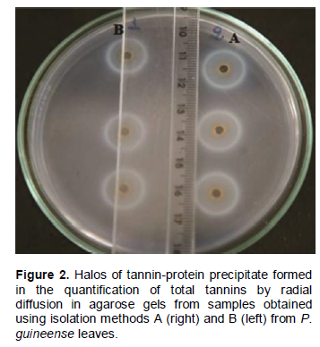
The concentration of total tannins present in the lyophilized samples was determined by radial diffusion in agarose gels (Hagerman, 1987). Five milligrams of the sample was dissolved in 100 µl of 50% methanol. Perforations were made in a previously prepared gel with the aid of a cylinder 2.8 mm in diameter. Twenty microliters of lyophilized sample dissolved in methanol was introduced into each perforation. After 96 h of incubation at 30°C, the diameters of the rings were measured in millimeters. The quantity of precipitated tannins was determined using a calibration curve for tannic acid (5 to 15 mg/ml). The data are presented as the area of the tannin-protein precipitate in cm2 per g of sample dry mass.
Antibacterial activity assay
The tests for bacterial growth inhibition were carried out with S. aureus (ATCC 25923) and Pseudomonas aeruginosa (ATCC 27853). Standard strains seeded in Brain Heart Infusion (BHI; Biobrás®) agar with mineral oil (Ideal®) were cultured in Müller-Hinton agar (Biobrás®) and incubated for 24 h in an oven at 35°C. Bacterial suspensions in 0.98% NaCl with turbidity equivalent to McFarland Scale 0.5 (1×10⁸ cells/ml) were prepared from recent cultures. The suspensions of the tested microorganisms were seeded in Petri dishes with Müeller-Hinton agar surface with the aid of a sterile swab. The disk diffusion test was based on the National Committee for Clinical Laboratory Standards (NCCLS M2-A8, 2003).
Lyophilized tannin samples obtained using both isolation methods were dissolved in dimethyl sulfoxide [DMSO (70 mg/ml)] and applied to sterile Blank discs 6 mm in diameter (Cecon). These discs were then deposited on the agar, and the dishes were incubated at 35°C for 24 h. After incubation, the halo of inhibition formed around the discs was measured in millimeters. Commercial discs with 30 µg chloramphenicol (Laborclin®) were used as a positive control for S. aureus, and discs with 30 µg tetracycline (Laborclin®) were used for P. aeruginosa. Discs infused with 10 µl DMSO were used as a negative control.
The broth microdilution test, based on the NCCLS standard M7-A6 (2003), was used to determine the minimum inhibitory concentration (MIC) of the isolated samples and raw extract. Con-centrations of isolates and raw extract from 0.5 to 1024 µg/ml were tested. The inoculated microdilution dishes were incubated at 35°C for 24 h in microplates. Resazurin, an indicator of microbial growth, was used to aid the analysis. The minimum inhibitory tannin con-centration was defined as the lowest concentration that resulted in no microbial growth, as indicated by the blue color of the resazurin. Then, the tannin dilutions that showed no microbial growth were divided into smaller increments to more precisely define the minimum bactericidal concentration (MBC). The antibacterial activity assay was performed in 5 repetitions.
Statistical analysis
The data were analyzed with the R statistical system version 2.8.0 using generalized linear models. The appropriate model was estimated from the elimination of non-significant variables tested from the full model, which is known as the backward method (Crawley, 2005).
RESULTS
The chromatograms as shown in Figure 1 track absorbances of the ethanolic wash fractions at 280 nm and the fractions eluted with acetone at 430 nm using a spectrophotometer for methods A (Asquith and Butler, 1985) and B (Hagerman and Klucher, 1986). The gel of the Sephadex LH-20 adsorbs tannins in alcohol and releases them in aqueous acetone. The chromatograms show that during elution with 95% ethanol, a discernable absorption region was observed up to approximately 400 min for method A (Figure 1a) and up to nearly 600 min for method B (Figure 1b). The other substances remained firmly adhered to the top of the column until they were eluted with 50% acetone, forming a single peak in both methods. The single peak eluted with aqueous acetone began to register after 700 min with method A (Figure 1a) and after 1500 min with method B (Figure 1b). For all the fractions eluted with 50% acetone (which correspond to the final peak), formation of a precipitate was observed in the gelatin precipitation test.
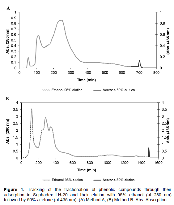
Regarding the yield of the fractions eluted from the column, there was no difference between the two isolation methods. The fractions eluted with ethanol had a yield of 43.68±3.8%, and those eluted with acetone showed a yield of 22.05±1.77%. Thus, approximately 66% of the total material applied to the column was recovered.
The equation of the tannin standard curve generated by radial diffusion in agarose gels (Figure 2) was y= 4.4924x + 0.3902, R2= 0.9974. The quantity of total tannins present in the sample obtained from isolation method A (312 ± 8 mg/g of dry material) was significantly greater than that from method B (257±8 mg/g of dry material) (ANOVA, P<0.01).
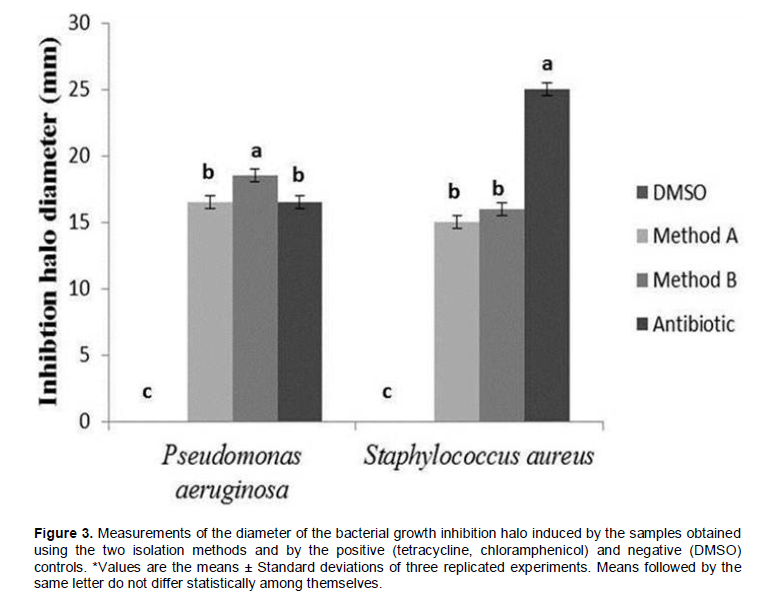
The results of the antibacterial disk diffusion assay are as shown in Figure 3 and are expressed as the diameter of the inhibition halo. The compounds isolated by me-thods A and B formed growth inhibition halos for both the tested bacterial strains. Despite showing lower tannin content, the sample originating from isolation method B presented a larger inhibition halo (18.2±0.10 mm) compared to method A (15.8±0.12 mm), against P. aeruginosa. The ethyl acetate used for the washing fractions in method A may have removed phenolic com-pounds, such as flavonoids, simple phenols and even polyphenols, which would act synergistically with tannins in the aqueous phase. Because the sample obtained by method B showed a larger inhibition halo, further analyses of its MIC and bactericidal level were performed.
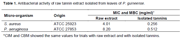
The minimum bactericidal and inhibitory concentrations of the raw extract and the tannin samples isolated by method B are as shown in Table 1. MBC was equal to the inhibitory concentration for both strains. These results demonstrate that this tannin isolation method effectively isolated compounds with antibacterial activity, because the isolated sample presented an MBC sixteen times smaller than the raw extract (personal data). According to the classification proposed by Aligianis et al. (2001), MIC values of ≤0.5 mg/ml are considered strongly inhibitory in the case of vegetable matter.
DISCUSSION
The percentages obtained by Wang and Lee (1996) for extracting tannins from Areca catechu fruits were 47 and 30% for the ethanol and acetone fractions, respectively, resulting in a total recovery of approximately 77%. The tannin content of the samples from both methods was higher than that obtained by Wang and Lee (1996) from Areca catechu fruits (160 mg/g of dry material).
The inhibitory effects of different classes of tannins against various microorganisms have been widely studied. According to Haslam (1996), tannins act on micro-organisms by binding to proteins and adhesins, inhibiting bacterial enzymes, rupturing the cell membrane and sca-venging microbial substrates. Kumar and Vaithiyanathan (1990) suggested that tannins directly inhibit microbial function in the rumen by complexing with bacterial cells or indirectly by reducing the availability of nitrogen and sulfur for microbial protein synthesis. Scalbert (1991) listed 33 studies documenting the antimicrobial properties of tannins. Sakanaka et al. (1996) reported the role of polyphenols in inhibiting growth and cellular adherence of the oral bacteria Porphyromonas gingivalis, which is responsible for the majority of acute periodontitis cases. De Bruyne et al. (1999) evaluated the antimicrobial activity of a series of proanthocyanidin dimers and very-fied that the minimum inhibitory concentration was >100 µg/ml for Escherichia coli, P. aeruginosa, Salmonella paratyphi, Enterobacter cloacae, Mycobacterium fortuitum, S. aureus and Candida albicans. Djipa et al. (2000) correlated the antibacterial activity demonstrated by Syzygium jambos (Myrtaceae) to its high tannin content, and Akiyama et al. (2001) iden-tified tannic acid as a possible adjuvant to antibiotics in the treatment of S. aureus infections. Panizzi et al. (2002) observed that fractions of tannins and other phenols ex-tracted from Rubus ulmifolius showed high antimicrobial activity. González et al. (2005) proved the antibacterial potential of the raw extract from the peel and pulp of P. guineense fruit, and they demonstrated that fractions isolated from this extract using petroleum ether, dichloromethane, ethyl acetate and water also had anti-microbial effects, which were attributed to the presence of tannins, flavonoids and terpenes in the fruits of this species.
Thus, P. guineense represents a promising source of molecules with pharmacological and antibacterial activities, and it contains high tannin content in its leaves, which exhibit a diverse set of biological activities, as extensive studies have shown.
ACKNOWLEDGEMENTS
This research was support by FAPEMIG and CNPQ. The authors thank State University of Montes Claros and Vallee S/A for logistical support.
CONFLICT OF INTEREST
Authors have not declare any conflict of interest.
REFERENCES
|
Akiyama H, Fujii K, Yamasaki O, Oono T, Iwatsuki K (2001). Antibacterial action of several tannin against Staphylococcus aureus. J. Antimicrob. Chemoth. 4(48):487-491. Crossref |
||||
|
Aligianis N, Kalpoutzakis E, Mitaku S, Chinou IB (2001).Composition and antimicrobial activity of the essential oil of two Origanum species. J. Agric. Food Chem. 9(49):4168-4170. Crossref |
||||
|
Asquith TN, Butler LG (1985). Use of dye-labeled protein as spectrophotometric assay for protein precipitants such as tannin. J. Chem. Ecol. 11(11):1535-1544. Crossref |
||||
|
Begum S, Hassan SI, Siddiqui BS, Shaheen F, Ghayur MN, Gilani AH (2002). Triterpenoids from the leaves of Psidium guajava. Phytochemistry 4(61):399-403. Crossref |
||||
|
Crawley MJ (2005). Statistical: An introduction using R. Jhon Wiley e Sons: New York,USA. P 761. Crossref |
||||
|
De Bruyne T, Pieters L, Deelstra H, Vlietinck AJ (1999). Condensed vegetable tannins: biodiversity in structure and biological activities. Biochem. Syst. Ecol. 4(27):445-459. Crossref |
||||
|
Djipa CD, Delmée M, Quetin-Leclercq J (2000). Antimicrobial activity of bark extracts of Syzygium jambos (L.) Alston (Myrtaceae). J. Ethnopharmacol. 1-2(71):307-313. Crossref |
||||
| Fernandes TG, Mesquita ARC, Randau KP, Franchitti AA, Ximenes EA (2012). In Vitro Synergistic Effect of Psidium guineense (Swartz) in combination with Antimicrobial Agents against Methicillin-Resistant Staphylococcus aureus Strains. Scientific World J. 2012:158237. | ||||
|
Ferreira PRB, Mendes CSO, Rodrigues CG, Rocha JCM, Royo VA, Valerio HM, Oliveira DA (2012) Antibacterial activity tannin-rich fraction from leaves of Anacardium humile. Ciên. Rural 10(42):1861-1864. Crossref |
||||
| González AMN, González MBR, Pinto NLS. 2005. Phytochemical study and antibacterial activity of Psidium guineense Sw (choba) against Streptococcus mutans, causal agent of dental caries. Rev. Cubana Med. Plant 10:3-4 | ||||
|
Gutiérrez RMP, Mitchell S, Solis RV (2008). Psidium guajava: A review of its traditional uses, phitochemistry and pharmacology. J. Ethnopharmacol. 17(117):1-27. Crossref |
||||
|
Hagerman AE (1987). Radial diffusion method for determination tannin in plant extracts. J. Chem. Ecol. 3(13):437-449. Crossref |
||||
|
Hagerman AE, Klucher KM (1986). Tannin-protein interation. In Plant Flavonoids in biology and Medicine: biochemical, pharmacological, and structure-activity relationships, Cody V, Middleton E, Harborne J (eds). Alan R. Liss: New York. pp. 67-76. Pubmed |
||||
|
Haslam E (1996). Natural polyphenols (vegetable tannins) as drugs and medicines: possible modes of action. J. Nat. Prod. 2(59):205-215. Crossref |
||||
|
Kumar R, Vaithiyanathan S. (1990). Occurrence, nutritional significance and effect on animal productivity of tannins in tree leaves. Anim. Feed Sci. Technol. 1-2(30):21-38. Crossref |
||||
|
Lee MH, Chiou JF, Yen KY, Yang LL (2000). EBV DNA polymerase inhibition of tannins from Eugenia uniflora. Cancer Lett. 2(154):131-136. Crossref |
||||
|
Matsuo T, Hanamure N, Shimoi K, Nakamura Y, Tomita I (1994). Identification of (+)- galoocatechin as a bio-antimutagenic compound in Psidium guajava leaves. Phytochemistry 4(36):1027-1029. Crossref |
||||
|
Meckes M, Calzada F, Tortoriello J, Gonzalez JL, Martinez M (1996). Terpenoids isolated from Psidium guajava hexane extract with depressant activity on central nervous system. Phytother. Res. 7(10):600-603. Crossref |
||||
| NCCLS (2003). Methods for Dilution Antimicrobial Susceptibility Tests for Bacteria That Grow Aerobically, 6th edn. Approved Standard, M7-A6. National Committee for Clinical Laboratory Standards: Wayne, PA. | ||||
| NCCLS (2003). Performance Standards for Antimicrobial Disk Susceptibility Tests, 8th edn. Approved standard, M2-A8. National Committee for Clinical Laboratory Standards: Wayne, PA. | ||||
|
Salib JY, Michael HN (2004). Cytotoxic phenylethanol glycosides from Psidium guaijava seeds. Phytochemistry 14(65):2091-2093. Crossref |
||||
|
Salminen JP, Ossipov V, Loponen J, Haukioja E, Pihlaja K (1999). Characterisation of hydrolysable tannins from leaves of Betula pubescens by high-performance liquid chromatography-mass spectrometry. J. Chromatogr. A 2(864):283-291. Crossref |
||||
| Santos SC, Mello JCP (1999). Taninos. In Farmacognosia: da planta ao medicamento, Simões CMO, Schenkel EP, Gosmann G, Mello JCP, Mentz LA, Petrovick PR (eds). Universidade Federal do Rio Grande do Sul: Porto Alegre. pp. 614-656. | ||||
|
Scalbert A (1991). Antimicrobial properties of tannins. Phytochemistry 12(30):3875-3883. Crossref |
||||
|
Schapoval EES, Silveira SM, Miranda ML, Alice CB, Henriques AT (1994). Evaluation of some pharmacological activities of Eugenia uniflora L. J. Ethnopharmacol. 3(44):137-142. Crossref |
||||
|
Strumeyer DH, Malin MJ (1975). Condensed tannins in grain sorghum: isolation, fractionation and characterization. J. Agric. Food Chem. 5(23):909-914. Crossref |
||||
|
Wang CK, Lee WH (1996). Separation, Characteristics, and Biological Activities of Phenolics in Areca Fruit. J. Agric. Food Chem. 8(44):2014-2019. Crossref |
||||
Copyright © 2024 Author(s) retain the copyright of this article.
This article is published under the terms of the Creative Commons Attribution License 4.0


