Full Length Research Paper

ABSTRACT
Acmella oleracea (L) RK Jansen is a typical plant species of Northern Brazil, used in gastronomy and in the Amazonian folk medicine of Para State as analgesic, to treat diseases of the mouth and throat. In industry, extracts of this genus have been used in oral hygiene products and in food compositions, as refreshing and flavoring agent, and is also used in cosmetics and toiletry. This paper reports the pharmacognostic characteristics of the herbal drug (flowers) and the in vitro antimicrobial effect of its ethanol extract (EEFAO) and fractions on pathogenic microorganisms present both in skin and in gastrointestinal tract of domestic animals. EEFAO Hexane, Chloroform, Ethyl Acetate and Methanol fractions at different concentrations (1000, 500, 250, 125, 62.5, 31.25, 15.62, 7.81 mg/mL) were tested against microorganisms (bacteria and fungus). The phytochemical characterization of A. oleracea extract and fractions indicates the presence of, probably, Spilanthol, detected by thin layer chromatography using Dragendorff Reagent. Chloroform fraction inhibited the growth of Salmonella typhi at a Minimum Inhibitory Concentration of 31.25 mg/mL and the microscopic analyses of young flowers demonstrates the presence of undifferentiated hypanthium and involucral bracts, cypselas and vascular bundles, structures also observed in other species of this genus. Quality parameters, including phytochemical description, reported in this work allow the identification and standardization of the flowers as herbal drug, whose microscopic description is very useful because it enables its micrographic characterization. The Chloroform fraction of EEFAO can inhibit the growth of Salmonella typhi making possible the use of A. oleraceae in phytomedicines or conservatives for foods.
Key words: Antimicrobial, phytotherapy, flowers pharmacognosy, Salmonella typhi.
INTRODUCTION
Acmella oleracea (L) RK Jansen, vernacular Jambu, has motivated the interest of researchers because of its therapeutic potential, inducing projects in different areas linked to health sciences, such as medicine, dentistry and pharmacy. A.oleracea is a perennial, herbaceous flowering shrub common in Northern Brazil, particularly in Para State (Coutinho et al., 2006; Silva and Santos, 2011). Its chemical constitution described in the literature includes the alkamide spilanthol, a and β-amyrinester, stigmasterol, miricilic alcohol glycosides, sitosterol, saponins and triterpenes (Lemos, 2012).
Although, the entire plant is medicinally used, it is mainly in the flowers where the highest amount of Spilanthol (Wongsawatkul, 2008) occurs (Cavalcanti, 2008; Nigrinis et al., 1986), using guinea pigs, confirmed the main biological activity of this plant organ; the local anesthetic effect. The same authors also reported the flavoring, insecticide, bactericide and healing action of the floral extract, both in oral mucosa and on the skin. No scientific report about the microscopic description of A. oleracea flowers was found in the surveyed literature. This work reports the result of the pharmacognostic investigation on the A. oleracea floral drug and its Ethanol extract. In addition, the in vitro antimicrobial activity of EEFAO and its fractions on different bacteria and fungi species were reported.
MATERIALS AND METHODS
Plant materials
Young leaves and flowers of A. oleracea ( L ) RK Jansen were acquired in Marituba, Para State, metropolitan region of Belem (01°21' latitude South and 48°20 longitude West). A voucher is deposited at the Herbarium of the Botany Laboratory, Embrapa Amazonia Oriental, Belem - Para, registered as IAN 188444, in October, 2012.
Macroscopic and microscopic description
Macroscopic characterization of the flowers and leaves was performed with bare eye, according to parameters described by Esau and Silva Provide YEAR (Nigrinis et al., 1986; Esau , 1976). For microscopic description, semi-permanent cuts were prepared using fresh leaves and young flowers. The plant material was sliced transverse in longitudinally by hand. The samples were clarified in 10% aqueous Sodium Hypochlorite and stained with Methylene Blue and Safranin. The cuts were observed using a Nikon optical microscope (Eclipse 50i) equipped with a Motic® camera (Moticam 2300) and the pictures were processed using Motic Image Plus 2.0® software. The photomicrographs obtained at 10X and 40X were analyzed in comparison to literature data.
Preparation of extract
To obtain the Ethanol extract, 3.8 kg of cleaned A. oleracea flowers were dried under circulating hot air (Quimis Q317B) at 40°C ± 2°C until constant weight of an aliquot. The dried plant material was grinded in a Wiley knives mill using a sieve of medium mash. The obtained herbal drug weighed 426.1 g, which was macerated in 2.5 L 70% Ethanol in a stainless steel container for seven days. The obtained tincture was then filtered and concentrated under reduced pressure on a rotatory evaporator (800 Fisatom). The aqueous residue was frozen and lyophilized (Freeze dryer L101, LIOTOP). The freeze-dried extract was stored under refrigeration until use.
Pharmacognostic essays
The physic-chemical quality control of the plant drug and its extract EEFAO was performed according to the Brazilian Pharmacopeia, 5th edition, and the following tests were performed: particle size distribution, solids contents, pH, ash content and moisture content, respectively (Silva, 2008).
Fractionation
An aliquot of 4.26 g of EEFAO was fractionated by solid-liquid partition using 5 to 7x 50 ml aliquots of increasing polarity solvents: Hexane, Chloroform, Ethyl Acetate and Methanol. This procedure is established in the routine of Phytochemical Analysis Laboratory, Faculty of Pharmaceutical Sciences, Federal University of Pará. The fractions were concentrated on a rotatory evaporator at low pressure (Fisatom 800).
Chromatographic analyses
The chromatographic profile of the samples was obtained by thin layer chromatography (TLC) using silica gel (SIGMA) as stationary phase, mixture of solvents of different polarities as the mobile phase ding according to Wagner and Bladt (2001), to detect Flavonoids, Tannins, Terpene and Alkaloids (ANVISA 2010).
Evaluation of antimicrobial activity
Microorganisms and growth conditions
Bacteria: Salmonella typhi ATCC00259, Enterobacterium faecalis ATCC29212 and Staphylococcus aureus ATCC00577.
Yeast: Candida albicans ATCC0175. The Laboratory of Microbiological Control, Faculty of Pharmaceutical Sciences provided the samples of microorganisms. The bacteria grown in Mueller-Hinton broth (Himedia) at 37°C were kept in Mueller Hinton agar plates at 4°C. The yeasts were grown and maintained in broth and Sabouraud Agar (Himedia).
Antimicrobial activity
The tests were performed using broth micro dilutions techniques and MIC of EEFAO and its fractions were determinate as described by Holetz et al. (2012) with modifications. Aliquots of 100 μL of broths and 10 μL of microorganisms adjusted McFarland scale (108 Colony Forming Units) were used to evaluate the activity of EEFAO and its fractions at a concentration of 100μg/Ml (Wagner and Bladt, 2001). minimal inhibitory concentration (MIC) was defined as the lowest concentration of the sample, which produces a marked reduction of at least 80% of the tested microorganisms (Wagner and Bladt, 2001).
Statistical evaluation
The data statistical evaluation was performed using Bioestat 5.3, and T Student test with 95% level of confidence.
RESULTS
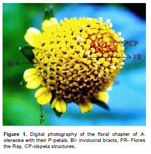
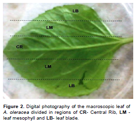
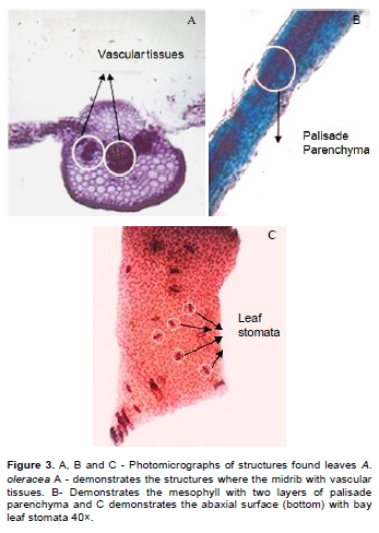
The macroscopic analysis of A. oleracea (L) RK Jansen leaves and fresh flowers was performed on the external surfaces of these organs (Figure 1). The microscopic analysis of A. oleracea leaves revealed simple mem-branous leaves showing wavy uniseriate epidermal cells, bicellular or tricellular trichome with basal cell showing rough cuticle and anomocytic stomata. The ventral mesophyll is well-organized showing two layers of palisade parenchyma and several layers of spongeous parenchyma. The cross-section of central rib shows a concave-convex profile and two vascular bundles are present in addition to the conducting vessels (Figure 2).
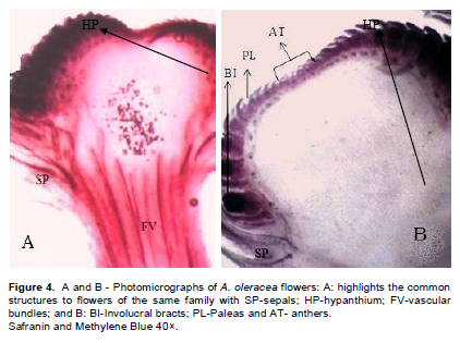
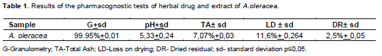
The presence of the undifferentiated hypanthium (HP) with various vascular bundles (FV) and differentiated laterally sepals (SP) can be microscopically observed in the longitudinal section of the immature flower buds. The central region of the hypanthium presents uniseriate epidermis, at its ends involucral bracts can be also observed in (BI), peleas (PL) and anthers (AT) (Figures 3 and 4). The pharmacognostic analyses show the following results: the granulometry of the A. oleracea herbal drug (dried and grinded flowers). The grinded dried flowers indicates that the sample is a coarse powder since its particles were predominantly retained on the sieve with the highest mash value (1.700 mm) reaching 99.95% of the sample weight (Table 1). The pH of the herbal drug was determined for decoction, after filtration and cooling in a calibrated potentiometer. The result, 5.33, is the middle value of three determinations with standard deviation of ± 0.24, using osmosed water as reference, pH of 6.25 (Table 1).
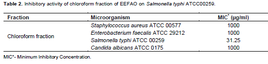
The determination of total ash present in the sample revealed as a middle value of three experiments 7.07%; standard deviation of ± 0.03% (Table 1). Completing the pharmacognostic analyses, moisture was determined in triplicate yielding, 11.6% standard deviation ± 0.264%. In addition, the middle value of dried residue of the extract is 2.5%; SD ±0.05 (Table 1). All the parameters were determined in triplicate. Chromatographic analyses of EEFAO and its Hexane, Chloroform and Ethyl Acetate fractions where performed using TLC on normal phase silica gel with Hexane/Acetone (80:20) as eluent. The obtained chromatograms, show orange colored bands due to reaction with Dragendorff’s reagent, at Rf value of 0,37. The antimicrobial test using EEFAO and its fractions resulted in significant reduction (p ≤ 0.05) of the colonies number of Salmonella typhi ATCC 00259 (Table 2); especially for the chloroform fraction, which inhibited the microorganism growth at a MIC of 31.25 µg/mL of the Salmonella typhi ATCC 00259.
DISCUSSION
As described in the literature, genus Acmella is part of the Asteraceae family, consisting of Angiosperm plants (Esau, 1976). The macroscopic description of leaves of this plant species discloses phylliform or membranous aspect of the largely oval leaves, being about 3 to 6 cm long and dark green in color (Coutinho et al., 2006; Silva and Santos, 2011). Mature capitula were macroscopically described as oval, irradiated or discoid; with conical re-ceptacle, golden or pink to reddish paleas and obtuse to acuminate apex; ray flowers when present, are ligulate, pistillate; corolla can be white yellow or orange, with two or three lacinios at the apex. The disc flowers are perfect, tubular, white, yellow or orange corolla, four or five acute lacinia and four to five anthers. Oval or ellipsoid cypselae disc is laterally compressed, sometimes with cortical margin present when mature; pappus absent or 1 to 10 weak bristles, tricosted, ellipsoid cypselae ray are usually usually present in mature cortical margin. Similar structures were found and described in Figure 1 (Coutinho et al., 2006).
No data about the microscopic structure of the flowers of this plant species was found in the literature. However, Flowers of Flowering Plants have similar microscopical structures like hypanthium, which is the region of the flower that receives seeds after differentiation (Figures 3 and 4). Comparing the microscopic structures of the flowers of this plant, species having similar structures were observed in Acmella brasiliensis and Acmella marajoensis (Holletz et al., 2002; Coutinho et al., 2006), it is possible to infer that A. oleraceae also has differentiated hypanthium for seeds accumulation, as well large caliber vascular bundles another to ensure the maintenance of these reproductive structures during prolonged periods.
The analyses of EEFAO show that the results here re-ported are in accordance with the parameters described in the Brazilian Pharmacopoeia 5th edition (Silva, 2008). In Brazil, the National Agency for Sanitary Surveillance (ANVISA) requires pharmacognostic tests for quality control, such as chromatographic profile by TLC or phytochemical screening, as criterion for notification or registration of traditional herbal medicines and notification of herbal product (Baccarin et al., 2009). This paper reports the TLC analysis, based on the methodology described by Wagner and Bladt (2010) (ANVISA, 2010), highlighting the substance at Rf 0.37, which reacts positively to Dragendorff’s reagent. Armond (2007) performed a phytochemical investigation of A. oleraceae using the same phytochemical methodology, the author reports about a substance with Rf 0.36, in the same chro-matographic condition (ANVISA, 2010), which reacts as Flavonoid with Sulfuric Vanillin and fail to react with Iodine and KOH like some Alkaloids he tested. In the present work the substance with Rf 0.36 reacts to Dragendorff’s reagent, as usual for substances containing
containing nitrogen, like alkamides. This observation raises the following question, can alkamides react both as N-containing substance with Dragendorff’s reagent or as bident structure common in Flavonoids allowing the complexation of a metal ion? (Marques et al., 2012). In fact, the reaction of alkamides with Sulfuric Vanillin may involve a nucleophilic attack of Nitrogen atom on the Aldehyde Carbonyl group of Vanillin.
Among the antimicrobial tests against Salmonella, typhi showed the best result. This genus of bacteria is usually found in the gastrointestinal tract of domestic and wild animals, especially birds and reptiles. Numerous Salmonella serotypes are pathogenic for both animals to humans (Anvisa, 2014; Armond, 2007). It is estimated that 36% of dogs are asymptomatic carriers of this bacteria; In witch, the clinical signs of the disease vary depending on the number of infective organisms and the immune status of the animal as well others adverse factors such as intercurrent illnesses. Young animals or elderly one are the most susceptible to the bacteria, increasing the severity of the infection (Armond, 2007).
There is evidence that besides frames of severe enteritis, bacteria can also cause this kind of generalized skin lesions redness, blistering and crusting in immuno-compromised patients (Anvisa, 2007). Considering that the antimicrobial susceptibility to plant extracts, the inhibitory action to be considered promising must show a minimum inhibitory concentrations less than or equal to 100 μg/mL. Samples with MIC extracts ranging from 100 to 500 μg/mL are considerate having moderate antimicrobial activity. However, MIC values greater than 1000 μg/mL characterize the samples microbiologically inactive (Wagner and Bladt, 2001). The crude extract of A. oleraceae does not show antimicrobial activity as re-ported by Holetz et al. (2002) (Wagner and Bladt, 2001).
This work reports the antimicrobial activity of the chloroform fraction prepared from the crude extract EEFAO. Prachayasittikul et al. (2009) (Carvalho et al., 2003) demonstrated that the chloroform fraction of A. oleracea was able to inhibit the growth of Saccharomyces cerevisiae ATCC 2601 and Streptococcus pyogenes II in a MIC 256 mg/mL. In this study Salmonella typhi ATCC00259, had their growth inhibited by a MIC of 31.25 mg/mL of chloroform fraction of EEFAO.
CONCLUSION
Quality parameters (particle size, total ash content, pH, loss on drying and dried residue) described in this work allow the identification and standardization of the herbal drug, the extract obtained from the flowers of A. oleracea and its fractions, where the presence of a substance reactive to the Dragendorff’ reagent can be detected, probably spilanthol.
The microscopic description of the flowers of this plant species is very useful because it enables the micro-graphic characterization of plant drug (Figures 3 and 4). No records of these data were found in the surveyed literature. The result of antimicrobial activity evaluation indicates that the chloroform fraction of EEFAO is able to reduce the visible growth of Salmonella typhi. This observation can justify the use of A. oleraceae in the development of therapeutic products or conservatives for foods.
CONFLICT OF INTEREST
All authors declare that they have no conflict of interest.
REFERENCES
| ANVISA (2010). Brazilian Pharmacopoeia, 5th edition Brasília. Fascicule 1; 2. | ||||
| ANVISA (2014). Law-RDC Nr. 26-13/05/2014. http://bvsms.saude.gov.br/bvs/saudelegis/anvisa/2014/rdc0026_13_05_2014.pdf. Accessed October 2014. | ||||
| Armond C (2007). Chemical indicators, plant growth and bioeletrography of jambu (Acmella oleracea L.), lemongrass (Cymbopogon citratus (Dc) Stapf) and leaf (Bryophyllum pinnatum (Lam. Oken) subjected to homeopathic treatments.142p. PhD Thesis. PPG Fitotecnia. Federal University of Viçosa, Minas Gerais, | ||||
| Baccarin T, Czepula AI, Ferreira RA, Lucinda-Silva RM (2009). Morfanatomic analysis of the aerial parts of Wedelia paludosa DC. (Acmela brasiliensis, Sphagneticola trilobata), Asteraceae. Braz. J. Pharmacogn. 19(2B):612-616. | ||||
|
Carvalho BTC, Iazzetti AV, Ferrarini MAG, Campos SO, Iazzetti MA, Carlesse FAMC (2003). Salmonella sepsis associated with interleukin-12 (IL-12Rß1) receptor deficiency. J. Pediatr. 79(3):273-6. Crossref |
||||
| Cavalcanti VMS (2008). Spilanthol extraction of Spilanthes acmella var acmella Oleracea with supercritical carbon dioxide. PhD Thesis. PPGEQ, Campinas State University, São Paulo. 144p | ||||
|
Coutinho LN, Aparecido CC, Figueiredo MB (2006). Galls and deformations in Jambu (Spilanthes oleracea) caused by Tecaphora spilanthes (Ustilaginales). Summa Phytopathol. 32(3):283-285. Crossref |
||||
| Esau K (1976). Anatomy of plants with seeds. Ed. Blucher. São Paulo. | ||||
|
Holetz FB, Pessini GL, Sanches NR, Cortez DAG, Nakamura CV, Dias Filho BP (2002). Screening of some plants used in the Brazilian folk medicine for the treatment of infectious diseases. Mem. Inst. Oswaldo Cruz 97(7):1027-31. Crossref |
||||
| Lemos VR (2012). Evaluation of in vitro Acmella cillata KUNTH extract: fluorescence microscopy techniques, cytotoxicity and infrared spectroscopy (FT-IR). MSc Dissertation. PPGEB, Vale do Paraíba University, São José dos Campos. São Paulo. 71p. http://biblioteca.univap.br/dados/000003/00000353.pdf. Accessed October 2014. | ||||
| Marques GS, Lyra MAM, Peixoto MS, Monteiro RPM, Leão WF, Xavier HS, Soares LAL, Rolim Neto PJ (2012). Phytochemical and physicochemical characterization of the leaves of Bauhinia forficata Link collected in two Brazilian regions. J. Braz. Appl. Pharm. Sci. 33(1):57-62. | ||||
| Nigrinis LSO, Caro JO, Olarte EM (1986). Phytopharmacological study of the fat-soluble fraction from the Spilanthes americana flowers (Mutis) part I: Phytochemical Study. Rev. Col. Ciên. Quím-Farm. 15: 37-47. | ||||
| Nigrinis LSO, Caro JO, Olarte EM (1986). Fitopharmacological study of the fat-soluble fraction from the Spilanthes americana flowers (Mutis) part II: Phytochemical Study. Rev. Colomb. Ciên. Quím-Farm. 16(2). | ||||
| Prachayasittikul S, Suphapong S, Worachartcheewan A, Lawung R, Ruchirawat S, Prachayasittikul V (2009). Bioactive Metabolites from Spilanthes acmella Murr. Int. J. Mol. Sci. 14:850-864. | ||||
| Silva GAR (2008). The subtribe Ecliptinae Less. (Heliantheae-Asteraceae) in the Brazilian Amazon. MSc Dissertation. PPGB, Federal University of Pará, Belém-Pará. 130p | ||||
|
Silva GAR, Santos JUM (2011). Acmella marajoensis: A new species of Asteraceae, native to the Brazilian Amazon. Acta Amaz. 41(2):191-194. Crossref |
||||
| Wagner H, Bladt S (2001). Plant drug analysis. 2nd ed. Springer, New York. | ||||
|
Wongsawatkul O, Prachayasittikul S, Isarankura-Na-Ayudhya C, Satayavivad J, Ruchirawat S, Prachayasittikul V (2008). Vasorelaxant and Antioxidant Activities of Spilanthes acmella Murr. Int. J. Mol. Sci. 9:2724-2744. Crossref |
||||
Copyright © 2024 Author(s) retain the copyright of this article.
This article is published under the terms of the Creative Commons Attribution License 4.0


