Full Length Research Paper

ABSTRACT
In the recent past, the importance of freshwater algae has grown enormously due to their antibiotic activity against certain species of bacteria known for several disease states like endocarditis, external otitis, skin rash, etc. Also, there is a growing concern among the immunocompromised individuals that they may be susceptible to antibiotics and anti-fungal resistant infections resulting in increased fatality rates. Hence, in this investigation, extraction of potentially bioactive compounds from natural resources like freshwater algae was performed along with the evaluation of the antimicrobial activities of these extracts against some opportunistic bacteria like Staphylococcus aureus, Escherichia coli, Staphylococcus xylosus and Pseudomonas aeruginosa using well diffusion method. The data showed a statistically significant (P < 0.05) antibacterial activity of the organic extracts (0.1 g/ml) obtained from freshwater algae against multi-drug resistant S. aureus and S. xylosus strains as compared to the control. Our data reinforce the importance of bioactive compounds from fresh algae as potential antimicrobial agents, and they could act as an alternative to conventional antibiotics.
Key words: Freshwater algae, Organic extracts, Antimicrobial activity, Opportunistic bacteria, Methicillin Resistant Streptococcus, Natural Antibiotics.
INTRODUCTION
Algae are plant-like organisms however with no roots, stem, or leaves, having photosynthesis process for converting light energy into chemical energy. Freshwater macroalgae Spirogyra is filamentous green algae of order Zygnematales, which contains novel biologically active compounds for antibiotic, antiviral, antioxidant, and anti-inflammatory purposes (Ramaraj et al., 2015; Mesbahzadeh et al., 2018). The genus Spirogyra belongs to the class Chlorophyceae, comprising 400 species and mostly free-floating algae. Commonly, they are found in freshwater ponds, lakes with few exceptions in slow running water (Naik et al., 2012; Mei et al., 2021). Furthermore, Spirogyra species have several biomedical applications due to their anti-viral, bacterial and fungal properties that could be exploited in human health. However, these beneficial effects of Spirogyra spp. are not fully realized particularly in the Kurdistan region due to the limited knowledge among the people about their edible nature, nutrient value as well as medicinal properties. Besides, Oedogonium species commonly found in the Red Mountain region, was known to play a vital role in the fixation of heavy metals in freshwater ecosystems and also proven to be good candidates for biomass applications. Also, the filamentous nature of the algae aids in adherence of it to stone, wood, and leaves (Phadnis and Iyer, 2016). The Oedogonium spp. has numerous advantages, for example, Oedogonium capillare was notably used in traditional medicine for the treatment of various human ailments such as dysentery, diarrhea, thrush on tongues of babies, wound healing, and as an antiseptic agent for various skin diseases (Perez-Gutierrez, 2006). Moreover, it could act as feed for many organisms such as frogs, fish as well as for biomass applications in a freshwater ecosystems (Lawton et al., 2014).
Endocarditis is an inflammation of the inner layer of the heart due to gaining access to the systemic circulation of certain bacteria and fungi thereby causing severe complications, if not intervened in time. Endocarditis due to bacteria are of two types, acute endocarditis is known as a progressive disease that rapidly damages the cardiac structure and spread via blood leading to death within weeks unless it is treated and subacute endocarditis, as compared, is a slow or gradual progressing disease possibly for weeks to months with a clinical outcome known as embolism (Vilcant and Hai, 2020; Hubers et al., 2020).
In particular, Staphylococcus aureus a Gram-positive bacteria and human pathogen, could cause infective endocardites, bacteremia, as well as skin and soft tissue infections. It was reported that the misusage or over usage of antibiotics such as penicillin against S. aureus could result in a resistant strain known as methicillin-resistant S. aureus (MRSA) (Lakhundi and Zhang, 2018; Karakonstantis and Kalemaki, 2019). Recently, our research group worked on the efficiency of the organic extracts from petals of Rosa damascena against methicillin resistant species of Staphylococcus and reported the importance of exploring the bioactives from natural resources as potential antibiotics (Thomas et al., 2020).
Moreover, infective endocarditis is also caused by another species of Staphylococcus called Staphylococcus xylosus found in human and animal normal skin. It is known to be resistant to antibiotics like novobiocin thereby eluding an effective treatment. Considering, freshwater algae could be effectively used to fight harmful pathogens as they could withstand stress conditions like osmotic pressure, salinity, and a high level of ultraviolet light. Since they thrive under stress conditions, they have the potential to release certain chemical substances with antimicrobial and biological activities (Shannon and Abu-Ghannam, 2016; Kogler et al., 2020).
The significance of naturally occurring algae in different ecosystems such as freshwater and marine was the focus of research from many ecologies and marine scientists because they have the potential biological effects against microbes including bacteria, fungi, and viruses. Indeed, a study by Naik et al. (2012) on Spirogyra spp. showed that it had significant antibiotic activity against Pseudomonas solanacearum, Escherichia coli, and Clavibacter michiganens. In particular, the organic compounds that acted as a solvent revealed a highly significant antibiotic effect against P. solancearum. Furthermore, the Antarctic freshwater microalgae, Choloromonas, was studied for the bioactivity in 20 ml ethanol extract. The residue obtained after removal of ethanol by vaccum evaporator had significant antimicrobial activity (Suh et al., 2017).
Present study aimed to evaluate the antimicrobial activity of freshwater algae Spirogyra spp. found in the Kurdistan region against S. aureus, E. coli, S. xylosus and Pseudomonas aeruginosa. In the last decade or so, many scientists from Kurdistan have published numerous articles on the significance of natural products and in coming years, we envisage that there would be burgeoning research interest and successful scientific articles in natural products due to their biological effects.
MATERIALS AND METHODS
Algal source
The freshwater algae were collected in slow-running water in Red Mountain near Qalachwalan, Sulaymaniyah, Kurdistan. Algae were transferred into two labeled plastic buckets. A similar sampling location was used for the entire study. The samples were collected by a wooden stick and transported to the phycology laboratory, College of Science, University of Sulaymaniyah, Kurdistan for microscopic examination during July 2019, October 2019, and December 2019.
Morphological identification of alga
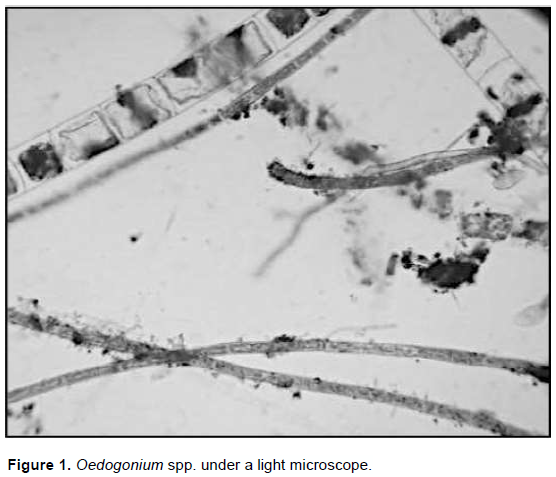
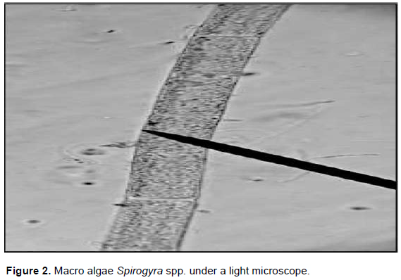
Oedogonium similar to spirogyra, was a filamentous green alga but it had cylindrical cells which some of them were ring shaped near the ends. Each cell had a parietal chloroplast and pyrenoids usually found in lakes, rivers, and ponds (Figure 1) (Piotrowski et al., 2020). The color of Spirogyra (Figure 2) was green due to the presence of chlorophyll a and chlorophyll b; however, they appeared yellow or orange under stress conditions because of the presence of secondary pigments (carotenoids) (Çelekli et al., 2017). The chlorophyll in Spirogyra was important for the photosynthesis process to convert light energy into chemical energy bound in biomass (Ramaraj et al., 2013).
Harvest and sample preparation
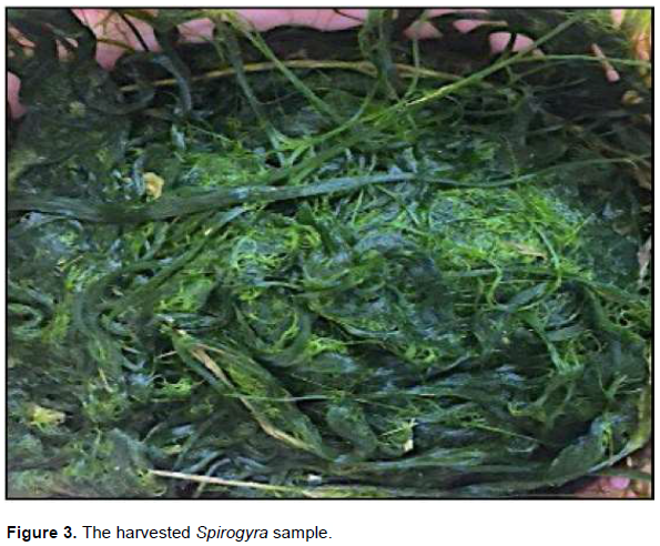
Five kilograms of wet Spirogyra were harvested (Figure 3), after they were exhaustively rinsed with fresh water to clean them from debris and sand. They were kept in a shady area for one week for drying. The dried algae were ground and crushed by a mortar and pestle; thus the resultant algae were further made into powder form using a mixer under sterile conditions. Subsequently, the powdered sample was ready for extraction (Kim et al., 2018).
Extract preparation
For the preparation of the organic extract was performed according to Wong and Shahirah (2019) with a slight modification. 20 g of powdered algae was mixed with 200 ml of ethanol and subjected to Soxhlet extraction. Then, the samples were incubated at 56°C for 96 h.
Antibacterial susceptibility assay
The antibacterial activity of three extracts was tested using the agar well diffusion technique on Mueller Hinton media (Redfern et al., 2014). Four different types of bacteria were used like E. coli, S. aureus, S. xylosus and P. aeruginosa obtained from the laboratory of Komar University of Science and Technology, Sulaymaniyah, Kurdistan. They were cultured in Petri plates for the testing antibacterial activity of the algae extract. Moreover, they were subcultured from the stocks maintained in nutrient broth. All the bacterial samples were maintained according to standard conditions.
Measurement of total phenolic content
The total phenolic content in the ethanol extracts of freshwater algae was determined using Folin-Ciocalteau reagent according to the method of Maliak and Singh (1980) using gallic acid as standard. 500 mg freeze-dried algal were homogenized in 80% ethanol using mortar and pestle and the homogenate was centrifuged at 10,000 × g for 20 min. Folin-Ciocalteau reagent (0.5 ml) was added to 3 ml of ethanolic extract and then 2 ml 20% sodium carbonate was added. The contents were incubated for 5 min at room temperature and the absorbance of blue colour was read at 650 nm. A calibration curve was prepared using gallic acid standards at concentrations of 100 μg to 1 mg L-1. The total content of phenolic compounds in plant extracts was calculated as gallic acid equivalents (GAE) using the formula:
c × V
C = M ?
where C-Total content of phenolic compounds g/g of freeze-dried algal biomass extract expressed in GAE; c- concentration of gallic acid (mg/ml), V- volume of plant extract (ml), and M ?- weight of tissue sample.
Extraction and quantification of carotenoids
The amount of total carotenoids was estimated following the method of Lichtenthaler and Buschmann (2001). 200 mL of a homogenous suspension of cultures was centrifuged at 5000×g for 10 min, and the cell pellet obtained was resuspended in 20 mL of acetone. The solvent biomass mixture was incubated at 50°C in a water bath for 2 h with manual shaking every 10 min. The mixture was centrifuged at 5000 × g and absorbance of the supernatant was noted at 661, 644, and 470 nm using UV-Visible spectrophotometer (Model 1280, Shimadzu, Japan). The amount of total carotenoids was determined using the following equations:
Chlorophyll a (µg/ml) = (11.24 A660) - (2.04 A645)
Chlorophyll b (µg/ml) = (20.13 A600) - (4.19 A645)
Carotenoids (µg/ml) = 1000 x (A470 - 1.9 Chl a - 63.14 Chl b)/214
where A660, A645, and A645 represent absorbance at 660, 645 and 470 nm, respectively. Simultaneously dry weight biomass of the algae was noted and the amount of carotenoids was equalized on dry weight biomass.
Statistical analyses
All the experiments were carried out thrice for the statistical analyses. The data were represented as means ± standard error of the mean. Group mean values of each experiment were compared by single-sided Student's t-test. The means were also compared by one-way analysis of variance and multiple pair-wise comparisons were done using the Tukey’s test. P < 0.05 was considered to be the level of significance. Statistical analyses were performed using Graph Pad Prism 6 Software package for Windows.
RESULTS
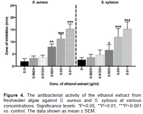
In the present study, the inhibition zones of each extracted sample were measured against four different types of bacteria such as E. coli, S. aureus, S. xylosus, and P. aeruginosa. The evaluation of the quality of each extract revealed the potential of the extract as antibacterial source. It was demonstrated that the ethanol extract of freshwater algae using Soxhlet extraction method had a statistically significant antibacterial effect against S. aureus and S. xylosus (Figure 4, P<0.05), however, it was not significantly effective against E. coli and P. aeruginosa as compared to the control (data not shown). In this study, the antibacterial activity of synergized freshwater algae was determined against different types of bacteria. The zone of inhibition of the freshwater algae extract against bacteria ranged between 5 and 19 mm at the concentration of 0.1 g/ml as compared to the controls. The crude ethanol extract of synergized freshwater algae at 0.1 g/ml concentration showed the highest zone of inhibition (19 mm) against Gram-positive S. aureus indicating a dose effect whereas it was 16 mm against S. xylosus at 0.05 g/ml dose in comparison to the controls. The lowest zone of inhibition was at the concentration of 0.0625 g/ml, that is, 5 mm in both S. aureus and S. xylosus (Figure 4).
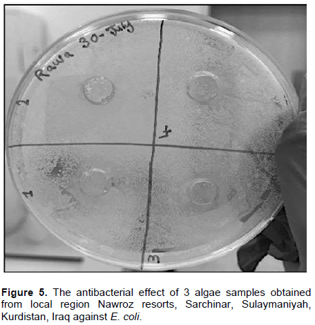
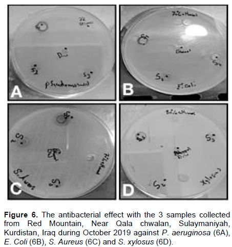
Furthermore, the antibacterial activity of the ethanol extract was carried out based on the collection of the algal samples at different time points and the results demonstrated varying antibacterial activity against different types of bacteria. The three samples of macroalgae collected during July, 2019 were tested against E. coli by agar well diffusion technique. The results revealed that the samples did not show any antibacterial activity against Gram-negative bacteria, E. coli when compared with the controls (ethanol and distilled water) (Figure 5). Similarly, the three samples of macroalgae obtained during October, 2019 were also tested against P. aeruginosa, E. coli, S. aureus and S. xylosus. The data divulged no significant antibacterial effect against both Gram-positive and Gram-negative bacteria as compared to the controls (Figure 6).
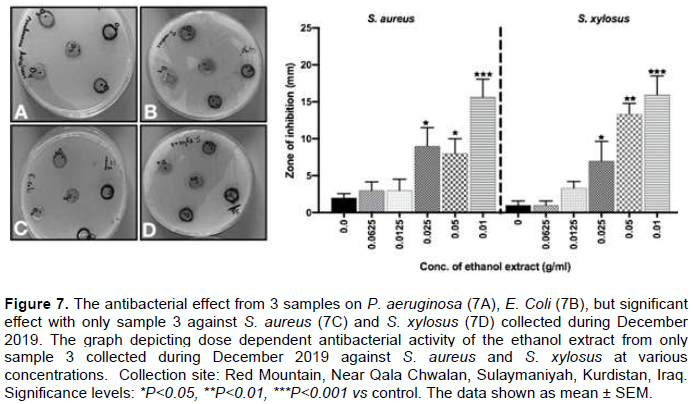
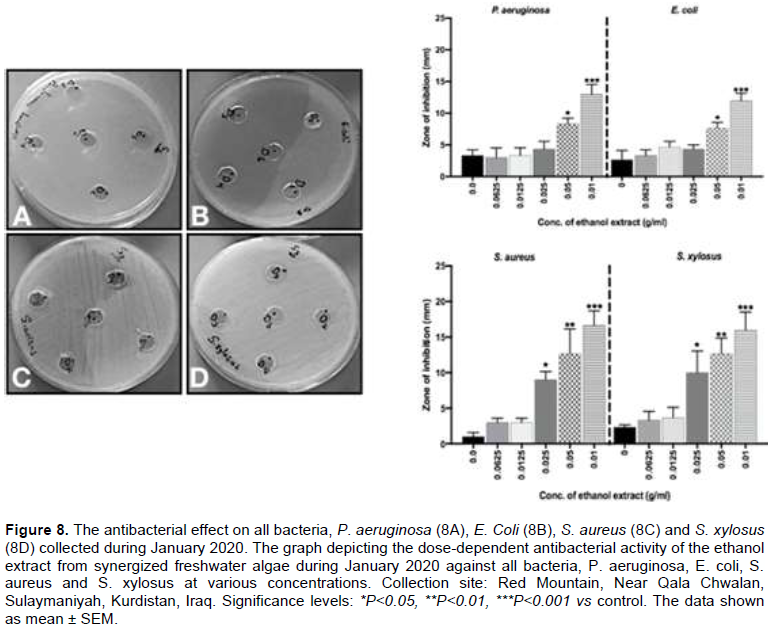
Moreover, the three different samples of macroalgae collected in December, 2019 were also evaluated against bacteria E. coli, S. aureus, P. aeruginosa and S. Xylosus. It was found that only one sample of freshwater macroalgae had a significant antibacterial effect with an increase in the concentration of the extracts on S. xylosus and S. aureus in comparison to the controls (P<0.05) (Figure 7). Finally, the two samples of freshwater macroalgae Spirogyra. and Oedogonium spp. collected during January 2020 were tested against E. coli, S. aureus, S. xylosus and P. aeruginosa as well. The data demonstrated that they were able to show a significant dose-dependent antibacterial effect against all types of bacteria like E. coli, S. aureus, S. xylosus and P. aeruginosa when compared with the controls (P<0.05) (Figure 8).
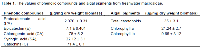
Besides, phenolic compounds detected in the samples in a gram/gram of freeze-dried algal biomass were protocatechuic acid (PA) 2.970 ± 0.312, epicatechin (E) 7.1 ± 0.401, chlorogenic acid (CA) 78 ± 5.2, syringic acid (SA), 22.12 ± 3.1 and catechins (C) 71.4 ± 6.1 μg/mg dry weight biomass. Similarly, the estimated total carotenoid content, chlorophyll a and chlorophyll b were 35 ± 3.1, 21.24 ± 2.7, and 9.66 ± 3.12 μg/mg dry weight biomass, respectively (Table 1).
DISCUSSION
The data from the present study indicated a dose-dependent antimicrobial activity of the ethanol extracts obtained from freshwater algae, Spirogyra and Oedogonium spp. against different types of bacteria, notably against S. aureus and S. xylosus. Furthermore, the results suggested that the harvesting period in terms of temperature and cell metabolism of the algae could play an important role in conferring the antibiotic effective against both Gram-positive and Gram-negative bacteria (Gacheva and Gigova, 2014) (Figure 8). The antibiotic activity of the ethanol extract of freshwater algae could be attributed to the presence of bioactive phenolics. In agreement with the present findings, several reports also revealed the potential antibiotic activity of a specific species of algae, Spirogyra spp. The studies tested the antibiotic effect of the ethanol, chloroform, petroleum ether, methanol, and acetone extracts from Spirogyra spp. against several bacteria like P. solanacearum, E. coli, and C. michiganens, showed a highly significant antibiotic activity against P. solanacearum (Naik et al., 2012; Dwaish et al., 2016; Daniel et al., 2019). Similarly, this study demonstrated a high zone of inhibition (19 mm) against Gram-positive S. aureus when the ethanol extract of Spirogyra spp. was tested. Inline, a study also found the antibacterial activity of solvent extracts of Spirulina platensis at 5.0 mg/ml against Gram-positive bacteria such as Streptococcus pyogens, S. aureus, Streptococcus epidermidis and Bacillus cereus (Usharani et al., 2015). They showed that the extract was able to show the highest mean zone of inhibition (20 ± 0.4 mm) against the Gram-positive cocci S. pyogenes, followed by S. aureus (19 ± 0.3 mm), S. epidermidis (18 ± 0.6 mm) and B. cereus (18 ± 0.2 mm). Moreover, for Gram-negative bacteria, the maximum zone of inhibition was recorded in methanol crude extract of S. platensis against Proteus mirabilis (19 ± 0.8 mm), followed by Kleibsella pneumoniae (19 ± 0.5 mm), Shigella flexneri (19 ± 0.3 mm), Salmonella Typhi (18 ± 0.6 mm). The mechanisms by which the freshwater algae as an antimicrobial agents including both macro and micro algae were depended on the function and structure of the bacteria and algae itself as an antimicrobial agent (Pradhan et al., 2014; Pina-Pérez et al., 2017). In general, the antibiotics were less effective against Gram-negative bacteria due to the rigid cell wall layers around them making them more complex than Gram-positive bacteria, hence there was difficulty of the active compounds like β-lactams, quinilons, colistins and other antibiotics to enter into bacteria thereby causing antibacterial effect (Breijyeh et al., 2020). In support, the data showed the significant antibacterial effect against Gram-positive bacteria but not in Gram-negative bacteria due to the cell wall structure and components of them and associated with the presence of short chain fatty acids, namely butanoic and methyl lactic acids (Santoyo et al., 2009). The mechanism by which the fatty acids were able to prevent the entry of the active compounds from algal extracts is still unknown, nevertheless, many findings agreed that the fatty acids and lipids were the cause of disruption of the cellular membrane (Leflaive and Ten-Hage, 2009; Al-Saif et al., 2014).
Furthermore, the potential of ethanol extract of Spirogyra spp. as an anti-viral agent against herpes simplex virus type 1 and 2 (HSV-1,2) infection was demonstrated in a recent study. The findings revealed that the ethanol and methanol extracts of Spirogyra spp. had highly significant inhibition of the viral infection on HSV-1 and HSV-2, respectively, when treated during the viral infection phase of Vero cells. The alkaloids, essential oils and terpenoids present in the freshwater macroalgae were regarded as the main active compounds responsible for anti-viral activity (Deethae et al., 2018).
Taken together, the study reiterated the significance of naturally occurring freshwater algae as a potential antibiotic against several debilitating and disease-causing bacteria. Nonetheless, it warrants further investigations to better understand the mechanisms by which they exert their antibacterial effect and could be an alternative to conventional antibiotics.
CONCLUSION
The antimicrobial activity of the ethanol extract from freshwater algae chiefly depended on the timing of harvesting of the samples as well as dose dependency against both Gram-positive and Gram-negative bacteria though the Gram-positive bacteria were more susceptible. Furthermore, the antibacterial effects due to the ethanol extract could be attributed to the significant presence of the bioactive phenolics as estimated in this study. It was envisaged that this study could pave a way for fully exploiting the freshwater algae of Sulaymaniyah for their antimicrobial properties in a cost-effective manner.
CONFLICT OF INTERESTS
The authors have not declared any conflict of interests.
ACKNOWLEDGEMENT
The authors are thankful to the Department of Medical Laboratory Science, Komar University of Science and Technology [KUST] and Komar Research Center, KUST, Sulaymaniyah, SU-46001, Kurdistan Region, Iraq for their support.
REFERENCES
|
Al-Saif SSA, Abdel-Raouf N, El-Wazanani HA, Aref IA (2014). Antibacterial substances from marine algae isolated from Jeddah coast of Red sea, Saudi Arabia. Saudi Journal of Biological Sciences 21(1):57-64. |
|
|
Breijyeh Z, Jubeh B, Karaman R (2020). Resistance of gram-negative bacteria to current antibacterial agents and approaches to resolve it. Molecules 25(6):1340. |
|
|
Çelekli A, Kap? E, Soysal Ç, Arslanargun H, Bozkurt H (2017). Evaluating biochemical response of filamentous algae integrated with different water bodies. Ecotoxicology and Environmental Safety 142:171-180. |
|
|
Daniel E, Girma T, VenkatesanJayakumar S (2019). Phytochemistry preparation of different crude extract and antimicrobial studies of Spirogyra rhizopus. Asian Journal of Pharmaceutical and Clinical Research 12(7):271-274. |
|
|
Deethae A, Peerapornpisal Y, Pekkoh J, Sangthong P, Tragoolpua Y (2018). Inhibitory effect of Spirogyra spp. algal extracts against herpes simplex virus type 1 and 2 infection. Journal of Applied Microbiology 124(6):1441-1453. |
|
|
Dwaish AS, Yousif DYM, Lefta SN (2016). Use of Spirogyra sp. extract against multi drug resistant bacterial pathogens. International Journal of Advanced Research 4(7):575-579. |
|
|
Gacheva GV, Gigova LG (2014). Biological activity of microalgae can be enhanced by manipulating the cultivation temperature and irradiance. Open Life Sciences 9(12):1168-1181. |
|
|
Hubers SA, DeSimone DC, Gersh BJ, Anavekar NS (2020). Infective Endocarditis: A Contemporary Review. Mayo Clinic Proceedings 95(5):982-997. |
|
|
Karakonstantis S, Kalemaki D (2019). Antimicrobial overuse and misuse in the community in Greece and link to antimicrobial resistance using methicillin-resistant S. aureus as an example. Journal of Infection and Public Health, 12(4):460-464. |
|
|
Kim T, Ren X, Chae KJ (2018). High-rate algal pond coupled with a matrix of Spirogyra sp. for treatment of rural streams with nutrient pollution. Journal of Environmental Management 213:297-308. |
|
|
Kogler W, Omar M, Zoltowska D, Sattiraju S (2020). Staphylococcus aureus infective endocarditis: role of transoesophageal echocardiography. BMJ Case Reports, 13(9), e236530. |
|
|
Lakhundi S, Zhang K (2018). Methicillin-Resistant Staphylococcus aureus: Molecular characterization, evolution, and epidemiology. Clinical Microbiology Reviews 31(4) e00020-18. |
|
|
Lawton RJ, de Nys R, Skinner S, Paul NA (2014). Isolation and identification of oedogonium species and strains for biomass applications. PLoS One 6;9(3):e90223. |
|
|
Leflaive J, Ten-Hage L (2009). Chemical interactions in diatoms: Role of polyunsaturated aldehydes and precursors. New Phytolologist 184(4):794-805. |
|
|
Maliak CP, Singh MB (1980). Estimation of polyphenols, In: CP. Malik, MB. Singh, (eds.). Plant enzymology and Histoenzymology. New Delhi ND: Kalyani Publishers P 286. |
|
|
Mei D, Ni M, Liang X, Hou L, Wang F, He C (2021). Filamentous green algae Spirogyra regulates methane emissions from eutrophic rivers. Environmental Science and Pollution Research International 28(3):3660-3671. |
|
|
Mesbahzadeh B, Rajaei S, Tarahomi P, Seyedinia S, Rahmani M, Rezamohamadi F, Kakar M, Moradi-Kor N (2018). Beneficial effects of Spirogyra Neglecta Extract on antioxidant and anti-inflammatory factors in streptozotocin-induced diabetic rats. Biomolecular Concepts 9(1):184-189. |
|
|
Naik AA, Hemavani C, Thippeswamy B (2012). Evaluation of antimicrobial property of Spirogyra species. International Multidisciplinary Research Journal 2(2):13-15. |
|
|
Pérez-Gutiérrez RM (2006). Isolation and identification of antibacterial compounds from Oedogonium capillare leaves. Boletín Latinoamericano y del Caribe de Plantas Medicinales y Aromáticas 5(1):15-19. |
|
|
Phadnis S, Iyer G (2016). Taxonomy of the green filamentous algae of the family Chaetophoraceae (order Chaetophorales) in Thane District, Maharashtra, India. International Journal. of Life Sciences 4(2):247-255. |
|
|
Pina-Pérez MC, Rivas A, Martínez A, Rodrigo D (2017). Antimicrobial potential of macro and microalgae against pathogenic and spoilage microorganisms in food. Food chemistry 235(11):34-44. |
|
|
Piotrowski MJ, Graham LE, Graham JM (2020). Temperate-zone cultivation of Oedogonium in municipal wastewater effluent to produce cellulose and oxygen. Journal of Industrial Microbiology and Biotechnology 47(2):251-262. |
|
|
Pradhan J, Das S, Das BK (2014). Antibacterial activity of freshwater microalgae: A review. African Journal of Pharmacy and Pharmacology 8(32):809-818. |
|
|
Ramaraj R, Tsai D, Chen PH (2013). Chlorophyll is not accurate measurement for algal biomass. Chiang Mai Journal of Science 40(4):547-555. |
|
|
Ramaraj R, Unpaprom Y, Whangchai N, Dussadee N (2015). Culture of macroalgae Spirogyra ellipsospora for long-term experiments, stock maintenance and biogas production. Life Sciences Research 1(1):38-45. |
|
|
Redfern J, Kinninmonth M, Burdass D, Verran J (2014). Using soxhlet ethanol extraction to produce and test plant material (essential oils) for their antimicrobial properties. Journal of microbiology & biology education 15(1):45-46. |
|
|
Santoyo S, Rodríguez-Meizoso I, Cifuentes A, Jaime L, Reina G, Señorans F, Ibáñez E (2009). Green processes based on the extraction with pressurized fluids to obtain potent antimicrobials from Haematococcus pluvialis microalgae. LWT- Food Science and Technology 42(7):1213-1218. |
|
|
Shannon, E, Abu-Ghannam N (2016). Antibacterial Derivatives of Marine Algae: An Overview of Pharmacological Mechanisms and Applications. Marine Drugs 14(4):81. |
|
|
Suh SS, Yang EJ, Lee SG, Youn UJ, Han SJ, Kim IC, Kim S (2017). Bioactivities of ethanol extract from the Antarctic freshwater microalga, Chloromonas sp. International Journal of Medical Sciences 14(6):560-569. |
|
|
Thomas NV, Jan GS, Klan RF (2020). Control of Opportunistic Bacteria Using Aqueous and Ethanol Extracts of Rosa damascena. International Journal of Microbiology Research, 12(2):1776-1778. |
|
|
Usharani G, Srinivasan G, Sivasakthi S, Saranraj P (2015). Antimicrobial activity of Spirulina platensis solvent extracts against pathogenic bacteria and fungi. Advances in Biological Research 9(5):292-298. |
|
|
Vilcant V, Hai O (2020). Bacterial Endocarditis. In: StatPearls, StatPearls Publishing, Treasure Island Finland. |
|
|
Wong YC, Shahirah R (2019). Effect of different solvent and ratio towards microalgae oil production by ultrasonic assisted soxhlet extraction techniques. Orient Journal of Chemistry 35(4):1377-1383. |
|
Copyright © 2024 Author(s) retain the copyright of this article.
This article is published under the terms of the Creative Commons Attribution License 4.0


