Full Length Research Paper

ABSTRACT
This study evaluated the anti-hyperglycemic potential of Stemonocoleus micranthus Harms. (Fabaceae) stem bark. Three models used in this study were: normoglycemic animal model, oral glucose tolerance test (OGTT) and alloxan-induced hyperglycemic model for acute and prolonged administration. Five (5) groups of rats (n=5) were used for all models; group 1 served as the control (received 2 ml/kg of distilled water; p.o.), groups 2, 3, and 4 received S. micranthus extract (SME) 100, 200, and 400 mg/kg, respectively, while group 5 received glibenclamide (GLI 0.2 mg/kg) as a reference drug. In the normoglycemic study, the % reduction in blood glucose concentration (BGC) was 22.24, 29.97, 30.03 and 37.28% for SME (100, 200 and 400 mg/kg) and GLI, respectively. In the OGTT study, suppression in BGC was statistically significant (p<0.05) at 120 min for the 400 mg/kg SME group. The glycemic changes (%) observed in SME (100, 200 and 400 mg/kg) treated rats were 3.4, 0.86 and 0.45%, respectively at the 120 min relative to 0 min values. Also, oral administration of SME (100, 200, 400 mg/kg) and GLI significantly (p<0.05) reduced the BGC to varying degrees in alloxan-induced hyperglycemic rats. The SME at 400 mg/kg produced the highest percentage diminution in BGC of 23.26 and 67.66% for the acute and the prolonged anti-hyperglycemic study respectively, whereas the standard drug, GLI, exhibited 73.55 and 66.10%, respectively. Histopathological studies revealed protection from the harmful effect of alloxan on the kidney and liver by SME-treatment after 28 days as against GLI treated group where there was evidence of mild hepatosis. From the results, it can be deduced that S. micranthus stem bark possesses anti-hyperglycemic effects, thus scientifically corroborating with the folkloric use.
Key words: Stemonocoleus micranthus, alloxan, glibenclamide, normoglycemia, hyperglycemia.
INTRODUCTION
The prevalence of diabetes is increasing worldwide, with the condition now recognized as one of the serious health problems affecting both developed and developing countries in the 21st century. This is evidenced by a growing awareness of the complications arising from public health issues, which has led to the prediction of about 69% increase in the total number of adults likely to be diabetics in developing countries compared to 20% for developed countries by the year 2030 (WHO, 2002; Shaw et al., 2010; Ezuruike and Prieto, 2014). Despite the use of several therapeutic approaches (such as sulfonylureas, meglitinides, biguanides and ?-glucosidase inhibitors etc.) to slow the progression of this disorder, diabetes mellitus and its associated complications continue to pose a major health challenge to people (Ravi et al., 2005; Kavishankar et al., 2011). In view of this, alternative approaches are therefore urgently needed to manage it (Osigwe et al., 2015).
In recent years, there have been renewed interests in curbing the menace of diabetes using herbal medicine possibly, due to the wide acknowledged potency of many herbal preparations. Ethnobotanical reports indicate that over 80, 000 species of higher plants have been used for a medicinal purpose globally (Duke, 1992; Jantan et al., 2015). Many of these plants have been studied experimentally to validate their antidiabetic activity with several human clinical studies indicating the beneficial effects of herbal medicinal products in the prevention and control of diabetes (Ota and Ulrih, 2017; Onyeji et al., 2017; Salehi et al., 2019).
In African traditional medicine, Stemonocoleus micranthus is used to treat various ailments. Its morphological characteristics have been described (Lemmens, 2010); and its analgesic, narrow-spectrum antibacterial, central nervous system (CNS) depressant, local anaesthetic (Anaga et al., 2010), anti-ulcer (Ezea et al., 2014), antioxidant/hepatoprotective (Mbaoji et al., 2016), hypolipidemic (Mbaoji et al., 2017), antimicrobial (Tchimene et al., 2018) and antimalarial (Orabueze et al., 2019) properties have been documented. However, to the best of our knowledge, in spite of its folkloric use in the management of diabetes mellitus, there has been no report of anti-hyperglycemic activity for the plant. The present study, therefore, investigated the effect on blood glucose concentration in normoglycemic and hyperglycemic animal models in order to verify its folkloric use.
MATERIALS AND METHODS
Plant material and extraction procedure
Fresh S. micranthus stem barks were collected from Orba in Nsukka, Nigeria. Botanical identification was confirmed by Mr A.O. Ozioko, International Centre for Ethnomedicine and Drug Development (Inter-CEDD), Nsukka, Nigeria. After careful separation of the bark from the woody parts, they were dried in a shade for 7 days and pulverized into a coarse powder using a milling machine (Lab mill, serial no. 4745, Christy and Norris Ltd, England). About 2 kg of the powdered sample was extracted with about 10 L of a mixture of methanol-dichloromethane (1:1) using a soxhlet apparatus. When extraction was completed, the resulting filtrate was concentrated under reduced pressure at a temperature of 40°C to obtain a dry S. micranthus extract (SME, 106.28 g). This was put in a clean sample bottle, well labelled and kept in the refrigerator at 4°C until further use.
Animals
Male Wistar albino rats (100-180 g; 8-12 weeks old) obtained from the Department of Pharmacology and Toxicology, University of Nigeria, Nsukka were used in this study. They were provided with standard laboratory pellets (Guinea Feeds Nigeria Limited) and water ad libitum. All animal experiments were conducted according to the institutional principles on the use of laboratory animals and, in compliance with the international guidelines for care and use of laboratory animals (Pub. No. 85 -23, revised 1985).
Normoglycemic animal study
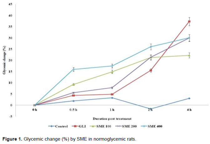
Twenty-five adult Wistar albino rats fasted for 18 h were grouped into 5 (n = 5) and treated as follows: Group 1 (control group) received 2 ml/kg of distilled water, Group 2 received the standard drug, 0.2 mg/kg glibenclamide (GLI) while Groups 3, 4 and 5 received 100, 200 and 400 mg/kg of SME respectively by gastric gavage. These treatments were done once as a single dose and blood samples collected from each of the rats through tail snipping at 0, 0.5, 1, 2 and 4 h. The blood glucose concentration (BGC) was measured using a One Touch Ultra® glucometer (Lifescan; Johnson & Johnson, Milpitas, CA, USA). Glycemic change (%) was calculated relative to 0 h values.
Oral glucose tolerance test (OGTT)
As previously described by Akunne et al. (2017), twenty-five Wistar albino rats were divided into five (5) groups (n = 5) and fasted for 18 h. The procedure, dosage of extract/standard drug/animal groupings were as described above. Furthermore, the rats were fed with glucose load (2 g/kg) and the BGC of each blood sample from the rats was measured immediately at time 0 (prior to glucose dosing) and at 30, 60, 90 and 120 min afterwards as described above. Glycemic change (%) was calculated relative to 0 min values.
Evaluation of anti-hyperglycemic activity
Induction of hyperglycemia
Hyperglycemia was induced using alloxan monohydrate (Sigma-Aldrich Co., St. Louis, MO, USA). Alloxan monohydrate was freshly prepared in ice-cold normal saline and administered at 130 mg/kg, i.p. The rats were weighed and fasted for 12 h before alloxanization, with free access to clean drinking water. After 5 days of stabilization, hyperglycemia was confirmed by determining the fasting blood glucose concentration (BGC) as described above. Animals having fasting BGC ≥ 200 mg/dL (11.1 mmol/L) were considered hyperglycemic and used for the study.
Acute anti-hyperglycemic study
In the experiment, twenty-five (25) alloxan-induced diabetic rats were grouped into five (5) (n = 5) as follows: Group 1 (diabetic control, DC) received only vehicle (2 ml/kg of distilled water; p.o.), Groups 2, 3, 4, and 5 received GLI (0.2 mg/kg), 100, 200 and 400 mg/kg of SME, respectively. The BGC was monitored at 0.5, 1, 2, 4 and 8 h respectively after administration of a single dose of the extract per oral.
Prolonged anti-hyperglycemic study
A total of twenty-five (25) fresh batches of animals were used for the study. The procedure, dosage of extract/standard drug/animal groupings were as described above. Treatment was administered by oral gavage once daily over a period of 28 days (4 weeks). The fasting BGC was monitored at weeks 1, 2, 3 and 4 respectively as described above. The percentage reductions in BGC were calculated relative to pre-treatment values using the formula:
PBGC = (BGCo - BGCT/BGCo) × 100
Where: PBGC = Percentage blood glucose concentration; BGCo = blood glucose conc. at 0 h/days; BGCT = blood glucose level at a particular hour or day.
Histopathological analysis
Histological studies were carried out on the kidney and liver of SME-treated diabetic as per the procedures described by Okoli et al. (2010) with minor modifications. Briefly, one animal from each group was sacrificed after twenty-eight days (four weeks) of extract administration and the kidneys and liver were sectioned in hematoxylin (H) and eosin (E) dyes for histological examination.
Statistical analysis
Data were analysed using One Way Analysis of Variance (ANOVA) (SPSS version 20) and the results presented as mean ± SEM. Differences between mean values were considered significant at p<0.05 (Dunnett’s Post Hoc test).
RESULTS
SME at all doses tested did not exert any significant (p<0.05) reduction in BGC of normoglycemic rats relative to the control group. Similar result was also observed with the standard (GLI, 0.2 mg/kg) at 0.5 h and 1 h, respectively (Table 1). However, at 2 and 4 h, administration of GLI produced significant (p<0.05) reduction in BGC relative to the control group. Relative to the baseline (0 h) values, the percentage reductions for SME (100, 200 400 mg/kg) were respectively 22.24, 29.97 and 30.03 % while GLI reduced the BGC by 37.28% (Figure 1) at 4 h.

Significant (p<0.05) increase in BGC of the rats was observed at 30 min of glucose load relative to 0 min values (Table 2). Administration of SME (100, 200 and 400 mg/kg) suppressed the postprandial rise in BGC throughout the experimental period. At 120 min, significant (p<0.05) reduction in BGC was observed in the 400 mg/kg treated group. The standard, GLI significantly reduced the BGC at 60, 90 and 120 min relative to the control. The glycemic changes (%) observed in SME-treated (100, 200 and 400 mg/kg) treated rats were 3.4, 0.86 and 0.45% respectively while the standard agent, GLI (0.2 mg/kg) produced the highest glycemic change of 42.11% relative to 0 min values at 120 min (Figure 2).


The result shows that the BGC of the DC (Group 1) increased from 446.21±72.45 at 0 h to 512.4±48.72 at 8 h, representing about a 15% increase. At the 8 h, SME (100 and 200 mg/kg) exhibited significant (p<0.05) reduction in BGC while the group treated with 400 mg/kg exhibited significant (p<0.05) reduction in the BGC of the rats from 0.5-8 h relative to the DC group. Similarly, GLI (0.2 mg/kg) significantly (p<0.05) reduced the BGC throughout the period of the study relative to the DC group and 0 h values (Table 3). The reduction (%) of blood glucose at 8 h was 5.62%, 20.06% and 23.26% for 100, 200 and 400 mg/kg, respectively (Figure 3).


The effects of repeated-dose administration of SME (100, 200, 400 mg/kg) and GLI (0.2 mg/kg) (Table 4) show a non-dose dependent reduction in BGC over the 28-day administration period, with all the values being lower than the basal (day 0) diabetic fasting BGC. The reduction in BGC was statistically significant (p<0.05) for the group treated with SME (100 and 200 mg/kg) and GLI (0.2 mg/kg) relative to the diabetic control (DC), and was sustained throughout the duration of the experiment. Similarly, for the group that received 400 mg/kg, the BGC was significantly (p<0.05) reduced at week 3 and week 4 respectively, relative to the control. With a value of 67.66% at week 4, the 400 mg/kg SME group exhibited the highest percentage reduction in BGC, a value which was higher than the standard, GLI (66.10%) (Figure 4).


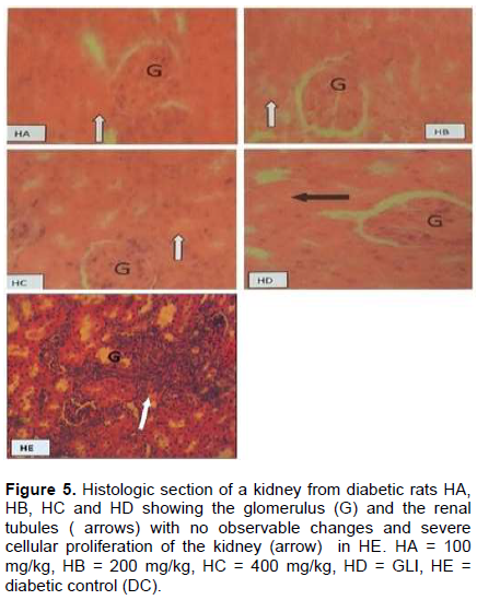
Histopathological studies revealed severe cellular proliferation of the kidney and epithelium of the bile duct in the diabetic control group. Rats treated with SME at different doses (100, 200 and 400 mg/kg) showed unaltered renal tubules of the kidney with normal glomerulus and apparently normal plates of the hepatocytes. However, a mild hepatosis (degeneration of hepatocytes) was observed in the standard control group (GLI; 0.2 mg/kg) (Figures 5 and 6).

DISCUSSION
The unprecedented increase in diabetes continues to attract wider interest in the quest for more efficient management of diabetes mellitus (Jebur et al., 2016). This study investigated the anti-hyperglycemic potential of S. micranthus Harms. (Fabaceae) stem bark using normoglycemic, oral glucose - loaded and alloxan-induced hyperglycemic rat models.
In the normoglycemic model, normal healthy animals were used in testing S. micranthus extract (SME) as a potential oral hypoglycemic agent. As a valid screening method, it tests the effect of drugs in animals with an intact pancreatic activity (Williamson et al., 2016). Since the SME at all doses tested did not exert any significant blood glucose reduction relative to the control group, it implies that SME, unlike the insulinotropic agent, glibenclamide used in this study does not precipitate hypoglycemia in non-diabetic animals. The oral glucose tolerance test (OGTT) is a research model used to verify the anti-hyperglycemic actions of medicinal drugs (Ernsberger and Koletsky, 2015). As SME suppressed the rise in BGC following a glucose load, it suggests that the extract may be effective in controlling the overt postprandial rise in blood glucose that increases the risk of chronic hyperglycemia in diabetes. This may be as a result of the reduced rate at which glucose is absorbed from the intestine; increased peripheral glucose utilization through a variety of metabolic pathways (Patel et al., 2011); or a possible incretin mimetic effect. Thus, postprandial hyperglycemia control in diabetes is considered beneficial in reducing the risk of micro and macrovascular complications (Balkau, 2000; Ceriello, 2000). Alloxan-induced hyperglycemia is widely accepted as a screening method for the study of antidiabetic agents (Etuk, 2010; Okoye et al., 2012). Alloxan monohydrate induces diabetes by selectively destroying the β-cells of the islets of Langerhans due to its selective accumulation through the glucose transporter 2 (GLUT2) and hence, minimizes the release of insulin and glucose uptake by peripheral tissues (Tafesse et al., 2017). It has been reported that insulin deficiency in animals, leads to the development of various metabolic aberrations (Gupta and Sharma, 2017). The observed increase in BGC corroborates with previous reports documenting elevated BGC in alloxan-induced hyperglycemic rats (Omonije et al., 2019). Treatment with SME even at the lowest dose of 100 mg/kg caused a marked reduction of BGC in the acute and prolonged study, an indication of higher potency and a possible non-pancreatic mechanism of anti-hyperglycemic action (Patel et al., 2011; Saxena and Argal, 2018).
The reduction in BGC correlates with that of the normoglycemic model, which showed that SME does not precipitate hypoglycemia in diabetic animals, an indication of its anti-hyperglycemic actions. The anti-hyperglycemic action of medicinal plants has been attributed to involving increase in peripheral glucose utilization; increase hepatic glycogen synthesis or decrease in glycogenolysis and gluconeogenesis; intestinal inhibition of glucose absorption and attenuation of glycemic response to carbohydrates (Andrade-Cetto and Wiedenfeld, 2004; Bnouham et al., 2006). In this context, SME may have acted via these mechanisms in maintaining glucose homeostasis. Several studies on medicinal plants have also reported similar anti-hyperglycemic mechanisms (Nyunaï et al., 2015; Ramu et al., 2016; Kumbhare and Sivakumar, 2019).
Data from scientific reports show that medicinal plants contain a large variety of bioactive constituents such as glycosides, alkaloids, terpenoids, flavonoids, carotenoids, etc., that are frequently implicated as having anti-hyperglycemic activity (Osadebe et al., 2014). S. micranthus Harms. (Fabaceae) stem bark extract has been reported to contain flavonoids, tannins, saponins and alkaloids (Ezea et al., 2014; Mbaoji et al., 2016). It has been reported that alkaloids have antidiabetic activity on alloxan-induced diabetic mice (Sharma et al., 2009). Flavonoids are known for their antidiabetic activity and hence they help to suppress glucose levels; decrease plasma cholesterol and triglyceride significantly, and increase hepatic glucokinase activity probably by stimulating release of insulin from pancreatic islets (Hossain et al., 2016; AL-Ishaq et al., 2019). Tannins are a very large group of plant-derived polyphenolic compounds that are known to exhibit antidiabetic effects mainly by inhibiting the activation of α-amylase and α-glycosidase activities (Kunyanga et al., 2011). Saponins have been shown to modulate insulin secretion and decrease glucose levels in alloxanized rats (Elekofehinti et al., 2013). It is suggested that these chemical constituents may be responsible for the anti-hyperglycemic activity either alone or in synergy with one another. This is in agreement with previous studies on other plants (Birru et al., 2015; Tomar et al., 2016, Akunne et al., 2017).
The severe cellular proliferation of the kidney and epithelium of the bile duct in the diabetic control rats reflects the pathological changes associated with chronic effects of alloxan-induced hyperglycemia (Lucchesi et al., 2015). The mild hepatosis (degeneration of hepatocytes) of the liver observed in the standard group may probably be due to post-mortem degeneration as studies have shown that glibenclamide can prevent the liver damage caused by severe hyperglycemia (Jelizaveta et al., 2012). Therefore, this damage could not have been as a result of the drug (glibenclamide). However, diabetic rats treated with SME (100, 200 and 400 mg/kg) showed unaltered renal tubules of the kidney with normal glomerulus and apparently normal plates of the hepatocytes. Previous study has reported no significant morphological or pathological changes in the kidney and liver tissues of rats treated with various doses of SME compared with the control group (Mbaoji et al., 2017). The organs had normal cellular architecture even in animals that received higher doses of the extract. This suggests that SME is not active or harmful and as such may have equally protected the liver and kidneys of the animals from the toxicity of alloxan which is known to cause organ damage by oxidative radical generation (Jebur et al., 2016). Consequent upon this histopathological investigation, the SME is considered safe and this, however, suggests that the active principles in the extract could be nephro or hepatoprotective in function. This is in tandem with the previous work on the antioxidant/hepatoprotective effects of SME as well as the LD50 which was estimated to be greater than 5000 mg/kg body weight (Mbaoji et al., 2016).
Based on the above investigation, S. micranthus stem bark extract had a pronounced effect in controlling hyperglycemic conditions without causing overt hypoglycemia, thus exhibiting anti-hyperglycemic activity. It may as well provide additional protection against hyperglycemic-related complications. This gives scientific evidence to the folkloric claims of the plant in the management of diabetes. Further research on the isolation and characterization of the active phytoconstituents is ongoing.
CONFLICT OF INTERESTS
The authors have not declared any conflict of interests.
REFERENCES
|
Akunne TC, Obi BC, Ofokansi MN, Nwonu PC, Okoli CO (2017). Antidiabetic activity and toxicological evaluation of the methanol-dichloromethane root bark extract of Nauclea diderrichii(De Wild) Merr. International Journal of Pharmacy Pharmaceutical Sciences 9(9):279-283. |
|
|
Al-Ishaq RK, Abotaleb M, Kubatka P, Kajo K, Büsselberg D (2019). Flavonoids and their anti-diabetic effects: cellular mechanisms and effects to improve blood sugar levels. Biomolecules 9(9):430. |
|
|
Anaga AO, Chah KF, Tchimene MK, Udeani IC, Anaele N, Akpa P, et al (2010). Investigation of the methanol extract of Stemonocoleus micranthus for pharmacological activities. Nigerian Journal of Experimental and Applied Biology 11(1):97-107. |
|
|
Andrade-Cetto A, Wiedenfeld H (2004). Hypoglycemic effect of Acosmium panamense bark on streptozotocin-diabetic rats. Journal of Ethnopharmacology 90(2-3):217-220. |
|
|
Balkau B (2000). The DECODE study, Diabetes epidemiology: collaborative analysis of diagnostic criteria in Europe. Diabetes and Metabolism 26(4):282-286. |
|
|
Birru EM, Abdel-Wahab M, Shewamene Z (2015). Effect of hydroalcoholic leave extract of Indigofera spicata Forssk on blood glucose level of normal, glucose loaded and diabetic rodents. BMC Complementary and Alternative Medicine 15(321):1-8. |
|
|
Bnouham M, Ziyyat A, Mekhfi H, Tahri A, Legssyer A (2006). Medicinal plants with potential antidiabetic activity-a review of ten years of herbal medicine research (1990-2000). International Journal of Diabetes Metabolism14(1):1-25. |
|
|
Ceriello A (2000). Postprandial hyperglycemia and diabetes complications: is it time to treat? Diabetes 54(1):1-7. |
|
|
Duke JA (1992). Handbook of biologically active phytochemicals and their activities. Boca Raton, Florida: CRC Press P 183. |
|
|
Elekofehinti OO, Kamdem JP, Kade IJ, Rocha JBT, Adanlawo IG (2013). Hypoglycemic anti-peroxidative and antihyperlipidemic effects of saponins from Solanum anguivi Lam. fruits in alloxan-induced diabetic rats. South African Journal Botany 88:56-61. |
|
|
Ernsberger P, Koletsky RJ (2015). The glucose tolerance test as a laboratory tool with clinical implications. In: Chackrewarthy S (Eds), Glucose tolerance. Croatia: InTech pp. 3-14. |
|
|
Etuk EU (2010). Animals models for studying diabetes mellitus. Agriculture and Biology Journal of North America 1(2):130-134. |
|
|
Ezea CC, Nnamdi-Okenwa CT, Ezugwu CO, Anowi FC, Ezea SC (2014). Investigation into the anti-ulcer activity of the methanol extract of Stemonocoleus micranthus Harms, bark (Leguminosae). World Journal of Pharmaceutical Research 3(3):3669-3675. |
|
|
Ezuruike UF, Prieto JM (2014). The use of plants in the traditional management of diabetes in Nigeria: Pharmacological and toxicological considerations. Journal of Ethnopharmacology 155(2):857-924. |
|
|
Gupta R, Sharma AK (2017) Anti-hyperglycemic activity of aqueous extracts of some medicinal plants on Wistar rats. Journal of Diabetes Metabolism 8(7):1-6. |
|
|
Hossain MK, Dayem AA, Han J, Yin Y, Kim K, Saha SK, Yang G, Choi HY, Cho SG (2016). Molecular mechanisms of the anti-obesity and anti-diabetic properties of flavonoids. International Journal of Molecular Sciences 17(4):569. |
|
|
Jantan I, Bukhari SNA, Mohamed MAS, Wai LK, Mesaik MA (2015). The evolving role of natural products from the tropical rainforests as a replenishable source of new drug leads. In: Omboon V, Suleiman O (eds.) Drug discovery and development from molecules to medicine. InTech pp. 3-27. |
|
|
Jebur AB, Mokhamer MH, El-Demerdash FM (2016). A review on oxidative stress and the role of antioxidants in diabetes mellitus. Austin Endocrinology and Diabetes Case Reports 1(1):1006. |
|
|
Jelizaveta S, Sergejs I, Olga S, Jelena S, Natalia P (2012). Comparison of the effects of glibenclamide on metabolic parameters, GLUT1 expression, and liver in rats with severe and mild streptozocin-induced. Medicina (Kaunas) 48(10):532-543. |
|
|
Kavishankar GB, Lakshanidevi N, Marthy SM (2011). Diabetes and medicinal plants - A review. International Journal of Pharmacy and Biomedical Sciences 2(3):65-80 |
|
|
Kumbhare MR, Sivakumar T (2019). Caesalpinia pulcherrima extracts on blood glucose in normal and alloxan monohydrate-induced diabetic rats. Journal of Biological Sciences19(1):34-39. |
|
|
Kunyanga CN, Imungi JK, Okoth M, Momanyi C, Biesalski HK, Vadivel V (2011). Antioxidant and antidiabetic properties of condensed tannins in acetonic extract of selected raw and processed indigenous food ingredients from Kenya. Journal of Food Science 76(4):C560-C567. |
|
|
Lemmens RHMJ (2010). Stemonocoleus micranthus Harms. In: Lemmens RHMJ, Louppe D, Oteng, AAA (eds.) PROTA (Plant Resources of Tropical Africa/Resources Vegetables de L' Afrique Tropicale), Netherlands: Wageningen <http:// database. Prota. Org/search.htm. |
|
|
Lucchesi AN, Cassettari LL, Spadella CT (2015). Alloxan-induced diabetes causes morphological and ultrastructural changes in rat liver that resemble the natural history of chronic fatty liver disease in humans. Journal of Diabetes Research vol. 2015, Article ID 494578, 11 pages. |
|
|
Mbaoji FN, Ezike AC, Nworu CS, Onyeto CA, Nwabunike IA, Okoli IC, Akah PA (2016). Antioxidant and hepatoprotective potentials of Stemonocoleus micranthusstem bark Harms (Fabaceae) extract. International Journal of Pharmacy and Pharmaceutical Sciences 8(7):47-51. |
|
|
Mbaoji FN, Ezike AC, Nworu CS, Onyeto CA, Obi BC, Akunne TC, Okoli AC, Okpalaeke CC, Akah PA (2017). Sub-chronic effect of methanol-dichloromethane stem bark extract of Stemonocoleus micranthus Harms. (Fabaceae) on lipid profile and histology of liver and kidney of rats. Journal of Pharmacognosy and Natural Products 3(2):1-6. |
|
|
Nyunaï N, Abdennebi EH, Bickii J, Manguelle-Dicoum MA (2015). Subacute antidiabetic properties of Ageratum conyzoides leaves in diabetics rats. International Journal of Pharmaceutical Sciences and Research 6(4):1378-1387. |
|
|
Okoli CO, Ibiam AF, Ezike AC, Akah PA, Okoye TC (2010). Evaluation of antidiabetic potentials of Phyllanthus niruri in alloxan-induced diabetic rats. African Journal of Biotechnology 9(2):248-259. |
|
|
Okoye TC, Akah PA, Ilogu CL, Ezike AC,Onyeto CA (2012). Anti-diabetic effects of methanol extract of the seeds of Buchholzia coriacea and its synergistic effects with metformin. Asian Journal of Biomedical and Pharmaceutical Sciences 2(12):32-36. |
|
|
Omonije OO, Saidu AN, Muhammad HL (2019). Anti-diabetic activities of Chromolaena odorata methanol root extract and its attenuation effect on diabetic induced hepatorenal impairments in rats. Clinical Phytoscience 5(1):1-10. |
|
|
Onyeji CO, Igbinoba SI, Olayiwola G (2017). Therapeutic potentials and cytochrome p450-mediated interactions involving herbal products indicated for diabetes mellitus. Drug Metabolism Letters11 (2):74-85. |
|
|
Orabueze CI, Ota DA, Coker HA (2019). Antimalarial potentials of Stemonocoleus micranthus Harms (Leguminosae) stem bark in Plasmodium berghei infected mice. Journal of Traditional and Complementary Medicine, |
|
|
Osadebe PO, Uzor PF, Omeje EO, Agbo MO, Obonga WO (2014). Hypoglycemic activity of the extract and fractions of Anthocleista vogelii (Planch) stem bark. Tropical Journal of Pharmaceutical Research13(9):1437-1443. |
|
|
Osigwe CC, Akah PA, Nworu CS, Okoye TC, Tchimene MK (2015). Antihyperglycemic studies on the leaf extract and active fractions of Newbouldia laevis (Bignoniaceae). Pharmacology and Pharmacy 6(11):518-532. |
|
|
Ota A, Ulrih NP (2017). An overview of herbal products and secondary metabolites used for management of type two diabetes. Frontiers in Pharmacology 8:1-14. |
|
|
Patel SK, Kumar R, Laloo D, Hemalatha S (2011). Evaluation of phytochemical and antioxidant activities of the different fractions of Hybanthus enneaspermu. Asian Pacific Journal of Tropical Medicine 4(5):391-396. |
|
|
Ramu R, Shirahatti PS, Nayakavadi S, Vadivelan R, Zameer F, Dhananjaya BL, Nagendra Prasad MN (2016). Effect of a plant extract enriched in stigmasterol and β- sitosterol on glycaemic status and glucose metabolism in alloxan-induced diabetic rats. Food and Function 7(9):3999-4011. |
|
|
Ravi K, Rajasekaran S, Subramanian S (2005). Antihyperlipidemic effect of Eugenia jambolana seed kernel on streptozotocin-induced diabetes in rats. Food and Chemical Toxicology 43(9):1433-1439. |
|
|
Salehi B, Ata A, Anil Kumar NV, Sharopov F, Ramírez-Alarcón K, Ruiz-Ortega A (2019). Antidiabetic potential of medicinal plants and their active components. Biomolecules 9(551):1-111. |
|
|
Saxena N, Argal A (2018). Evaluation of antidiabetic activity of a herbal formulation. EC Pharmacology and Toxicology 65:349-355. |
|
|
Shadli S, Alam M, Haque A, Rokeya B, Ali L (2014). Antihyperglycemic effect of Zingiber officinale Roscoe bark in streptozotocin-induced type 2 diabetic model rats. International Journal of Pharmacy and Pharmaceutical Sciences 6(1):711-716. |
|
|
Sharma B, Salunke R, Balomajumder C, Daniel S, Roy P (2009). Anti-diabetic potential of alkaloid-rich fraction from Capparis deciduas on diabetic mice. Journal of Ethnopharmacology 127(2):457-462. |
|
|
Shaw JE, Sicree RA, Zimmet PZ (2010). Global estimates of the prevalence of diabetes for 2010 and 2030. Diabetes Research and Clinical Practice 87(1):4-14. |
|
|
Tafesse TB, Hymete A, Mekonnen Y, Tadesse M (2017). Antidiabetic activity and phytochemical screening of extracts of the leaves of Ajuga remota Benth on alloxan-induced diabetic mice. BMC Complementary and Alternative Medicine 17(1):1-9. |
|
|
Tchimene MK, Chah KF, Anaele N, Okunji CO, Iwu MM (2018). Antimicrobial activity of crude extract, fractions and isolated compound from Stemonocoleus micranthus. International Journal of Microbiological Research 9(3):70-72. |
|
|
Tomar V,Tonpay SD, Shashi A (2016). Effect of ethanolic extract of leaves of Passiflora insarnata Linn in streptozotocin-induced diabetes in rats. International Journal of Pharmaceutical Sciences and Research 7(10):4246-4252. |
|
|
Williamson EM, Okpoko DT, Evans FJ (2016). Pharmacological Methods. In: Williamson EM. Phytotherapy Research. USA: John Wiley and SonsInc, pp. 155-167. |
|
|
World Health Organization (WHO) (2002). WHO launches the first global strategy on traditional and alternative medicine, press release. Geneva; WHO/38/16. |
|
Copyright © 2024 Author(s) retain the copyright of this article.
This article is published under the terms of the Creative Commons Attribution License 4.0


