Full Length Research Paper

ABSTRACT
The toxicity and effects of Ficus carica leaf aqueous extract on the haematology and some biochemical parameters were investigated in wistar albino rats for 4 weeks. Various doses (100, 200 and 400 mg/kg body weight) of the extract were administered orally to the rats. The haematological parameters were determined using a Neubauer-type hemocytometer with Toisson’s solution as the diluting fluid for red blood cell (RBC) and Turk’s solution for white blood cell (WBC). The biochemical parameters and glucose level were estimated using O-toluidine method. The acute toxicity LD50 could not be established from the range of 50 to 6000 mg/kg doses of the extract. The phytochemical analysis showed the presence of flavonoid, tannins, cardiac glycosides, steroids, and saponins, in plant with flavonoid and tannins in high and moderate abundance. Oral LD50 was therefore not determined because mortality was not observed. The packed cell volume (PCV), RBC and hemoglobin (Hb) were significantly increased with prolonged administration of the extract. There was dose and time dependent significant decrease (p < 0.05) in neutrophils when compared with the control in the experiment. The results indicated dose-related decreases in serum activities of alanine aminotransferase (ALT) and aspartate aminotransferase (AST) in the test groups (9.88±3.14b to 24.67±6.43a and 19.00±3.00a to 99.33±25.32a U/L), respectively when compared with those of the ALT and AST control 15.03±0.04a to 39.67±0.58b and 30.33±0.58d to 126.0±5.29a, respectively. F. carica aqueous extract has hepatoprotective capability and could be used at an average and prescribed dosage in consumption and drug production for enhancement of erythrocytes and haemoglobin concentration.
Key words: Ficus carica, biochemical indices, haematology, albino rats, phytochemical screening.
INTRODUCTION
Plants have been the major source of drugs for medicine in most developing and established countries in the world (Obadoni and Ochuko, 2001). The use of plant extracts and phyto-chemicals, both with its known toxicity effects on haematological and biochemical parameters have been identified by the World Health Organization as alternative sources of therapeutic treatments (Ghasemi, 2014). Medicinal plants therefore remain significant as therapeutic remedies in many developing countries (Sibel et al., 2005). The beneficial medicinal effects of plant materials typically result from the combinations of their secondary metabolites; their medicinal actions are unique to particular plant species or groups of distinct taxonomy (Wink, 1999).
For many centuries, humans relied on medicinal plants to cure various diseases (Ramaa et al., 2006). Most plant parts such as the leaves and roots have been used for curative, antipyretic, purgative and treatment of other diseases. The acceptance of herbal remedy is gradually increasing worldwide (Inoue et al., 2011).
There is also growing concern about the toxicity and adverse effects of several hundreds of dietary herbs/vegetables which have remained either uninvestigated or poorly investigated and are increasingly used by patients with different diseases (Ghasemi, 2014). Therefore, in contradiction to their usefulness, some herbal remedies used in treatment of some diseases (liver diseases inclusive) may as well induce some toxic effects on the patient (Ghasemi, 2014). This specific toxic component of the plant most times is not known because of difficulty in proper analysis of the plant material especially if unsuspected (Ekpenyong et al., 2012).
According to several reports, other factors such as active and toxic components overdose/abuse, prolonged use, and allergies can also be attributed to the major causes of hepatotoxicity and other forms of toxicity. Apart from the safety and the acclaimed therapeutic efficacy, other reasons for the recent surge in consumption of herbal remedies included easy accessibility, more acceptability from cultural and spiritual perspectives and inadequacy of primary health care services. The importance of herbs and plant leaves remain highly significant in medicine. Thus, proper research on leaves and herbs used in herbal and normal medicine is important to check the adversity and usefulness of the leaves and herbs (Ekpenyong et al., 2012).
Plant-derived drugs thus served as a prototype to develop more effective and less toxic medicines. Ficus carica is one of the medicinally important plants that belong to the mulberry tree (Moraceae). The Moraceae leaves are well known because of its high antioxidant character but the actual active constituents responsible for this have not been determined despite several experiments. It is used in the traditional system of medicine for healing various diseases (Guarrera, 2003). It has been reported that the leaves are used in traditional medicine as laxatives, stimulant, against throat diseases, emollient and haemorrhoid treatment (Sibel et al., 2005).
The importance of blood in maintaining good health cannot be overstated. The Chinese described blood as the ‘mother of energy’ in the sense that it provides the basic materials and fluid substances that are required to nourish the life essence of our being; thus, blood is represented as a receptacle for sustaining life energy (Sheng, 2003). In addition, because the lifespan of blood cells is relatively short, the blood needs to be constantly replenished (Sheng, 2003; Nebedum et al., 2010).
In South Eastern part of Nigeria, the leaves of F. carica among others are considered as excellent natural herbal blood boosters used especially for debilitating conditions, acute blood loss and blood deficiency diseases (Ahmad et al., 2013). Aqueous extract of F. carica leaves were further found to induce a significant hypoglycemic effects in rats but the mechanism involved in this effect was not elucidated (Sibel et al., 2005).
Most research works carried out on F. carica have focused on its hypoglycemic, antioxidant and other therapeutic effects. Research works on the F. carica toxicity effects on haematology and liver enzymes (biochemical parameters) are scanty thus signifying the importance of this study. The objective of this study was to determine the toxicity of F. carica leaf aqueous extract and its effects on the haematology and some biochemical parameters (liver enzymes) on albino rats (Rattus norvegicus).
MATERIALS AND METHODS
Plant
F. carica leaves were collected from International Centre for Ethno-medicine and Drug Preparation in Nsukka, Enugu State, Nigeria. The leaves were identified and authenticated by Dr Nzekwe at the Department of Plant Science and Biotechnology, University of Nigeria. A voucher number UNN-EGACC, protocol no. 0330/2013 specimen was deposited in the herbarium.
Preparation of crude extract
F. carica leaves were thoroughly washed, shade-dried for 7 days and ground in an electric blender, Moulinex Master Chef Delicio (Type DFB1). Each crude extract was filtered through Whatman No. 3 filter paper and the filtrate concentrated in vacuo using a rotor evaporator for solvent evaporation. Dried extract was reconstituted in 10 ml dimethyl sulphoxide (DMSO) prior to use. The F. carica leaves extracts were prepared according to the method described by Nebedum et al. (2010). The homogenate was suspended in absolute ethanol for extraction, and then centrifuged, filtered through Whatman No. 1 filter paper and further re-centrifuged through Whatman No. 3. This was then rotor-evaporated to remove the ethanol before suspending in DMSO. The final concentration of F. carica extract in DMSO was determined by subtracting the weight of insoluble material from the weight of original sample to give 64% (w/v) of extract. The filtrate was then kept at -70°C until used.
Phytochemical screening
Qualitative phytochemical analysis of plant extracts was carried out according to the method described by Trease and Evans (1978) as the following.
Flavonoids
Lead acetate test: briefly, 2 .0 ml of each ethanolic extract was added 10% lead acetate solution and observed for a coloured precipitate indicative of flavonoids.
Tannin
To 2.0 ml of distilled water was added 0.1 g of each extract, boiled for 2 min, filtered and allowed to cool. To this was then dispensed 5% ferric chloride (FeCl3) solution drop-wise and observed for colour change.
Saponins
The presence of saponins was assayed by the frothing test. Approximately, 5 mg of extract was added into 5 ml of distilled water, shaken vigorously and observed for persistent frothing.
Alkaloid
Approximately 2.0 ml of each ethanolic extract was added to 5 ml of 2% hydrochloric acid and allowed to steam in a water bath for 20 min, and then filtered. To 1 ml of each filtrate was dispensed 2 drops of Dragendroff’s reagent (bismuth potassium iodide solution) and observed for precipitation.
Steroids
The Salkowski’s test was used in testing for steroids. Approximately, 5 ml of each ethanol extract was evaporated to dryness and the residue re-dissolved in 5.0 ml of anhydrous chloroform and then filtered. To each filtrate in a 10 ml test tube was carefully dispensed 2.0 ml of concentrated sulphuric acid until a bi-layer was formed. The interface was then observed for a reddish brown coloration indicative of steroid. About 2 ml of each extract was further dried by evaporation, and to this was dispensed 2 ml of anhydrous chloroform and filtered. To a 2 ml filtrate was simultaneously added 2 ml acetic anhydride and 2 ml concentrated H2SO4 and carefully observed for the appearance of a blue-green ring indicating the presence of terpenoids.
Cardiac glycosides
The Keller-Kiliani test was adopted; to a 2 ml filtrate was dispensed 1 ml glacial acetic acid, FeCl3 and concentrated H2SO4, and observed for green-blue coloration indicative of cardiac glycosides.
Experimental animals
A total of 180 wistar healthy albino rats of both sexes weighing 100 and 185 g used in this research work were obtained from the animal house of Department of Zoology, University of Nigeria, Nsukka. The animals were acclimatized for 2 weeks in their respective cages. The cages were kept in a well-ventilated and conducive room in the animal house inside the zoological garden. The animal were housed within the facility and maintained on grower’s vital feeds and water ad libitum. Care and handling of the albino rats were done in accordance to the protocols described by the Animal Care Committee (UNN-EGACC) of University of Nigeria, Nsukka.
Toxicity and lethality (LD50) test
The oral toxicity study was carried out using fixed dose method. The LD50 of aqueous extracts of the plant was determined using a total of 132 wistar rats of both sexes. The rats were fasted for 12 h and divided into the first groups of 2, 3, 4 and 5 which received an administration of 50, 100, 500 and 1000 mg/kg body weight of the extract, while in an another first phase of 2, 3, 4 and 5 groups were administered with 2000, 3000, 4000 and 5000 mg/kg body weight, respectively for 24 h. In another second phase that was observed for 48 h, the dosages were increased to 5500, 5750 and 6000 mg/kg. The rats were closely observed for toxic symptoms and behavioural changes for the first 2 h after extract administration. The LD50 was calculated as the geometric mean of the dose that resulted in 50% mortality (Shrivastava, 2012).
Experimental design
The wistar rats were divided into four groups and each group had 3 replicates of 4 rats: Group 0: control received distilled water orally only; Group 1: received 100 mg/kg of body weight of the extract; Group 2: were administered with 200 mg/kg body weight; and finally Group 3: were fed with 400 mg/kg body weight.
The animals were housed within the facility and maintained on grower’s vital feeds and water ad libitum. The administration of this extract lasted for four weeks while haematological and biochemical studies were carried out weekly for 4 weeks of the experiment as well.
Estimation of haematological parameters
The haematological parameters were determined using a Neubauer-type hemocytometer with Toisson’s solution as the diluting fluid for red blood cell (RBC) and Turk’s solution for white blood cell (WBC) (Saravanan et al., 2012). The haemoglobin level of blood was estimated following the cyanmethaemoglobin method with some modifications. Each blood sample (0.02 ml) was mixed with 4 ml Drabkin’s solution and allowed to stand for 10 min for proper colour development after which absorbance was read at 540 nm in Unican spectrophotometer against a blank. Haematocrit was analyzed by centrifugation of the blood using a microhaematocrit centrifuge (Hawkesley and sons, Ltd., Lancing, UK) at room temperature. The haematocrit was read after centrifugation using the micro haematocrit reader and the result expressed as the percentage of the whole blood. Haematological parameters such as MCHC, MCH and MCV were calculated according to Dacie and Lewis (2001):
MCHC (g/dl) = Hb (g/dl) PCV (%) × 100
MCH (pg/cell) = Hb (g/dl) × 10 RBC count in millions/mm3
MCV (fl/cell) = PCV (%) × 10 RBC count in millions/mm3
where MCHC is the mean corpuslar haemoglobin concentration, MCH is the mean corpulsar haemoglobin, PCV is the packed cell volume, Hb is the haemoglobin concentration, RBC is the red blood cell, pg is the pictograms, and fl is the femtolitres.
Estimation of biochemical parameters and glucose level
These were estimated using O-toluidine method (Sadauskas et al., 2011). Blood samples were collected from the eyes using haematocrit tubes by carefully inserting the tube into the median cantus to puncture the vein thus enabling the flow of blood into plain tubes (no EDTA anticoagulant) and these were allowed to clot and the serum separated by centrifugation. The serum collected is then subjected to biochemical assays. This was carried out according to the protocols described by Sadauskas et al. (2011) which is internationally accepted.
The following liver biochemical parameters analyzed were aspartate aminotransferase (AST), alanine aminotransferase (ALT) and alkaline phosphatase (ALP) from the serum obtained from the blood sample. The ALT was determined by monitoring the concentration of pyruvate hydrazone formed with 2, 4-dinitrophenyl hydrazine, while AST was determined using concentrations of oxaloacetate hydrazone formed with 2, 4-dinitrophenyl hydrazine. ALP was determined by the method described by Englehardt (Sadauskas et al., 2011).
Glucose level was estimated using Otoluidine method (Nwani et al., 2014), while total protein was estimated spectrophotometrically using bovine serum albumin as a standard following the methods (Sadauskas et al., 2011).
Statistical analysis
All values were expressed as mean ± standard deviation (SD). The differences were compared using analysis of variance (ANOVA) followed by Dennett’s test. P values< 0.05 were considered as significant.
RESULTS
Phytochemical screening
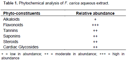
The flavonoid contents were high in abundance (+++), while other constituents such as the cardiac glycosides, steroids, saponins, and tannins were lower in abundance (++). Similarly, the alkaloids were the lowest (+) as elucidated in Table 1.
Toxicity studies
The acute toxicity LD50 could not be established from the range of 50 to 6000 mg/kg body weight doses of the extract. There was no mortality recorded when all the doses of the extract were administered orally to the rats. The rats showed negative behavioural changes at 5000,5500,57250 and 6000 mg/kg dosages. Oral LD50 was therefore not determined, because mortality was not observed. The LD50 obtained was higher than 6000 mg/kg by implication.
Haematological and morphological parameters
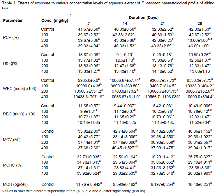
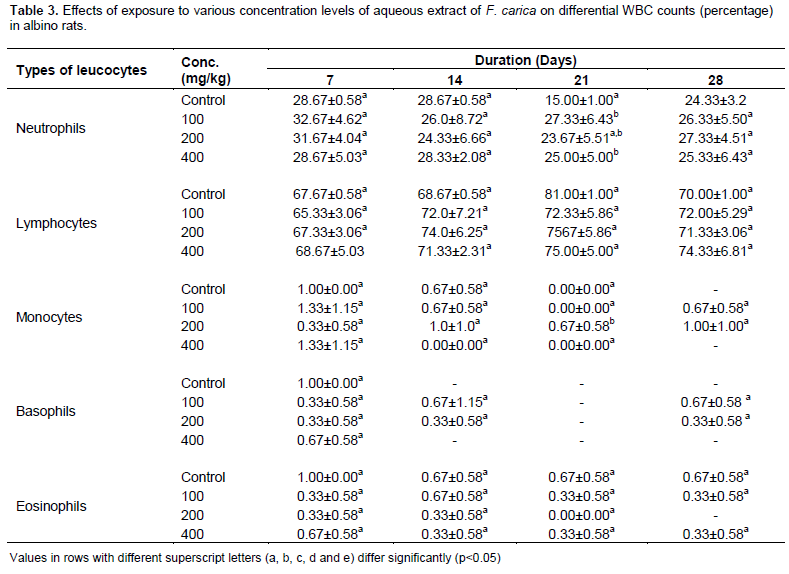
Results of the haematological parameters of the control and experimental groups are presented in Table 2. The RBCs count and haemoglobin in the experimental groups was significantly different from the control (p< 0.05) throughout the duration of the experiment and was significantly increased (p<0.05). There was significant difference in PCV values between the control and treated rat on day 7 which subsequently increased significantly (p<0.05) from day 14 of exposure. F. carica induced dose and time dependent significant increase in WBC count from day 14 onward (p<0.05) were observed, while values of blood parameters (MCV, MCH and MCHC) in the experimental rat were not significantly different (p < 0.05) from the control group throughout the duration of the experiment. Changes in the mean values of the leukocyte differentials are presented in Table 3. There was dose and time dependent significant decrease (p < 0.05) in the levels of neutrophils when compared with the control throughout the experimental duration. The lymphocyte levels were significantly elevated (p< 0.05) from day 7 onward, but the values of the monocytes, basophils and eosinophils were not significantly different (p<0.05) from the control.
Biochemical parameters
The activities of the enzymes assayed as well as the concentrations of other biochemical parameters are shown in Table 4. The results indicated dose-related decreases in serum activities of ALT and AST in the test groups (9.88±3.14b to 24.67±6.43a and 19.00±3.00a to 99.33±25.32a U/L), respectively when compared with those of the ALT and AST control 15.03±0.04a to 39.67±0.58b and 30.33±0.58d to 126.0±5.29a, respectively.

However, the groups fed 200 mg kg-1 body weight and 400 mg kg-1 body weight extracts had significantly lower ALT activities of 22.67±9.87a and 20.67±14.47ab U/L, respectively relative to those of the control 39.67±0.58b and 15.03±0.04a. There were mild variations in the ALP activities, 73.60±31.87a to 305.6±24.66a U/L of the experimental rats when compared with the ALP control of 38.07±0.16b to 332.1±0.90a as were also variations in the ALT/AST ratios of the experimental animals (0.22±1.557c to 0.55±0.445a) when compared with the control of 0.12±0.07c to 0.56±0.051a, respectively.
There were mild alterations in the serum concentrations of total proteins (85.08±0.09a to 92.00±5.0a gL-1). There were very slight dose-dependent decreases (p>0.05) in the serum glucose concentrations of the test rats (3.01±0.07a to 3.58±0.154.68a mmoL-1) when compared with those of control rats 4.69±0.90a to 4.90±0.07a mmoL-1.
DISCUSSION
Phytochemical screening
Preliminary phytochemical investigations revealed the presence of alkaloids, saponins, tannins, and cardiac glycosides similar to the findings of such extract of F. carica (Janardhanan, 2000). Some of the biological functions of flavonoids include protection against allergies, free radicals, platelet aggregation of microorganisms, ulcer, hepatotoxins and tumours (Okwu, 2004; Nebedum et al., 2010). Saponins on the hand have the properties of precipitation of proteins, cholesterol-binding, and haemolysis (Fatemi et al., 2007). Other phyto components such as alkaloids and glycosides found in this plant also did not have properties relating to increased haemapotesis. Furthermore, the behavioural components observed during exposures to the extract in this investigation were dose-dependent somnolence and sedation which could easily be likened to side effects of the active constituents (flavonoid and saponins) of the extract (Ekpenyong et al., 2012). However, studies are needed to determine the effect of the extract on humans since it is popularly used in folk medicine for seizure control, level of consciousness and blood pressure.
Haematological parameters
Time-dependent increases in haemoglobin red blood cells and PCV imply that the extracts may enhance the populations of red blood cells produced from the bone marrow, as well as increase the oxygen-carrying capacityof the whole blood because of the increased number of red blood cells in the blood (Fatemi et al., 2007).
From the results obtained during the experiment, the PCV and the Hb were observed to have increased significantly. This indicated that the F. carica aqueous extract is not toxic because decrease in PCV and Hb concentration shows toxicity to the red blood cells. The RBC count was observed to have increased significantly from the second week of extract administration to the last day of blood analysis. This showed that the F. carica extract increased the RBC and this agreed with Nebedum et al. (2010) who reported on F. carica as an excellent blood builder. It also supports the traditional use of the F. carica as a blood enhancer.
Biochemical parameters
The decrease in the serum AST activities and ALT/AST of the test groups when compared with the control are not significant (p>0.05). Serum ALT/AST has been used as an index to monitor liver pathology (Eteng et al., 1998; Akinloye and Olorede, 2000). Ratios higher than unity are indicative of adverse pathological effect on the liver. From the studies, it has been shown that infusion of F. carica leaves maintain the ALT/AST ratios at favourable levels.
Serum ALP is a sensitive detector for intrahepatic and extrahepatic bile obstruction, the presence of infiltrative diseases of the liver and all bone diseases associated with osteoblastic activity, for example, osteomalcia and rickets among others (Mayne, 1994; Vasudevan and Sreekumari, 2005). From the results obtained, it is likely that the concentrations of F. carica leaf extract used in this study did not adversely interfere with the calcification and/or metabolic activities involving the liver.
There were decreases in the serum glucose concentrations of the test rats (in a dose-dependent manner) when compared with those of control rats. Aqueous infusions of some medicinal plants have been reported to cause hypoglycemia in rats by increasing the level of insulin in the blood (Edem and Usoh, 2009). Previous reports have indicated a significant reduction in blood glucose when crude metanolic extracts of F. carica leaves were administered to rats (Osadebe and Ukweze, 2004) and a significant reduction in blood had also been reported (Lamela et al., 1985, 1986; Eno and Itam, 1996; Kako et al., 1996; Akinloye and Olorede, 2000; Svetlov et al, 2006). The observed differences in glucose-lowering effects could be attributed to the type of solvent used in preparing the extracts in these reports.
It can be stated that F. carica aqueous extract does not have any adverse effect on the liver or blood constituents; rather it could be a blood enhancer and builder.
CONCLUSION
Researches in herbal medicine have attained an incredible level in recent past. The applications have received greater attention as an alternative to clinical therapy leading to increasing demand. The results obtained from analysis of some haematological and biochemical profile (some plant constituents) in pharmaceutical industries have gone a long way in the elevation of the status of the traditional herbal medicine in Africa and Nigeria in particular. Hence, herbal medicines’ MCH, MCV, HB, ALT and AS activities on F. carica treated rats have shown that this species has no adverse effect on the liver or blood constituents and possess no or low hepatotoxic activity.
CONFLICT OF INTERESTS
The authors have not declared any conflict of interests.
REFERENCES
|
Akinloye OA, Olorede BR (2000). Effect of Aamaranthus spinosa leaf extract on haematology and serum chemistry of rats. Niger. J. Nat. Prod. Med. 4:79-81. |
|
|
Dacie JV, Lewis SM (2001). Practical Hematology, 6th ed. University press, London. P 633. |
|
|
Edem DO, Usoh IF (2009). Biochemical Changes in Wister rats on oral doses of Mistletoe (Loranthus micranthus). Am. J. Pharm. Toxicol. 4(3):94-97. |
|
|
Ekpenyong CE, Akpan EE, Udoh NS (2012). Phytochemistry and toxicology studies of Telfaira Occidentalis Aqueous leaves extract on liver Biochemical indices in Wister rats. Am. J. Med. Med. Sci. 2(5):103-110. |
|
|
Eno AE, Itam EH (1996). Hypoglycemic agents in leaves of Elephorbia drupifera. Phytother. Res. 10:680-682. |
|
|
Eteng MU, Ebong PE, Ettarh RR, Umoh IB (1998). Aminotransferae activity in serum, liver and heart tissue of rats exposed to threobromine. Indian J. Pharm. 30:339-342. |
|
|
Fatemi A, Rasouli A, Asadi F (2007). Effect of Fig (Ficuscarica) Leaf Extract on the Secretion and Content of Cholesrerol in Hepg2 Cell. J. Anim. Vet. Sc. 2(4):104-107. |
|
|
Ghasemi M, Azhnaz M, Tahamtani Y (2014). Protective effects of Ephedra pachyclada extract on mouse models of carbon tetrachloride-induced chronic and acute liver failure. Tissue Cell 46(1):78-85. |
|
|
Guarrera P (2003). Food medicine and minor nourishment in the folk traditions of central, Italy. Fitoterapia 74:515-544. |
|
|
Inoue H, Yamazaki S, Shimizu M, Uozaki H, kioke K (2011). Liver injury induced by the Japanese Herbal Drug KaMishoyo Sun. Gastroenterol. Hepatol. 7(10):692-694. |
|
|
Janardhanan VVK (2000). Nutritional and anti-nutritional composition of velvet bean: an under-utilized food legume in south India. Int. J. Food Sci. Nutr. 51(4):279-287. |
|
|
Kako MT, Miura Y, Nishiyama M, Ichimaru M (1996). Hypoglycemic effect of the rhizomes of Polygala senega in normal and diabetic mice and its component, the triterpenoidglycoside Senegin-II. Planta Med. 62:440-443. |
|
|
Lamela M, Cadavid I, Gato A, Callega MJ (1985). Effects of Lythrumsalicaria in normoglycemic rats. J. Ethnopharmacol. 14(1):83-91. |
|
|
Lamela M, Cadavid I, Gato A, Callega MJ (1986). Effects of Lythrumsalicaria in normoglycemic rats and mice. J. Ethnopharmacol. 15:153-160. |
|
|
Nebedum JO, Udeafor PC, Okeke CU (2010). Comparative effects of ethanolic extracts of Ficus carica and Mucuna pruriens leaves on haematological parameters in albino rats. Biokemistri 22(2):77-84. |
|
|
Nwani CD, Ifo CT, Nwamba HO, Ejere VC, Onyishi G, Ikwuagwu EO, Odo GE (2014). Oxidative stress and biochemical responses in the tissues of African catfish Clarias gariepinus juvenile following exposure to primextra herbicide. Drug Chem. Toxicol. 41:1-8. |
|
|
Obadoni BO, Ochuko PO (2001). Phytochemical studies and comparative efficacy of the crude extracts of some haemostatic plants in Edo and Delta states of Nigeria. Glob. J. Pure Appl. Sci. 8:203-208. |
|
|
Okwu DE (2004). Phytochemicals and vitamin content indigenous species of South- Eastern Nigeria. J. Sustain Agric. Environ. 6:30-34. |
|
|
Osadebe PO, Ukweze SE (2004). A comparative study of the phytochemical and antimicrobial properties of the Eastern Nigeria species of African mistletoe (Loranthus micranthus) sourced from different host areas trees. Bio-Research 2(1):18-23. |
|
|
Ramaa CS, Shirode AR, Mundada AS, Kadam VJ (2006). Nutraceuticals: An emerging era in the treatment and prevention of cardiovascular disease. Curr. Pharm. Biotechnol. 7(1):15-23. |
|
|
Sadauskas-Henrique H, Sakuragui MM, Paulino MG, Fernandes MN (2011). Using condition factor and blood variable biomarkers in fish to assess water quality. Environ. Monit. Assess. 181(1-4):29-42. |
|
|
Saravanan M, Usha DK, Malrvizhi A (2012). Effects of Ibuprofen on haematological, biochemical and enzymological parameters of blood in an Indian major carp Cirrhinus mirigala. Environ. Toxicol. Pharm. 34:14-22. |
|
|
Sheng SJ (2003). Chinese Herb Products. Int. J. Food Sci. Nutr. 16:49-51. |
|
|
Shrivastava S (2012). Amelioration of aluminium induced toxicity by Allium sativum. Scientific Res. Essays 8(4):168-177. |
|
|
Sibel K, Hüsniye S, Bijea K (2005). α-Tocopherol, Flavonoid and Phenol contents and Anti-oxidant activity of Ficus carica leaves. Pharm. Biol. 43(8):683-686. |
|
|
Svetlov SI, Xiang Y, Oli MW, Foley DP, Huang G, Hayes RL, Ottens AK, Wang KW (2006). Identification and preliminary validation of novel biomarkers of acute hepatic helmia/reperfusion injury using dual platform proteomic/degradomic approaches. Biomarkers 11(4):355-369. |
|
|
Trease GE, Evans WC (1978). Phytochemistry :Introduction and general Methods. Pharmacgnosy. 11th Edition. pp. 227-247. |
|
|
Vasudevan DM, Sreekumari S (2005). Textbook of Biochemistry (For Medical Students). 4th Edn., Jaypee Brothers Medical Publishers(P) Ltd., New Delhi, India, ISBN: 81-8061-369-0. pp. 502-503. |
|
|
Wink M (1999). Introduction Biochemistry, role and biotechnology of secondary, products In: Biochemistry of secondary product Metabolism Wink (Ed.), CRC Press. Boca Raton, FL. pp. 1-16. |
|
Copyright © 2024 Author(s) retain the copyright of this article.
This article is published under the terms of the Creative Commons Attribution License 4.0


