ABSTRACT
Adiantum capillus veneris (ACV) and Pteris quadriureta (PQ), two common ferns belonging to Pteridophyta family, has been used in traditional Ayurvedic and Unani medicine against numerous human ailments since ancient times. This study was designed to analyse the presence of various phytochemicals in the ACV and PQ leaves and their pharmacological activities. The methanol extract of ACV and PQ leaves was screened for the presence of various primary and secondary metabolites such as proteins, lipids, phenols, flavonoids, alkaloids, saponins, and tannins. Anti-oxidant, anti-bacterial, and anti-fungal activities were also analysed for methanolic extracts of ACV and PQ leaves using various methods. Various metabolites such alkaloids, phenols, flavonoids, saponins and tannins in the ACV and PQ leaves were found. Phenols and flavonoids were present in high concentration when compared with other metabolites. The results also showed that methanolic extracts of ACV and PQ leaves have anti-oxidant, anti-haemolytic, anti-bacterial, and anti-fungal activities. The pharmacological activities such as anti-oxidant, anti-haemolytic, anti-bacterial, and anti-fungal activities of ACV and PQ leaves might be due to the presence of phenols and flavonoids.
Key words: Adiantum capillus veneris, Pteris quadriureta, anti-bacterial, anti-fungal, anti-oxidant, phytochemicals.
Abbreviation: ABTS, 2,2'-Azino-bis(3)-ethylbenzothiazoline-6-sulphonic acid; ACV, Adiantum capillus veneris; DPPH, 1,1-diphenyl-2-picrylhydrazyl; NA, nutrient agar; NBT, nitro-blue tetrazolium; PDA, potato dextrose agar; PQ, Pteris quadriureta; UV, ultraviolet.
INTRODUCTION
Inverse correlations between antioxidant status and human diseases such as cancer, aging, neuro-degenerative disease and atherosclerosis have been reported (Halliwell, 1997; Fusco et al., 2007; Malliaraki et al., 2003; Rajendran et al., 2014). Many plant-derived non-nutritive compounds and dietary natural compounds present in food materials have been reported to possess antioxidant properties. Advantages of using phytochemicals include their abundance, less toxicity and low cost (Lee et al., 2017). Therefore, in recent years, the researchers are more interested to investigate the pharmacological behaviour of medicinal plants including antioxidant and antimicrobial properties. Adiantum capillus veneris (ACV), a common fern belonging to Pteridophyta family, has been used in traditional Ayurvedic and Unani medicine against numerous human ailments since ancient times (Pandey and Rizvi, 2009; Pandey et al., 2013; Ahmed et al., 2012). ACV contains various secondary metabolites including triterpenes, flavonoids, phenylpropanoids, carotenoids, quercetin, rutin, shikimic acid, violaxanthin, and zeaxanthin (Ibraheim et al., 2011; Hussein et al., 2016; Vadi et al., 2017). ACV has been used as anti-fertility, anti-candidal, anti-viral, contraceptive, cough suppressant, blood cleanser, diaphoretic, diuretic, expectorant, hepatoprotective, menstrual stimulant and wound healer (Singh et al., 2008; Abbasi et al., 2010). Pteris quadriureta (PQ), another common fern from the same Pteridophyta family, is known for its anti-helmintic activity (Nayar, 1959). This plant is also used as a phytoremediation which removes toxic contaminants from soil and water. It removes heavy metals like arsenic and selenium (Singh and Upadhyay, 2014; Feng et al., 2015). The present study was designed to investigate the antioxidant and antimicrobial activities of the methanolic extract of ACV and PQ leaves. These two plants have been analysed for the presence of various phytochemicals. Also, the antioxidant and anti-haemolytic activities of ACV and PQ extracts have been assessed. In addition, ACV and PQ extracts were also tested for their anti-bacterial and anti-fungal activities.
MATERIALS AND METHODS
Collection, identification and processing of plants
ACV and PQ plants were collected from Kodaikanal hills, Tamil Nadu on the 15 July, 2016 and identified by Regional Plant Resource Centre, Odisha Biodiversity Board (No. 2175). The leaves were washed thoroughly under running tap water and dried in hot air oven at 50 to 60°C for 3 to 4 h. The dried leaves were then powdered using the blender and stored at 4°C in air tight bottles.
Preparation of plant extract
Plant extraction was carried out using various solvents such as petroleum ether, chloroform, acetone, methanol and water with 20 g of powdered sample and 250 ml of respective solvent using a Soxhlet apparatus for 48 h. The extract was then filtered using Whatman No.1 filter paper and the filtrate was kept in a hot-air oven at 37°C to allow the solvents to evaporate and stored at 4°C.
Phytochemical analysis
Methanolic extracts of ACV and PQ leaves were screened for the presence of various bioactive compounds such as phenols, tannins, flavonoids, steroids, alkaloids, terpenoids, triterpenoids, phytosterols, glycosides, cardiac glycosides, anthraquinone glycosides, phlobatannins, quinine, coumarins, and saponins.
Quantification of chlorophyll
About 100 mg powdered sample was soaked in 10 ml of dimethyl sulfoxide (DMSO): acetone mixture (1:1) for overnight in the dark and absorbance was read at 663 and 645 nm. Total chlorophyll content was calculated using the following equations (Harborne, 1973):
Chlorophyll a (Ca) = (12.25 × OD at 663) - (2.79× OD at 645) × 10 / (1000×wt.)
Chlorophyll b (Cb) = (21.50 × OD at 645) - (5.10× OD at 663) × 10 / (1000×wt.)
Total Chlorophyll (C) = (7.15 × OD at 663) + (18.71× OD at 645) × 10 / (1000×wt.)
Estimation of protein
Protein estimation of the samples was done by using the extraction of dried, fresh, or frozen plant material in 0.1 sodium hydroxide (NaOH) for 30 min. 100 μl aliquots of centrifuged supernatant were analysed with 5 ml Bio-Rad Bradford dye reagent (Coomassie brilliant blue G-250) diluted 1:4 and containing 3 mg/ml soluble polyvinyl pyrollidone. Absorbance was recorded at 595 nm after 15 min against a NaOH blank and the samples were calibrated against a BSA standard in NaOH (Jones et al., 1989).
Quantification of lipids
About 10 g of dried powdered sample was taken for the lipid extraction using 150 ml of petroleum ether for 16 h at a solvent condensation rate of 2 to 3 drops/s according to American Association for Clinical Chemistry (AACC) method 30 to 25 with minor modifications of sample size and extraction time. The extract achieved was concentrated and evaporated at room temperature. Then, the weight of extract was taken which is the total lipid content and expressed as mg/g dry matter (Harborne, 1973).
Quantification of saponins
To 50 mg of methanol extract, 100 ml of 20% ethanol was added and placed on a boiling water bath at 55°C with continuous stirring for 4 h. Then, the solution was diluted with 20 ml of diethyl ether and 5 ml of 5% sodium chloride and sent for centrifuge at 10000 rpm for 10 min. The obtained pellet was dried and saponins were estimated as percentage of the dried fraction (Harborne, 1973).
Quantification of alkaloids
Alkaloids were estimated by the method of Harborne with slight modifications (Harborne, 1973). Dried fraction (50 mg) of each fraction was mixed with 200 ml of 10% acetic acid in ethanol and the beaker was kept for incubation for 4 h. The mixture was concentrated up to one third of its total volume and then the ammonium hydroxide was added dropwise to precipitates the mixture. The precipitate was then washed with ammonium hydroxide and filtered. Alkaloids in the filtrate were calculated as percentage of the dried fraction.
Estimation of total phenol content
The total phenolic content was determined according to McDonald et al. (2001). To 1 ml of plant extract or standard, 5 ml of Folin Ciocalteau reagent and 4 ml of 7.5% sodium carbonate were added. The mixture was kept for 15 min under room temperature and eventually there was a formation of blue colour, read at 765 nm using UV/visible spectrophotometer. The total phenolic content was calculated against the calibration curve of gallic acid and the results were expressed as gallic acid equivalent (mg/g).
Estimation of total flavonoid content
The total flavonoid content was determined according to Chang et al. (2002). To 0.5 ml of plant extract or standard, 4.5 ml of methanol, 0.1 ml of 10% aluminium chloride and 0.1 ml of 1 M sodium acetate were added. Hence, the reaction mixture was kept at room temperature for 30 min and the absorbance was read at 415 nm using UV/visible spectrophotometer. The flavonoid content was calculated by calibration curve of quercetin.
Estimation of total tannin content
Total tannin content was determined by the method of Schanderl (1970). To 1 ml of the plant extract or standard, 0.5 ml Folin-Ciocalteu phenol (FCP) reagent and 5 ml of 35% sodium carbonate was added and then the mixture was sent for incubation for 5 min at room temperature. Hence, there was a formation of the blue colour that occurred which was read at 640 nm using UV visible spectrophotometer. The tannin content was calculated by calibration curve of tannic acid and the results were expressed as gallic acid equivalent (mg/g).
Measurement of 1, 1- diphenyl-2-picrylhyorazyl (DPPH) radical scavenging activity
This assay of the methanolic extracts was performed by the scavenging activity of stable DPPH free radical by the method of Brand-Williams et al. (1995) with slight modifications. 1 ml of plant extracts of different concentrations including 50, 100, 150, 200 and 250 µg/ml were mixed with 0.1 mM DPPH solution in methanol. L-Ascorbic acid (1-100 µg/ml) was taken as standard with different concentrations and a blank was also used. Mixture of 1 ml methanol and 1 ml DPPH solution was used as control. The reaction mixture incubated for 30 min in dark and then the decrease in absorbance was measured at 517 nm using UV-Vis spectrophotometer. The reaction was carried out in triplicate manner. The inhibition % was calculated using the following formula:
Inhibition (%) = Ac - As / Ac × 100
where Ac is the absorbance of the control and As is the absorbance of the sample.
Measurement of total antioxidant assay (Phosphomolybdate assay)
This assay was carried out on the basis of the transformation of Mo6+ to Mo5+ to form phosphomolybdenum complex (Prior et al., 2005). In this assay, 300 μl of extract was incubated with a mixture of 0.6 M sulfuric acid, 28 mM sodium phosphate, and 4 mM ammonium molybdate and the complete mixture incubated for 90 min. Hence, the absorbance was read at 695 nm and the results were expressed as AAE/100 mg dry weight of extract.
Measurement of ABTS·+ radical scavenging activity
The ability of antioxidant molecules to quench ABTS radical cation (ABTSN+) was determined according to the method of Okamoto et al. (1992). A stable stock solution was prepared by adding 7 mM aqueous solution of ABTS with 2.45 mM potassium persulfate (final concentration) and then incubated the mixture to stand in the dark at room temperature for 16 h. 1 ml of ABTSN+ stock solution was added to the 3 ml of sample solutions at various concentrations (2, 4, 6, 8, and 10 mg/ml). The contents were mixed properly and incubated at 3°C exactly for 30 min. Then, the absorbance was determined at 534 nm and the ABTSN+ radical scavenging activity was calculated as follows:
ABTSº+ scavenging effect (%) = Control abs534 – Sample abs534 / Control abs534 × 100%
Determination of anti-haemolytic activity
Anti-haemolytic activity was assessed by the spectrophotometric method of Yang et al. (2005) with slight modifications. From a normal healthy individual, 5 ml of blood was taken and centrifuged at 1500 rpm for 3 min (Institutional Human Ethics Committee No. 2189). Pellet of blood was washed three times with sterile phosphate buffer saline solution at pH 7.2. The pellet was re-suspended in normal 0.5% saline solution and 0.5 ml of the extract and various fractions (10, 50, 100, 200, 250 μg/ml in saline) were added in 0.5 ml of cell suspension. After incubation at 37°C for 30 min, the mixture was centrifuged at 1500 rpm for 10 min and absorbance was measured for the supernatant at 540 nm. For positive and negative controls, distilled water and phosphate buffer saline were used, respectively.
Estimation of superoxide radical scavenging assay
Superoxide radicals were generated by a modified method of Beauchamp and Fridovich (1971). The assay was based on the capacity of the sample to inhibit formazan formation by scavenging the superoxide radicals generated in riboflavin-light-nitroblue tetrazolium (NBT) system.
Each 3 ml reaction mixture contained 50 mM sodium phosphate buffer (pH 7.6), 20 mg riboflavin, 12 mM EDTA, 0.1 mg NBT and various concentrations (50 to 250 μg) of sample extracts. Then the reaction mixture was incubated for 90 s. Immediately after incubation, the absorbance was measured at 590 nm. The mixture was covered with aluminium foil. The reaction mixture without extracts kept in dark served as blank. The percentage inhibition of superoxide anion generation was calculated as:
% Superoxide radical scavenging activity = (Control OD - Sample OD / Control OD) × 100
The analysis was performed in triplicate. The sample concentration providing 50% inhibition (IC50) under the assay condition was calculated from the graph of inhibition percentage against sample concentration.
Measurement of reducing power
The reducing power of the extracts was determined according to the method of Oyaizu (1986). The reaction mixture was made by adding 1 ml of extract with 2.5 ml of phosphate buffer and 2.5 ml of 1% potassium ferricyanide. The reaction mixture was incubated for 20 min at 50°C and 2.5 ml of 10% TCA was added and centrifuged. Hence, the supernatant was mixed with 2.5 ml of distilled water and 0.5 ml of FeCl3, and the absorbance was read at 700 nm. The assay was carried out in triplicate, and the results are expressed as mean ± standard error (SE). Increase in absorbance of sample with concentrations indicates high reducing potential of the samples.
Control abs534 – Sample abs534 / Control abs534 × 100%
Assay of antimicrobial activities
Bacteria such as Escherichia coli, Pseudomonas aeruginosa, Salmonella enteric, Staphylococcus aureus, and Bacillus subtilis and fungi such as Trichophyton rubrum, Scedosporium apiospermum, Aspergillus fumigates, Aspergillus niger, and Aspergillus flavus were collected and clinically isolated. Each bacterial strain was suspended in a nutrient broth and incubated for 18 h at 37°C. Nutrient agar (NA) and potato dextrose agar (PDA) were used for the study of anti-bacterial activity and anti-fungal activity, respectively. The nutrient broth cultured bacteria were spread over NA plate, whereas a 24 h cultured fungi was spread on PDA by using cotton swab. A 5 mm disc was dipped in each extract as well positive control solution such as ampicillin and itraconazole (10 μg) for bacteria and fungi, respectively and placed on the swabbed agar plate. Each disc absorbs 15 μl of sample which is made up of 50 and 100 mg/ml concentration. The plates were then incubated at 37°C for 24 h for bacterial and 72 h for fungal pathogens. The antimicrobial activity was evaluated by measuring the diameter of inhibition zone.
Statistical analysis
The data of various analyses were expressed as mean ± standard deviation. All tests were carried out in triplicate to improve the accuracy. The data were analysed using one-way analysis of variance (ANOVA) followed by Dunnet’s test. P<0.05 were considered significant.
RESULTS
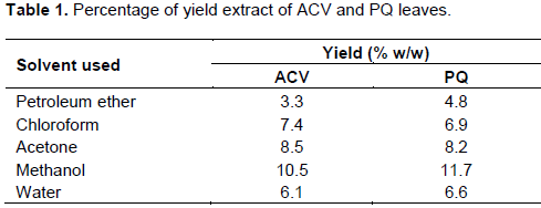
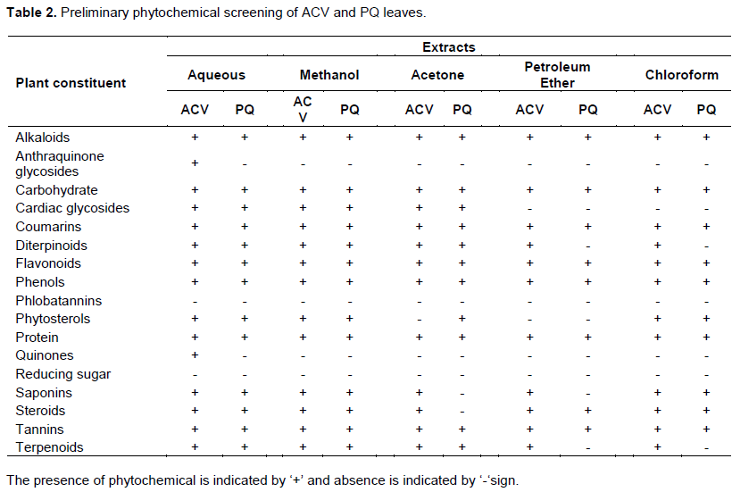
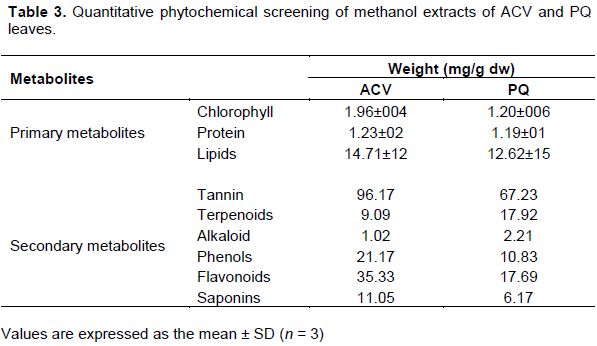
The percentage extraction yield of different extracts is shown in Table 1. The yield percentage of methanol extract of ACV and PQ was 10.5 and 11.7, respectively to that of dry powder. The yield percentage of methanol was higher than that of other solvents, and in the following order methanol>acetone>chloroform>water>petroleum ether. Since the yield percentage of methanol was higher than that of other solvents used, methanolic extracts of ACV and PQ leaves were used for further experiments. A large number of biologically active compounds were found in aqueous, methanol, acetone, diethyl ether and chloroform extracts of ACV and PQ. Several primary metabolites such as carbohydrates, proteins, and alkaloids, and secondary metabolites including coumarins, terpenoids, diterpenoids, flavonoids, phenols, tannins, saponins and steroids were found in the extracts of ACV and PQ (Table 2).
The free radical scavenging activity of the methanol extracts of ACV and PQ leaves was determined by the DPPH method to evaluate the antioxidant activity of plant extracts. The extracts of each plant examined in the present study exhibited free radical scavenging activities and the highest activity was shown by ACV followed by PQ. At concentrations 10 to 200 μg/ml, the scavenging activities of ACV were 14.52 to 84.64%, while the scavenging activities of PQ were 8.71 to 71.78%. Percentage DPPH radical scavenging activities of both the extracts were dose dependent (Figure 1A). Further, ABTS radical cation scavenging activity of methanol extracts of ACV and PQ was analysed. The ABTS± scavenging activity of ACV was significantly higher than the PQ. At concentrations 10 to 200 μg/ml, the scavenging activities of ACV were 10.49 to 90.55%, while the scavenging activities of PQ were 2.36 to 68.74% (Figure 1B).
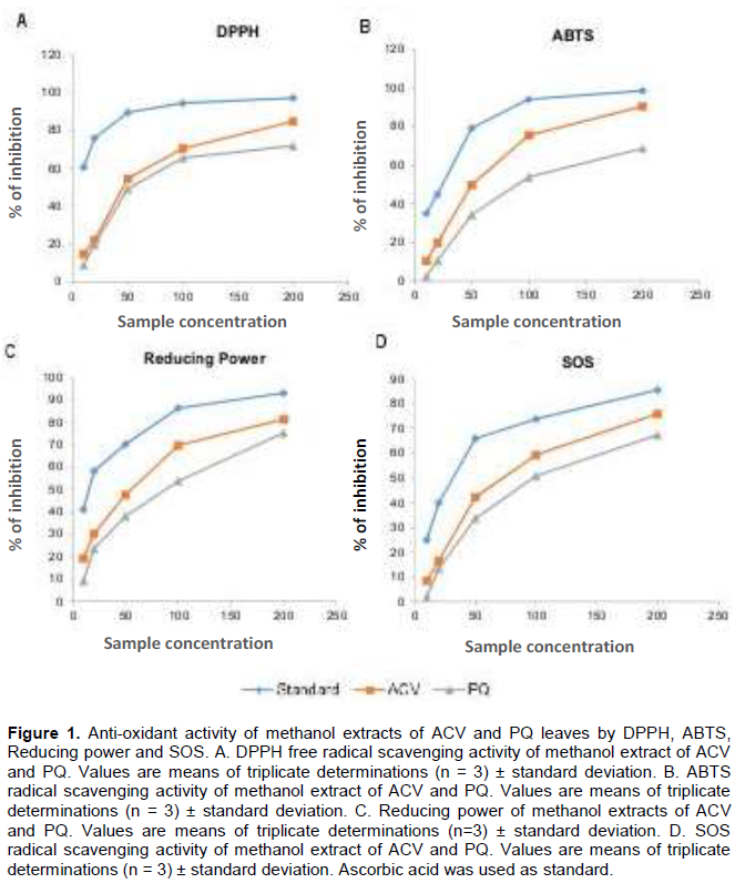
Antioxidant potential of the methanol extract of ACV and PQ was further estimated using potassium ferric cyanide reduction method. The presence of reductants (antioxidants) in the plant extract causes the reduction of Fe3+/Ferric cyanide complex to Fe2+ form. Therefore, the Fe2+ complex can be monitored by measuring the formation of Perl’s
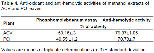
Prussian blue at 700 nm. It was observed that the reducing power of ACV and PQ was increased from 19.08 to 81.41% and 9.13 to 75.31%, respectively at concentrations 10 to 200 μg/ml. This may be due to the presence of secondary metabolites in the extract (Figure 1C). Further, ACV (42.24% at 50 μg/ml concentration) also showed potent superoxide activity as compared to PQ (32.68% at 50 μg/ml concentration) (Figure 1D). The phosphomolybdate assay was used to determine the total antioxidant capacity of samples. In this assay, Mo6+ is reduced to Mo5+ by antioxidant potential of the extract. The antioxidant capacity of methanolic extract of ACV was more than that of PQ. The percentage of activities of ACV and PQ were 53.16±.3 and 40.55±1.2, respectively (Table 4).
Then, the anti-haemolytic activity of methanolic extracts of ACV and PQ leaves using a biological test based on free radical-induced erythrocytes lysis in human blood was analysed. Lipid oxidation of human blood erythrocyte membrane mediated by H2O2 induces membrane damage and subsequently haemolysis. The results showed that ACV exhibited a maximum anti-haemolytic activity followed by PQ. The percentage of activities of ACV and PQ were 79.07±1.05 and 70.78±7, respectively (Table 4). Moreover, the RBC haemolysis is a more sensitive system for evaluating the antioxidant properties of the phytochemicals. The anti-haemolytic activity of ACV and PQ may be due to the presence of phenols and flavonoids in the extracts.
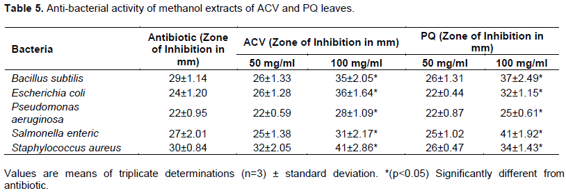
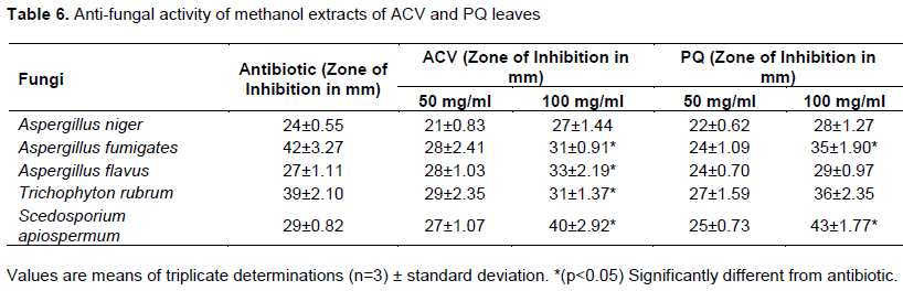
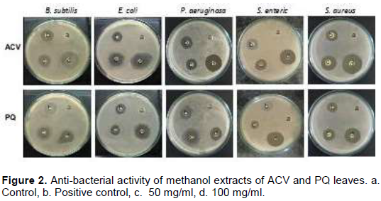
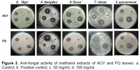
Tables 5 and 6 show the anti-bacterial and anti-fungal activities of methanol extracts of ACV and PQ leaves. Two concentrations (50 and 100 mg/ml) of extracts were tested against five different bacteria including B. subtilis, E. coli, P. aeruginosa, S. enteric, and S. aureus and five different fungi including A. niger, A. fumigates, A. flavus, T. rubrum, and S. apiospermum. Zone of inhibition for the following was measured in mm. It has been observed that there was a significant increase in the zone of inhibition, on increasing the concentration of extracts (Figures 2 and 3).
DISCUSSION
Medicinal plants are very much in demand because of their biological properties and bioactive compounds which are well known to act against various diseases (Misra, 2013; Atanasov et al., 2015; Pandey and Rizvi, 2009). In the present study, it has been shown that methanolic extracts of ACV and PQ leaves possess anti-oxidant, anti-bacterial, anti-fungal, and anti-haemolytic activities.
Phytochemical analysis gives the basic information about the bioactive components present in the plant extract (Hosseinzadeh et al., 2015). In the present study, the qualitative and quantitative analysis of methanol extracts of ACV and PQ leaves showed the presence of various secondary metabolites such as alkaloids, anthraquinones, cardiac glycosides, phenols, flavonoids, saponins, tannins and terpenoids. Several researchers reported that secondary metabolites including alkaloids, phenols and flavonoids, contribute to the biological activities of the plant (Dipankar et al., 2011; Oliveira et al., 2014). Quantitative analysis revealed that the extracts contained a high concentration of flavonoids, phenols and tannins. It is well known that phenols and flavonoids possess various biological activities such as anti-viral, anti-inflammatory, anti-cancer, anti-haemolytic and anti-oxidative potential (Beg et al., 2011; Bertrand Sagnia et al., 2014; Ameni et al., 2015). The anti-oxidant and anti-microbial activity observed in the present study may be due to the presence of phenols and flavonoids in ACV and PQ extracts.
As the scavenging of DPPH radical depends on electron transfer/donating ability, the radical scavenging activity of extracts could be related to the presence of phenols, thus contributing to their electron transfer/ hydrogen donating ability (Bab and Malik, 2015; Diemdo et al., 2014; Saha and Verma, 2016). Both ACV and PQ showed a less percentage of inhibition for DPPH radical scavenging activity as compared to well-known antioxidant ascorbic acid. However, methanol extracts of ACV leaves exhibited a higher antioxidant capacity than PQ. Similarly, Hamid et al. (2017) reported that Adiantum venustum extracts exerted DPPH radical scavenging activity. ACV and PQ methanolic extract also showed effective scavenging activity of superoxide and ABTS radical. It has been reported that phenols and flavonoids have anti-radical and anti-oxidant activities (Agarwal, 2011; Saxena et al., 2012). It also has been studied by Sowndhararajan et al. (2013) that tannins are more capable to reduce free radicals (ABTS_+) due to their molecular weight, the number of aromatic rings and nature of hydroxyl group’s substitution than the specific functional groups.
The presence of phenolic compounds in the extracts causes the reduction of Fe3+/Ferric cyanide complex to ferrous form. Similar observation between the polyphenolic constituents in terms of dose dependent and reducing power activity have been reported for several plant extracts including ferns (Lai et al., 2009). Superoxide radical can lead to the formation of hazardous hydroxyl radicals as well as singlet oxygen which results in oxidative stress and DNA damage (Lobo et al., 2010; Khanna et al., 2014; Rahal et al., 2014). In the present study, ACV and PQ showed significant superoxide scavenging activity and the scavenging potential may be due to the presence of bioactive phytoconstituents such as phenols and flavonoids. Similarly, Kaur et al. (2017) reported that fern extract showed significant superoxide radical scavenging activity. Recent studies proved that phenolic compounds reduce the Mo6+ into Mo5+ leading to the formation of a green phosphomolybdate complex. The phosphomolybdate has the hydrogen and electron donating ability that helps to detect the antioxidants such as ascorbic acid, α-tocopherol, and some phenolic, cysteine, and aromatic amines (Malliaraki et al., 2003; Prior et al., 2005). The methanolic extracts of ACV and PQ showed significant total antioxidant capacity which may be due to the presence of phenols.
Lipid peroxidation can injure every molecule of the biological system and can break the DNA strands which lead to mutation and cancer (Barrera, 2012; Zhong and Yin, 2015). Due to the heavy accumulation of poly-unsaturated fatty acids and haemoglobin, the erythrocytes can be damaged severely such that it can lead to oxidative damage resulting in haemolysis (Asgary et al., 2005; Pandey and Rizvi, 2010). The compounds present in ACV and PQ extracts are capable of anti-haemolytic and anti-lipid peroxidation activities, which is evident from inhibition of erythrocyte lysis with increasing concentration of extracts. In line with the present findings, Kaur et al. (2017) reported that fern extract showed significant anti-haemolytic activity. The methanol extracts of ACV and PQ were more effective in inhibiting microbial growth and this may be due to the presence of sterols and secondary metabolites. Similarly, Ishaq et al. (2014) reported that fern extract shows significant antimicrobial activity against various strains of bacteria and fungi.
The present investigation suggests that bioactive compounds from ACV and PQ leaves possess potential anti-oxidant and anti-microbial activities. However, isolation and preparation of phytochemicals from ACV and PQ and assessment of their impact on various health improvements/control of free radical mediated diseases through in vitro and in vivo studies are needed. Such identified potential and natural constituents could be exploited as cost effective food/feed additives for human and animal health.
CONFLICT OF INTERESTS
The authors have not declared any conflict of interests.
REFERENCES
|
Abbasi AM, Khan MA, Ahmad M, Zafar M, Jahan M, Sultana S (2010). Ethnopharmacological application of medicinal plants to cure skin diseases and in folk cosmetics among the tribal communities of North-West Frontier Province. Journal of Ethnopharmacology 128(2):322-335. |
|
|
Agarwal AD (2011). Pharmacological activities of flavonoids: A review. International Journal of Pharmaceutical Sciences and Nanotechnology 4.2:1394-1398. |
|
|
Ahmed A, Jahan N, Wadud A, Imam H, Hajera S, A Bilal A (2012). Physicochemical and biological properties of Adiantum capillus-veneris Linn: An important drug of unani system of medicine. International Journal of Current Research 4(21). |
|
|
Ameni D, Baghiani A, Boumerfeg S, Dahamna S, Khennouf S, Zarga MHA (2015). Phytochemical profiles, antioxidant capacity and protective effect against APPH-induced mouse erythrocyte damage by Daphne gnidium L. Shoots extracts. International Journal of Pharmacy and pharmaceutical Sciences 7:148-156. |
|
|
Asgary S, Naderi GH, Askari N (2005). Protective effect of flavonoids against red blood cell hemolysis by free radicals. Experimental & Clinical Cardiolog 10(2):88. |
|
|
Atanasov AG, Waltenberger B, Pferschy-Wenzig EM, Linder T, Wawrosch C, Uhrin P (2015). Discovery and resupply of pharmacologically active plant-derived natural products: A review. Biotechnology Advances 33(8):1582-1614. |
|
|
Bab SA, Malik SA (2015). Determination of total phenolic and flavonoid content, antimicrobial and antioxidant activity of a root extract of Arisaema jacquemontii blume. Journal of Taibah University for Science 9(4):449-454. |
|
|
Barrera G (2012). Oxidative stress and lipid peroxidation products in cancer progression and therapy. ISRN oncology 2012. |
|
|
Beauchamp C, Fridovich I (1971). Superoxide dismutase: improved assays and an assay applicable to acrylamide gels. Analytical Biochemistry 44(1):276-287. |
|
|
Beg S, Swain S, Hasan H, Barkat MA, Hussain MS (2011). Systematic review of herbals as potential anti-inflammatory agents: Recent advances, current clinical status and future perspectives. Pharmacognosy Reviews 5(10):120. |
|
|
Bertrand Sagnia B, Fedeli D, Casetti R, Montesano C, Falcioni G, Colizzi V (2014). Antioxidant and anti-Inflammatory activities of extracts from Cassia alata, Eleusine indica, Eremomastax speciosa, Carica papaya and Polyscias fulva medicinal plants collected in Cameroon. PloS One 9(8):e103999. |
|
|
Brand-Williams W, Cuvelier ME, Berset C (1995). Use of a free radical method to evaluate antioxidant activity. LWT-Food science and Technology 28(1):25-30. |
|
|
Chang C, Yang M, Wen H, Chem J (2002). Estimation of flavonoids total content in propolis by two complimentary colorimetric methods. Journal of Food and Drug Analysis 10(3). |
|
|
Diemdo Q, Tran-Nguyen PL, Huonghuynh L, Edisoetaredjo F, Suryadiismadji, Hsuju Y (2014). Effect of extraction solvent on total phenol content, total flavonoid content, and antioxidant activity of Limnophila aromatic. Journal of Food and Drug Analysis 22(3):296-302. |
|
|
Dipankar C, Murugan S, Devi PU (2011). Review on medicinal and pharmacological properties of Iresine herbstii, Chrozophora rottleri and Ecbolium linneanum. African Journal of Traditional, Complementary and Alternative Medicines 8(5S). |
|
|
Feng R, Wang X, Wei C, Tu S (2015). The accumulation and subcellular distribution of arsenic and antimony in four fern plants. International Journal of Phytoremediation 17(4):348-354. |
|
|
Fusco D, Colloca G, Lo Monaco MR, Cesari M (2007). Effects of antioxidant supplementation on the ageing process. Clinical Interventions in Aging 2(3):377. |
|
|
Halliwell B (1997). Antioxidants and human disease: a general introduction. Nutrition Reviews 55(1):S44. |
|
|
Hamid J, Ahmed D, Waheed A (2017). Evaluation of anti-oxidative, antimicrobial and anti-diabetic potential of Adiantum venustum and identification of its phytochemicals through GC-MS. Pakistan Journal of Pharmaceutical Sciences 30(3). |
|
|
Harborne JB (1973). Phytochemical methods: A Guide to Modern Techniques of Plant Analysis. Great Britain: Chapman & Hall Google Scholar. |
|
|
Hosseinzadeh S, Jafarikukhdan A, Hosseini A, Armand R (2015). The application of medicinal plants in traditional and modern medicine: a review of Thymus vulgaris. International Journal of Clinical Medicine 6(09):635-642. |
|
|
Hussein HM, Hameed IH, Ibraheem OA (2016). Antimicrobial activity and spectral chemical analysis of methanolic leaves extract of Adiantum capillus-veneris using GC-MS and FTIR spectroscopy. International Journal of Pharmacognosy and Phytochemical Research 8(3):369-385. |
|
|
Ibraheim ZZ, Ahmed AS, Gouda YG (2011). Phytochemical and biological studies of Adiantum capillus-veneris L. Saudi Pharmaceutical Journal 19(2):65-74. |
|
|
Ishaq MS, Hussain MM, Afridi MS, Ali G, Khattak M, Ahmad S (2014). In vitro phytochemical, antibacterial and antifungal activities of leaf, stem, and root extracts of Adiantum capillus veneris. The Scientific World Journal 2014. |
|
|
Jones CG, Hare JD, Compton SJ (1989). Measuring plant protein with the Bradford assay. Journal of Chemical Ecology 15(3):979-992. |
|
|
Kaur P, Kumar M, Singh AP, Kaur S (2017). Ethyl acetate fraction of Pteris vittata L. Alleviates 2-acetylaminofluorene induced hepatic alterations in male Wistar rats. Biomedicine and Pharmacotherapy 88:1080-1089. |
|
|
Khanna RD, Karki K, Pande D, Negi R, Khanna RS (2014). Inflammation, free radical damage, oxidative stress and cancer. Interdiscip Journal of Microinflammation 1(109):2. |
|
|
Lai HY, Lim YY, Tan SP (2009). Antioxidative, tyrosinase inhibiting and antibacterial activities of leaf extracts from medicinal ferns. Bioscience, Biotechnology, and Biochemistry 73(6):1362-1366. |
|
|
Lee MT, Lin WC, Yu B, Lee TT (2017). Antioxidant capacity of phytochemicals and their potential effects on oxidative status in animals - A review. Asian-Australasian Journal of Animal Sciences 30(3):299. |
|
|
Lobo V, Patil A, Phatak A, Chandra N (2010). Free radicals, antioxidants and functional foods: Impact on human health. Pharmacognosy Reviews 4(8):118. |
|
|
Malliaraki N, Mpliamplias D, Kampa M, Perakis K, Margioris AN, Castanas E (2003). Total and corrected antioxidant capacity in hemodialyzed patients. BMC Nephrology 4(1):4. |
|
|
McDonald S, DPrenzler P, Antolovich M, Robards K (2001). Phenolic content and antioxidant activity of olive extracts. Food Chemistry 73(1):73-84 |
|
|
Misra L (2013). Traditional phytomedicinal systems, scientific validations and current popularity as nutraceuticals. International Journal of Traditional and Natural Medicines 2:27-75. |
|
|
Nayar BK (1957). Medicinal Ferns of India. Bulletin of National Botanic Garden 58:1-38. |
|
|
Okamoto G, Hayase F, Kato H (1992). Scavenging of active oxygen species by glycated proteins. Bioscience, Biotechnology, and Biochemistry 56(6):928-931. |
|
|
Oliveira LL, Carvalho MV, Melo L (2014). Health promoting and sensory properties of phenolic compounds in food. Revista Ceres 61:764-779. |
|
|
Oyaizu M (1986). Studies on products of browning reactions: antioxidative activities of products of browning reaction prepared from glucosamine. Japanese Journal of Nutrition 44(6). |
|
|
Pandey KB, Rizvi SI (2009). Plant polyphenols as dietary antioxidants in human health and disease. Oxidative Medicine and Cellular Longevity 2(5):270-278. |
|
|
Pandey KB, Rizvi SI (2010). Markers of oxidative stress in erythrocytes and plasma during aging in humans. Oxidative Medicine and Cellular Longevity 3(1):2-12. |
|
|
Pandey MM, Rastogi S, Rawat AK (2013). Indian traditional ayurvedic system of medicine and nutritional supplementation. Evidence-Based Complementary and Alternative Medicine 2013. |
|
|
Prior RL, Wu X, Schaich K (2005). Standardized methods for the determination of antioxidant capacity and phenolics in foods and dietary supplements. Journal of Agricultural and Food Chemistry 53(10):4290-4302. |
|
|
Rahal A, Kumar A, Singh V, Yadav B, Tiwari R, Chakraborty S, Dhama K (2014). Oxidative stress, prooxidants, and antioxidants: The Interplay. BioMed Research International 2014. |
|
|
Rajendran P, Nandakumar N, Rengarajan T, Palaniswami R, Gnanadhas EN, Lakshminarasaiah U (2014). Antioxidants and human diseases. Clinica Chimica Acta 436:332-347. |
|
|
Saha S, Verma RJ (2016). Antioxidant activity of polyphenolic extract of Terminalia chebula Retzius fruits. Journal of Taibah University for Science 10(6):805-812. |
|
|
Saxena M, Saxena J, Pradhan A (2012). Flavonoids and phenolic acids as antioxidants in plants and human health. International Journal of Pharmaceutical Sciences Review and Research 16(2):130-134. |
|
|
Schanderl SH (1970). Method in food analysis. Academic Press, New York, 1970. |
|
|
Singh BP, Upadhyay R (2014). Medicinal pteridophytes of Madhya Pradesh. Journal of Pharmacognosy and Phytochemistry 3(3):173-176. |
|
|
Singh M, Singh N, Khare PB, Rawat AK (2008). Antimicrobial activity of some important Adiantum species used traditionally in indigenous systems of medicine. Journal of Ethnopharmacology 115(2):327-329. |
|
|
Sowndhararajan K, Kang SC (2013). Free radical scavenging activity from different extracts of leaves of Bauhinia vahlii Wight & Arn. Saudi Journal of Biological Sciences 20(4):319-325. |
|
|
Vadi R, Manisha V, Swati K (2017). Hansraj (Adiantum capillus veneris Linn.): A systematic review on its ethnobotany, phytochemical and pharmacological profile. International Journal of Ayurveda and Pharma Research 5(6). |
|
|
Yang ZG, Sun HX, Fang WH (2005). Haemolytic activities and adjuvant effect of Astragalus membranaceus saponins (AMS) on the immune responses to ovalbumin in mice. Vaccine 23(44):5196-5203. |
|
|
Zhong H, Yin H (2015). Role of lipid peroxidation derived 4-hydroxynonenal (4-HNE) in cancer: Focusing on mitochondria. Redox Biology 4:193-199. |
|
Copyright © 2024 Author(s) retain the copyright of this article.
This article is published under the terms of the Creative Commons Attribution License 4.0


