Full Length Research Paper

ABSTRACT
The use of medicinal plants as alternative therapy for the treatment of microbial infections is significant in the maintenance of good health, especially in developing countries. The aim of this study was to determine the phytochemical composition and antimicrobial activity of Buchholzia coriacea and Psychotria microphylla leaf extracts on bacteria isolated from aquatic environments in Nigeria. Exactly 736 water samples from boreholes, ponds, rivers, streams, and wells respectively were collected for this study. Antimicrobial activity of B. coriacea and P. microphylla leaf extracts were determined using standard microbiological techniques. Phytochemical and chemical constituents of the herbal extracts were determined using standard analytical techniques. Bacteria isolated from the water samples were Aeromonas hydrophila (n = 103), Escherichia coli (n = 118), and Vibrio cholerae (n = 87). extracts of B. coriacea and P. microphylla showed appreciable antibacterial activities. B. coriacea and P. microphylla leaf extracts showed the presence of minerals, alkaloids, protein, terpenoids, phenols, flavonoids, glycosides, steroids, tannins, and vitamins. Plant extracts used in this study exhibited an amazing antibacterial activity against bacterial isolates from the water bodies. Thus, B. coriacea and P. microphylla plants should be further explored to determine the active component(s) responsible for their antibacterial activity.
Key words: Antimicrobial activity, B. coriacea, P. microphylla, leaf extracts, bacteria, aquatic environments, phytochemical composition.
INTRODUCTION
Plants are important for human existence and they are the major sources of foods and medicine (Olowe et al., 2017). Medicinal plant preparations obtained from common traditional herbs and medicinal plants, has been linked to the existence of natural products in plants with medicinal properties (Hoareau and Dasilva, 1999).
UNESCO (1996) reported that the application of traditional medicine and medicinal plants in most developing countries is a major basis for the maintenance of good health. The dependence on the utilization of medicinal plants in western societies has been traced to the development and extraction of several drugs from these plants as well as from traditionally used rural herbal therapies (UNESCO, 1998).
It has been estimated that about 25% of all modern prescription medicines are directly or indirectly (CF)can diminish the use of expensive artificial fertilizers. The optimal useof combination of organic andinorganic sourced from plants (Chinyere et al., 2017). Such drugs that have long been used include quinine from back of chinchona tree, reserpine from root of Rauwolfia vomitoria, ephedrine from ephedra sinica, morphine from opium poppy (Papaver somniferum) and more recently adopted compounds such as the anti-malaria artemisinin from the plant Artemisia annua (sweet wormwood) (Balandrin et al., 1993). There is an emerging trend in research to establish the biological activities of medicinal plants. Many scientific researchers have reported on the efficacies and chemotherapeutic roles of medicinal plants in the treatment of diverse ailments such as malaria, diarrhea, dysentery, convulsion, asthma, diabetes, pains, ulcer and epilepsy. Many plants have shown their tremendous capability in lowering raised level of liver enzymes in viral hepatitis (Oshima et al., 1995), to possess immense potential as anti-peptic ulcer (Ibrahim et al., 2008), antimicrobial and antioxidant properties (Ali et al., 2009). Such important medicinal plants suspected to have some activity and could serve as a possible solution in treatment of bacterial infections include Buchholzia coriacea and Psychotria microphylla. Antibiotic resistance genes are emerging contaminants posing a potential worldwide human health risk (Allen et al., 2010; Wellington et al., 2013; Yang et al., 2014). Aeromonas hydrophila, Escherichia coli, and Vibrio cholerae are among a myriad of bacteria commonly found in natural water bodies, and are potential invasive pathogens for those who suffer trauma while submerged in water or waterborne diseases while consumed in drinking water sources especially in rural communities in third world countries (Stokes and Gillings, 2011). The search for medicinal plants as alternative therapeutic options against multidrug-resistant bacteria causing infections is very significant, especially with the increasing bacterial resistances to commonly used antibiotics. This study was designed to determine the phytochemical composition and antimicrobial activity of B. coriacea and P. microphylla leaf extracts on bacteria isolated from aquatic environments in ebonyi state, South Eastern Nigeria.
MATERIALS AND METHODS
Study site and description of study area
The study area of this research is Ebonyi State, Nigeria. Ebonyi State is located in the south-eastern part of Nigeria which lies approximately within longitude 7°301 and 7°E and, latitude 5°401 and 6°451 N. It has a population of 149,683 and a land mass of about 5,935 square kilometers (NPC Census, 2006). Ebonyi State is bounded to the north by Benue State, to the south by Abia State and to the west by Enugu State and east by Cross River State respectively. Two seasons are distinguishable in Ebonyi State: a dry season (November to March) and a wet season (April to October). For the purpose of this study, water samples were collected from three local government areas which include: Abakaliki, Ebonyi and Ikwo LGA.
Sample collection
B. coriacea and P. microphylla leaves were collected between November, 2017 and January, 2018. Water samples were collected between a period of eight months (February to September, 2018) covering both dry and rainy seasons. Water samples from five sampling sources namely: stream (n=117), river (n=114), well (n=149), borehole (n=215) and pond (n=111) from designated areas were collected for this study. A total of 736 water samples were collected.
The grab sampling procedure was adopted in accordance to standard methods for examination of water as recommended by Standard Method for microbiological analysis (APHA, 2015). The collection of water samples was altogether made in early hours of the morning and transported to the Applied Microbiology Laboratory Complex of Ebonyi State University (EBSU) within one hour of sample collection for laboratory analysis.
Physicochemical parameter studies
Physicochemical characteristics which were studied include: alkalinity, biochemical oxygen demand (BOD), chemical oxygen demand (COD), conductivity, dissolved oxygen (DO), odour, pH, salinity, taste, temperature, total dissolved solid (TDS), total hardness, total organic carbon, total suspended solid (TSS) and turbidity. Each parameter was measured in triplicates from which the mean and standard deviation counts were recorded.
Isolation of bacteria
Discrete colonies obtained from the plate count agar plates were further sub-cultured onto freshly prepared plates of selective and media such as Aeromonas isolation agar base, MacConkey agar, and thiosulphate citrate bile salts sucrose agar plates. The Petri dishes was placed in an inverted position in the incubator for 24 h at 37°C to obtain pure cultures. Presumptive morphological identification of the colonies was done by observing their individual appearance on the selective media used. The colonies were stored in test tubes containing peptone water for cultural, morphological, and biochemical identification (Cheesebrough, 2006). Further characterizations of bacterial isolates was done by using the API 20E (Biomerieux S.A., Marcy-l`Etoile/France) identification system.
Antimicrobial susceptibility studies using plant leaves extracts
This was carried out to determine the effects of aqueous, ethanolic and ethyl-acetate leaf-extracts of B. coriacea and P. microphylla against bacteria isolated from water bodies by methods described by Orji et al. (2015).
Collection of plant materials
B. coriacea and P. microphylla plants were identified and authenticated by a taxonomist (Dr. Nwankwo Ephraim) in the Department of Applied Biology, Faculty of Science, Ebonyi State University, Abakaliki, Nigeria. Fresh leaves of B. coriacea were collected from free growing B. coriacea trees in Mgbabor village of Izza North Local Government Area of Ebonyi state. The leaves of P. microphylla used in this study were collected from Afikpo South Local Government Area of Ebonyi State, Nigeria. Plant samples were collected and washed with running tap water and transported to the Department of Applied Microbiology, Ebonyi State University, Abakaliki, Nigeria for further analysis (Orji et al., 2015). After plant identification, the leaves were washed with distilled water to reduce the bacterial load, and also were chopped into small sizes with sterile knife in order to facilitate drying. The plant parts were dried at room temperature in order to prevent loss of active constituents which may be thermo-liable and drying continued. After drying, the leaves were ground using sterile mortar and pestle and electric grinder into fine powder and was stored for further use (Orji et al., 2015).
Preparation of plant extracts
Twenty grams of each of the dried grounded leaves was weighed separately using weighing balance. This was transferred into conical flasks containing 80 ml of water, ethanolic and ethyl-acetate. The different mixtures were placed on a mechanical shaker and allowed to macerate for 24 h, filtration was done using sterile filter cloth and the filtrate collected. The total filtrate collected was evaporated to dryness by pouring into sterile stainless plate and was kept in hot air oven at 50°C until whole moisture evaporates completely leaving powder behind (Orji et al., 2015).
Determination of mineral, phytochemical, proximate, and vitamin composition
The different leaf extracts was used for the analysis of the mineral, phytochemical, proximate and vitamin composition of the different leaves extracts.
Determination of mineral composition of leaves extract
The mineral composition of the extracts was determined for the presence of Ca, Cu, Fe, Mg, Zn, P, As and Pb in the different extracts using the methods previously described (AOAC, 1995; Pearson, 1976).
Determination of phytochemical composition of leaves extract
Phytochemical analysis was carried out to determine the quantity of alkaloids, flavonoids, glycosides, phenols, phlobatannins, steroids, tannins, and terpenoids present in the extracts using standard methods (AOAC, 1995; Harborne, 1973; Trease and Evans, 1989).
Determination of proximate composition of leaves extract
The proximate parameters studied include: Ash, carbohydrates, fats, fibre, moisture, and proteins. This was carried out in accordance to the methods of AOAC (1995).
Determination of vitamin composition of leaves extract
The vitamin content of the extracts was determined to ascertain the level of vitamin C, E, B12, B2, D, B1, K, B7, A, and B3 present in the extracts (Pearson, 1976; Baraket et al., 1973; Okwu and Josiah, 2006).
Antibacterial susceptibility testing using plant extracts
A standardized inoculum of 0.5 McFarland standard was introduced onto the surface of sterile agar plates, and a sterile glass spreader was used for even distribution of inoculums. A sterile paper disc (8 mm in diameter) soaked in known concentration of extracts (20 µg/ml per disc) was carefully placed at the centre of the seeded labelled agar. The plates were incubated aerobically at 37°C and examined for zone of inhibition after 24 h. Each zone of inhibition was measured with a ruler and compared with the control (disc containing only physiological saline) in accordance to the method of Akinyemi et al. (2006) and Udu-ibiam et al. (2015).
Determination of minimum inhibitory concentration (MIC) and minimum bactericidal concentration (MbC)
The MIC of extracts was determined by diluting the various concentrations (0.0-36, 0.0-41, and 0.0-37 µg/ml). Equal volume of the extracts and nutrient broth was mixed in the test tube. Specifically, 0.1 ml of standardized inoculum of 0.5 McFarland standard was added to each tube. The tubes were incubated aerobically at 37°C for 18 - 24 h. Two control tubes were maintained for each test batch. This was as follows: tube containing extracts and the growth medium without inoculum (antibiotic control) and the tube containing the growth medium, physiological saline and the inoculums (organism control). MIC was determined as the lowest concentration of the extracts with no visible growth (no turbidity) when compared with the control tubes. The MBC was determined by sub-culturing the test dilution on fresh solid medium and further incubated at 37°C for 18 - 24 h. The lowest concentration of MIC tubes with no visible bacterial growth on solid medium was regarded as MBC as described by Akinyemi et al. (2006).
Statistical analysis
The raw data obtained were presented as mean ± standard deviation in tables and bar charts while relevant data were interpreted using simple descriptive statistics such as minimum, maximum, and one way analysis of variance (ANOVA) with the aid of IBM Statistical Package for Social Sciences (SPSS) version 22 and Microsoft Excel 2013 software.
RESULTS
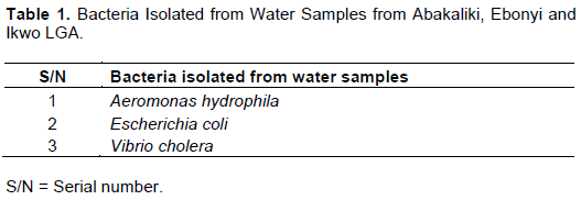
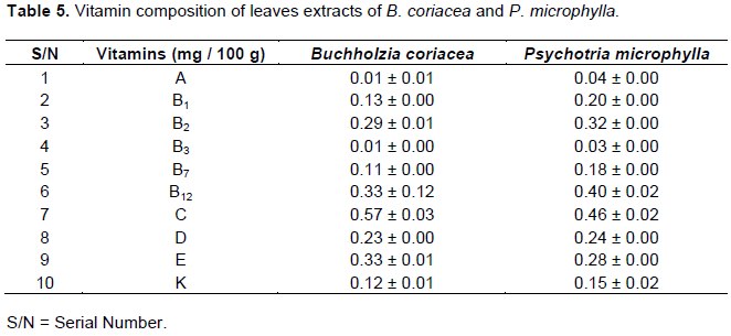
According to the different tests carried out three differentorganisms were identified and include: A. hydrophila, E. coli and V. cholera (Table 1). The result of chemical composition (mineral, phytochemical, proximate and vitamin) of leaves extracts of B. coriacea and P. microphylla is presented in Tables 2 to 5.
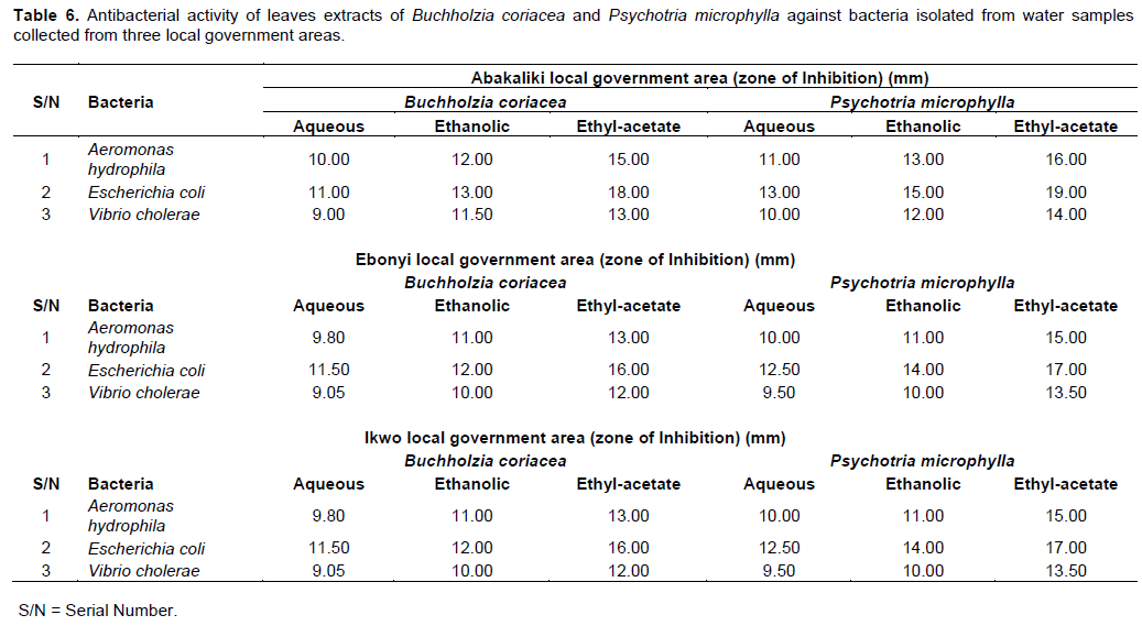
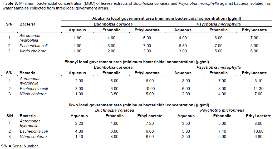
Tables 6 to 8 shows the antibacterial activity of leaves extracts of B. coriacea and P. microphylla against bacteria isolated from water samples collected from different water sources in the study locations.

DISCUSSION
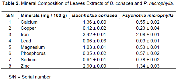
The use of traditional medicine and medicinal plants as alternative therapy for the treatment of microbial infections in most developing countries is a major basis for the maintenance of good health. Analysis of the mineral, phytochemical, proximate, and vitamin composition of B. coriacea and P. microphylla leaf extracts used in this study revealed that the calcium, copper, iron, lead, magnesium, sodium, and zinc contents were abundant in B. coriacea when compared to those of P. microphylla in contrary to copper and phosphorus concentrations. The mineral composition of P. microphylla and B. coriacea obtained in this study is in agreement with the findings of Ibrahim and Fagbohun (2012) who reported the presence of sodium (1.22 ppm), potassium (1.34 ppm), phosphorous (0.22 mg/g), calcium (0.19%), magnesium (1.62%), zinc (0.18%) iron (1.11%) and manganese (0.46%) in extracts of B. coriacea.
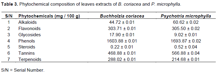
Alkaloids, flavonoids, phenols, steroids, and tannins were observed to be more abundant in Psychotria microphylla than in Buchholzia coriacea, in contrary to glycosides and terpenoids which were less. Ibrahim and Fagbohun (2012) reported the detection of phytochemicals such as alkaloids (3.16 and 3.32%), glycosides (2.16 and 2.46%), saponin (2.10 and 2.23%), steroids (0.14 and 0.16%), tannin (6.46 and 6.73%), flavonoids (0.68 and 0.79%), terpenes (0.22 and 0.16%), reducing sugars (1.14 and 1.71%) and phenol (1.83 and 1.26%) in ethanol and methanol extract respectively. The phytochemical components detected in the present study have been documented by other researchers for different antibacterial properties (Ajani, 2015; Balogun, 2016; Chinyere et al., 2017).
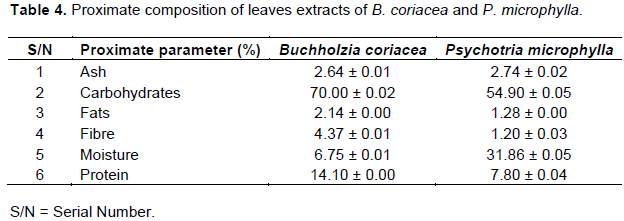
Ash and moisture were observed to be more abundant in P. microphylla than in B. coriacea, while carbohydrate, fats, fibre, and protein contents of B. coriacea were higher than that of P. microphylla. Proximate analysis carried out by Ibrahim and Fagbohum in 2012 showed that B. coriacea contained moisture (1.30%), crude fat (2.30%), crude protein (13.34%), ash content (6.6%), crude fibre (2.19%) and carbohydrate (75.43%).
Vitamin A, Vitamin B1, Vitamin B2, Vitamin B3, Vitamin B7, Vitamin B12, Vitamin D and Vitamin K content of P. microphylla was higher than in B. coriacea, while Vitamin C and E and content of B. coriacea were more abundant than in P. microphylla. The contribution of these compounds to human health cannot be overstated. There was a significant difference (p < 0.05) in the antibacterial activity of the extracts.
Our study showed that ethyl acetate extracts of the two plants have the highest antimicrobial activity, followed by the ethanolic extracts while aqueous extracts have the lowest activity against the tested organisms in this study. The chemical composition of P. microphylla extracts could be responsible for their antibacterial activity, owing to the fact that extracts of P. microphylla expressed a lethal activity on aquatic organisms as was demonstrated by Orji et al. (2015). Several workers have reported bioactivity of crude extracts of medicinal plants within the inhibition zone diameter range obtained in their study (Ogbeche et al., 1997; Akinyemi et al., 2005).
Phytochemical constituents were further reported to be responsible for many antimicrobial activities of different plant species (Ghoshal et al., 1996; Iwu et al., 1999; Adetunji et al., 2013). For instance, flavonoids have been reported to be synthesized by plants in response to microbial infections and are good antibacterial agents; tannins have been demonstrated to have antibacterial activities (Akiyama et al., 2001). The result obtained in this study is in agreement with the findings of Udu-ibiam et al. (2015) in a study which determined the antimicrobial effects of the aqueous and ethanol extracts of edible mushroom; Pleurotus species and P. microphylla against E. coli, Pseudomonas aeruginosa, Salmonella Typhi, Klebsiella pneumoniae, and S. aureus.
In this study, the highest antibacterial activity was recorded against E. coli in water samples from Abakaliki local government area with an inhibition zone diameter (IZD) of 19 mm with ethyl acetate extracts of P. microphylla while the lowest antibacterial activity was recorded against V. cholerae in water samples from the same location with an IZD of 9 mm with aqueous extracts of B. coriacea. Udu-ibiam et al. (2015) in their study reported that the highest sensitivity was recorded against P. aeruginosa with IZD of 7 mm, followed by Salmonella Typhi (5 mm), and then E. coli and S. aureus (4 mm each). In their study, it was observed that ethanol extract of P. microphylla was the most efficient of all the extracts against test organisms used.
Other reports have shown that extracts of B. coriacea can be applied externally to treat pleurisy, rheumatism, conjunctivitis, smallpox, scabies, skin complaints, fever, ulcers, boils and haemorrhoids (Akiyama et al., 2001). The highest minimum inhibitory concentration (MIC) was obtained with ethyl acetate extracts, followed by the ethanolic extracts while aqueous extracts showed the lowest MIC against the organisms studied.
This is comparable to the result of Adetunji et al. (2013) who reported similar MIC values of plant extracts on P. aeruginosa, S. aureus, and E. coli. In their study, it was observed that ethanolic extract recorded the highest MIC against test isolates. Ezekiel and Onyeoziri (2009) carried out a study on the effect of the fresh kola, hexane, and methanol extracts of B. coriacea on food borne pathogens (E. coli, Enterococcus faecalis, S. aureus, Trichoderma viride, and Aspergillus niger). The fresh kola showed IZD with the test bacteria: E. coli (62 mm), Enterococcus faecalis (40 mm), and S. aureus (50 mm). It was observed that the highest minimum bactericidal concentration (MBC) was obtained with ethyl acetate extracts, followed by the ethanolic extracts and aqueous extracts.
This present observations on MBC are similar to the report of Adetunji et al. (2013). Studies carried out by Ajaiyeoba et al. (2003) on fractions prepared from the methanol extract of B. coriacea stem bark showed higher concentration-dependent antibacterial and antifungal activity when compared to standard antibiotics; ampicillin, and ticonazole.
CONCLUSION
In conclusion, this investigation has revealed that extracts of B. coriacea and P. microphylla showed appreciable antibacterial activity on the isolates. The extracts were rich in minerals, alkaloids, terpenoids, phenols and vitamins. Plant extracts should be further explored to elucidate the active components responsible for its antimicrobial activity. Elucidating the active components will greatly reduce the pressure on the use of synthetic drugs in the treatment of bacterial infections and also curtail the development and spread of antibiotic resistance among bacterial species.
CONFLICT OF INTERESTS
The authors have not declared any conflict of interests.
REFERENCES
|
Adetunji CO, Olaniyi OO, and Ogunkunle AT (2013). Bacterial Activity of Crude Extracts of Vernonia amygdalina on Clinical Isolates. Journal of Microbiology and Antimicrobials 5(6):60-64. |
|
|
Ajaiyeoba EO, Patricia O, Nwozo SO, Woquan S (2003). Antimicrobial and Cytotoxicity Evaluation of Buchholzia coriacea Stem Bark. Fitoterapia 74(7-8):706-9. |
|
|
Ajani EO (2015). Evaluation of the Acute and Sub-acute Toxicity Effects of Ethanolic Leaves Extract of Lagenaria brevifolia (Bitter gourd) on Hepatic and Renal Function of Rats. European Journal of Medicinal Plants 5(2):210-219. |
|
|
Akinyemi KO, Oladapo O, Okwara CE, Ibe CC, Fasure AK (2005). Screening of Crude Extracts of some Medicinal Plants used in South-West Nigerian Unorthodox Medicine for Anti-methicilin Resistant Staphylococcus aureus. BMC Complementary and Alternative Medicine 5:6-8. |
|
|
Akinyemi KO, Oluwa OK, Omomigbehin EO (2006). Antimicrobial Activity of Crude Extracts of Three Medicinal Plants used in South-west Nigerian Folk Medicine on some Food Borne Bacterial Pathogens. African Journal of Traditional, Complementary and Alternative Medicines 3(4):13-22. |
|
|
Akiyama H, Fujii K, Yamasaki O, Oono T, Iwatsuki K (2001). Antibacterial Action of Several Tannins against Staphylococcus aureus. Journal of Antimicrobial Chemotherapy 48(4):487-491. |
|
|
Ali I, Rubina N, Wahib NK, Rukhsana G, Choudhary MI (2009). Biological Screening of Different Root Extracts of Euphorbia wallichi. Pakistan Journal of Botany 41(4):1737-1741. |
|
|
Allen HK, Donato J, Wang HH, Cloud-Hansen KA, Davies J, Handelsman J (2010). Call of the Wild: Antibiotic Resistance Genes in Natural Environments. Nature Reviews Microbiology 8:251-259. |
|
|
American Public Health Association (APHA) (2015). Standard Methods for the Examination of Water and Wastewater, 16th ed. American Public Health Association, Washington, D.C, 32. |
|
|
Association of Official Methods of Analysis (AOAC) (1995). Official Methods of Analysis. I5th Edition. AOAC Incorporation, Virginia, USA, pp. 249-260. |
|
|
Balandrin MF, Kinghorn AD, Farnsworth NR (1993). Plant-derived Natural Products in Drug Discovery and Development. An overview. In: Human Medicinal Agents from Plants. Eds: Kinghorn, A.D. and Balandrin, M.F. ACS Symposium Series. American Chemical Society, Washington, pp 2-12. |
|
|
Balogun ME (2016). Gongronema Latifolium: A Phytochemical, Nutritional and Pharmacological Review. Journal of Physiology and Pharmacology Advances 6(1):811-824. |
|
|
Baraket MZ, Shehab SK, Darwish N, Zahermy EI (1973). Determination of Vitamins from Plants. Analytical Biochemistry 53:225-245. |
|
|
Chinyere MO, Malachy CU, Charity CE, Ugochukwu O, Chika PE (2017). Antibacterial Evaluation of Ethanolic Leaf Extract of Gongronema Latifolium Benth on MDR Bacteria from Clinical Specimens. Clinical Biotechnology and Microbiology 1(4):156-164. |
|
|
Ezekiel OO, Onyeoziri NF (2009). Preliminary studies on the Antimicrobial Properties of Buchholzia coriacea (Wonderful Kola). African Journal of Biotechnology 8(3):472-474. |
|
|
Ghoshal SK, Prasad BN, Lakshmi V (1996). Antiamoebic Activity of Piper longum Fruits against Entamoeba histolytica In-Vitro and In-Vivo. Journal of Ethnopharmacology 50:167-170. |
|
|
Harborne JB (1973). Phytochemical Methods: A Guide to Modern Technique of Plant Analysis. Chapman and Hall, London, 113. |
|
|
Hoareau L, Dasilva EJ (1999). Medicinal Plants: A Re-emerging Health Aid. Electronic Journal of Biotechnology 2:56-70. |
|
|
Ibrahim AA, Abdulqader AA, Jaber SM, Mohammed AA, Mohammed OA, Syed R, Shaffi S (2008). Gastroprotective Effect of an Aqueous Suspension of Black cumin Nigella Saliva on Necrotizing Agents - Induced Gastric Injury in Experimental Animals. Saudi Journal of Gastroenterology 14(3):128-134. |
|
|
Ibrahim TA, Fagbohun ED (2012). Phytochemical and Nutritive Quality of Dried Seeds of Buchholzia coriacea. Greener Journal of Physical Sciences 2(5):185-191. |
|
|
Iwu MW, Duncan AR, Okunji CO (1999). New Antimicrobials of Plant Origin. In: Perspectives on New Crops and New Uses. J. Janick (Ed). ASHS Press, Alexandria, V.A., pp. 457-462. |
|
|
National Population Commission (NPC) (2006). Report of Nigeria National Population Commission on the 2006 census. Population Commission on the 2006 Census. Population and Development Review 33:206-210. |
|
|
Ogbeche AK, Ajayi GO, Onyeneta P (1997). Antibacterial Activities of the Leaf Extract of Ageratum conyzoides. Nigerian Quarterly Journal of Hospital Medicine 7:397-399. |
|
|
Okwu DE, Josiah C (2006). Evaluation of the Chemical Composition of Two Nigerian Medicinal Plants. African Journal of Biotechnology 5(4):357-361. |
|
|
Olowe BM, Oluyege JO, Famurewa O, Ogunniran AO, Adelegan O (2017). Molecular Identification of Escherichia coli and New Emerging Enteropathogen, Escherichia fergusonii from Drinking Water Sources in Ado-Ekiti, Ekiti State, Nigeria. Journal of Microbiology Research 7(3):45-54. |
|
|
Orji OU, Ibiam UA, Aja PM, Uraku AJ, Ezeani N, and Alum EU (2015). Hepatotoxic Effects of Aqueous Extract of Psychotria Microphylla Leaves on Clarias giriepinus Juveniles. Journal of Pharmacy and Biological Sciences 10(4):60-68. |
|
|
Oshima Y, Namao K, Kamijou A, Matsuoka S, Nakano M, Terao K, Ohizumi Y (1995). Powerful Hepatoprotective and Hepatotoxic Plant Oligostilbenes Isolated from the Oriental Medicine Plant Vitis coignetiae (Vitaceae). Cells and Molecular Life Science 51(1):63-69. |
|
|
Pearson D (1976). The Chemical Analysis of Foods. 7th Edition. Churchhill Livingstone, Edingburg, 23 p. |
|
|
Stokes HW, Gillings MR (2011). Gene Flow, Mobile Genetic Elements and the Recruitment of Antibiotic Resistance Genes into Gram-negative Pathogens. Federation of European Microbiological Societies Microbiol Review 35:790-819. |
|
|
Trease GE, Evans WC (1989). Textbook of Pharmacognosy. 12th Edition. Balliese Tindall and Company Publisher, London pp. 343-383. |
|
|
Udu-ibiam OE, Ogbu O, Ibiam UA, Nnachi AU (2015). Synergistic Antibacterial Activity of Pleurotus species (Mushroom) and Psychotria microphylla (Herb) against some Clinical Isolates. British Journal of Pharmaceutical Research 7(1):1-8. |
|
|
UNESCO (1996). Culture and Health Orientation Texts. Paris, France pp. 129-134. |
|
|
UNESCO (1998). Terminal Report: Promotion of Ethnobotany and Sustainable Use of Plant Resources in Africa, Paris, France pp. 60-70. |
|
|
Wellington EM, Boxall AB, Cross P, Feil EJ, Gaze WH, Hawkey PM (2013). The Role of the Natural Environment in the Emergence of Antibiotic Resistance in Gram-negative Bacteria. The Lancet Infectious Disease 13(2):155-165. |
|
|
Yang Y, Li B, Zou S, Fang HH, Zhang T (2014). Fate of Antibiotic Resistance Genes in Sewage Treatment Plant Revealed by Metagenomic Approach. Water Research 62:97-106. |
|
Copyright © 2024 Author(s) retain the copyright of this article.
This article is published under the terms of the Creative Commons Attribution License 4.0


