Full Length Research Paper
ABSTRACT
Azadirachta indica, used in many parts of the world as herbal medicine, has been reported to be antioxidative and hepatoprotective. This study investigated the effect of leaves aqueous extract of A. indica on plasma levels of vitamins C and E, liver enzymes alanine transaminase (ALT), aspartate transaminase (AST) and alkaline phosphatase (ALP) in Wistar rats with hepatotoxicity. Four groups of twenty Wistar rats each was used. Group A was given normal saline, group B 800 mg/kg body weight of paracetamol, group C 800 mg/kg body weight paracetamol and 400 mg/kg body weight of leaves extract of A. indica and group D 800 mg/kg body weight paracetamol and 1000 mg/kg of leaves extract of A. indica. The animals were weighed before and after the experiment and the levels of ALT, AST, ALP and vitamins C and E were estimated. There were significant differences between the initial and final mean weights of the animals (ρ<0.05). Plasma liver enzymes were significantly increased in group B, while these enzymes were significantly decreased in group D when compared with B (ρ< 0.05). There was no change in ALP levels (ρ>0.05). Vitamins C and E in liver homogenate were decreased in group B and increased in group D (ρ<0.05) while group C showed no change (ρ>0.05). Vitamins C and E were decreased in group B and increased in group D (ρ<0.05). The observed decrease in liver enzymes and increase in vitamins C and E in group D suggest that the extract enhances vitamins C and E levels, and may be hepatoprotective.
Key words: Azadirachta indica, hepatotoxicity, vitamins C and E, paracetamol, Wistar rats.
INTRODUCTION
Several pharmacological activities and medicinal application of various parts of Azadirachta indica A. Juss (Meliaceae) are well known and the biological activities of its leaves aqueous extract have been reported (Pennington and Styles, 1975). According to Boeke et al. (2004), the medicinal potential of the extracts of A. indica can be preventive, curative and/or protective. The chemical constituents found in the leaves of neem are nimbin, nimbanene, 6-desacetylnimbinene, nimbandiol, nimbolide, ascorbic acid, n-hexacosanol and amino acid, 7-sdesacetyl-7-benzoylazadiradione, 7-desacetyl-7-benzoylgedunin, 17-hydroxyazadiradione and nimbiol (Hossain et al., 2013). The leaves aqueous extract of A. indica has many bioactive components which are useful in the treatment of human diseases. It possesses potent immunostimulant activity which is evidenced by both humoral and cell-mediated responses (Sen et al., 1992). The hepatocellular activity of leaves aqueous extract of A. indica has demonstrated that it offers protection against paracetamol induced liver necrosis in rats (Bhanwra et al., 2000). This cellular damage could be a result of oxidative stress, which is caused by an imbalance between the production of reactive oxygen and biological system’s ability to readily detoxify the reactive intermediates or easily repair resulting damage (Ihim et al., 2013). The free radicals and reactive oxygen species (ROS) attack lipids, proteins, carbohydrates, and DNA to induce oxidation, cleavage, cross-linking, and modification which eventually cause cell damage (Halliwell and Glutteridge, 1989).
The attack by these free radicals leads to changes in membrane permeability, membrane lipid bilayer disruption and functional modification of various cellular proteins (Valko et al., 2007; Molavi and Mehta, 2004). Reactive oxygen species cause cellular damage leading to many diseases, including cancer, autoimmune disease, and immunodegenerative disease. Such toxic insults are normally detoxified by phase II detoxification enzymes and antioxidant proteins. These antioxidant proteins are modulated by nuclear factor (Nrf2) (erythroid-derived 2)-like 2). Nrf2 is a potent protein and transcription factor that turns on and off the genes that produce antioxidants. It assists in protecting the liver through increased sensitivity to acetaminophen-induced hepatocellular necrosis and hepatotoxicity (Thomson, 2013).
The elevated levels of liver enzymes, indicative of liver damage, were found to be significantly reduced on the administration of A. indica leaves aqueous extract in rats (Biswas et al., 2002). The commonest enzymes regarded as indicators of liver damage are aspartate transaminase (AST), alanine transaminase (ALT) and alkaline phosphatase (ALP). Hepatic cell damage results in the increase of these enzyme activities (Alimba et al., 2012). Livers of paracetamol-induced stress rats were normal in appearance and histology after the administration of leaves aqueous extract of A. indica (Biswas et al., 2002). The extract was observed to cause a reduction of paracetamol induced high serum levels of transaminases (AST and ALT) as reported by Bhanwra et al. (2000).
Vitamins C and E are chain breaking antioxidants and could individually halt the chain of oxidative reactions that ultimately lead to pathology (Niki, 1991). Vitamin C is a water-soluble antioxidant that reacts rapidly with superoxide and peroxyl radicals, and even more rapidly with hydroxyl radicals to give semi dehydroascorbate (Rao et al., 2005). Vitamin C acts as the primary defence
in the blood against aqueous radical attack (Frie et al., 1988). The use of these vitamins combined with moderate exercise has been shown to counteract oxidative stress and also lower the level of malondialdehyde (MDA), a critical marker of oxidative stress (Nwanjo and Orjiako, 2006; Kutlu et al., 2005). Antioxidant defences are classified into three groups namely, (i) the preventive antioxidants, such as superoxide dismutase (SOD), glutathione peroxidase(GPx) and metal chelating proteins; (ii) the radical-scavenging antioxidant,such as vitamins C and E, and (iii) the repairs and de novo enzymes, such as lipase and DNA repair enzymes (Willcox et al., 2004).
Vitamin E is the most important lipid soluble chain breaking natural antioxidant in mammalian cells and is able to cross the blood-brain barrier and accumulate at therapeutic levels in the brain, where it reduces lipid peroxidation (Veinbergs and Mallory, 2000). It blocks the production of ROS when fats undergo oxidation (Ha et al., 2010). Vitamin E levels also negatively correlate with the production of oxidative stress products and indirectly correlate with the extent of liver damage (Masalkar and Abhang, 2005). Vitamin C or E alone or in combination can facilitate scavenging of free radicals generated in liver tissues (Zaidi et al., 2005).
This study aimed to elucidate the role of A. indica leaves aqueous extract in potentiating the antioxidative activity of vitamins C and E using paracetamol induced hepatotoxicity in Wistar rats.
MATERIALS AND METHODS
The experimental design and laboratory techniques for this study were approved by the Research Ethics Committee of Faculty of Health Sciences and Technology, Nnamdi Azikiwe University, Nnewi Campus.
Plant materials
Procurement
Fresh matured leaves of A. indica were obtained from a local neem tree in Ihiala, Anambra State, Nigeria, and identified in the Department of Plant Science and Biotechnology, Imo State University, Owerri, Nigeria.
Extract preparation
The leaves were thoroughly washed and dried in carbolated moisture extraction drying oven (Grant instruments, Cambridge, England) at 45 – 50°C for 3 h. Grinding was done using Thomas contact Mills (PyUnicam, Cambridge, England). The powder was sieved through 1 mm sieve and 200 g soaked in 1000 ml of water and allowed to stand for 48 h. The extract was filtered and the filtrate dried using a hot air oven (Grant instrument, Cambridge,
England) at 45-50°C.The residue yield was 52 g and appropriate concentrations made for the experimental design using distilled water (Nunomura et al., 2006).
Paracetamol
Paracetamol tablets (manufactured by Emzor Pharmaceuticals Nigeria Limited) were purchased from a registered pharmacy shop in Ihiala, Anambra State, Nigeria. The tablets were dissolved in distilled water according to the required concentration (w/v) for the administration to the Wistar rats on the basis of body weight.
Experimental animals
Wistar rats weighing 150 – 250 g were procured from the Animal House of College of Medicine and Health Science, Imo State University, Owerri. They were maintained under controlled conditions of light (12/24 h) and temperature. The animals were fed with standard pellet diet (product of Pfizer, Nigeria Ltd) and allowed free access to water ad libitum throughout the period of the experiment (Challopadhyay and Bandyopadhyay, 2005).
Experimental design
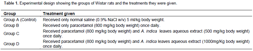
Eighty Wistar rats were used in this study. They were randomly divided into four groups of twenty animals each and given different treatments as shown in Table 1. Leaves extract, paracetamol, and normal saline were administered with the aid of a feeding cannula.
Sample collection
After 14 days of treatment, all the animals were weighed and sacrificed via euthanasia using chloroform after a fasting of 16 h following the last administration. Blood was collected by cardiac puncture, allowed to clot and then centrifuged at 10,000 revolutions per minute for 5 min using Wisperfuge model 1384 (Tamson, Holland). Serum was separated for various biochemical analyses and stored at -20°C prior to use. The livers were dissected from all the animals, cleared of blood using normal saline and immediately transferred into blood ice-cold container of normal saline. The livers were homogenized in 0.1 N tris-HCL buffer (7.4) and used for the estimation of vitamins C and E (Challopadhyay and Bandyopadhyay, 2005).
Acute toxicity testing
The acute toxicity of A. indica leaves aqueous extract was done using 30 mice divided into 5 groups of 6 mice each. Each group received graded doses (200 – 1000 mg/kg body weight) of the extract and the animals observed for toxic effects after 48 h of treatment. The toxicological effect was observed in terms of mortality expressed as LD50. The number of animals that died during the period was noted. The LD50 of the extract was estimated from the graph of percentage (%) mortality, converted to probity, against log-dose of the extract, probit 5 being 50% (Litch and Wilcoxon, 1959).
Laboratory methods and procedures/biochemical analysis
All reagents were commercially procured and with strict adherence to the manufacturer’s Standard Operation Procedures (SOP) in carrying out the analysis.
Liver enzymes
Both AST and ALT were estimated using the method of Reitman and Frankel (1957). AST was measured by monitoring the concentration of oxaloacetate hydrazone formed with 2,4-dinitrophenylhydrazine (2,4-DNPH) read spectrophotometrically at 546 nm.
0.1 ml of serum was added to 0.5 ml of buffered AST substrate and incubated at 25°C for 20 min. 5.0 ml of sodium hydroxide (0.4 mol/l) was added at the end of 20 min, mixed and allowed for 5 min. The blank was set up in the same manner except for the addition of distilled water in the place of serum. The absorbance of the sample was read against the blank at 546 nm wavelength using a spectrophotometer.
ALT was measured by monitoring the concentration of pyruvate hydrazine formed with 2,4-DNPH read spectrophotometrically at 546 nm. The procedure is the same as AST except that 0.5 ml of buffered ALT substrate was used in the place of 0.5 ml of buffered AST substrate. AST and ALT activities were then obtained from the respective tables provided by the manufacturer in the SOP.
Alkaline phosphatase
The substrate, p-nitrophenyl phosphate (colourless), on hydrolysis catalyzed by ALP, produces phosphate and p-nitrophenol (yellow). The production of p-nitrophenol was monitored and measured spectrophotometrically at 405 nm (King and King, 1954).
To a tube containing 1.0 ml of buffered ALP substrate was added 0.02 ml of serum sample and mixed, the initial absorbance was spectrophotometrically read at 405 nm and absorbance reading repeated at 1, 2 and 3 min. ALP activity was calculated using the formula: u/l = 2760 x ∆ 405/min.
Vitamin C (ascorbic acid)
Ascorbic acid is converted to dehydroascorbic acid by cupric ion with 2,4-DNPH in the presence of thiourea as a mild reducing agent, sulphuric acid then converts DNPH to a red coloured compound which is read spectrophotometrically at 530 nm (Omaye et al., 1979).
0.5 ml each of serum and liver homogenate were added to different tubes and 1.5 ml of the standard add to another tube and to all the tubes were added 0.5 ml of DNPH reagent (92% DNPH in 0.9 N sulphuric acid, 4% thiourea and cupric sulphate solution), mixed and incubated at room temperature for 3 h. Thereafter, 2.5 ml of 8.5% sulphuric acid was added to each tube and the colour developed read spectrophotometrically at 530 nm after 30 min. The values were then calculated.
Vitamin E
Vitamin E reduces ferric to ferrous ions which then forms a red complex with x-x-dipyridyl. Vitamin E and carotenes were first extracted into xylene and extinction read at 460 nm to measure the carotenes. A correction is made for these after adding ferric chloride and reading at 520 nm (Quaife et al., 1949).
Statistical analysis
All values were expressed as mean ± SD and then subjected to analysis of variance (ANOVA) using the Statistical Package for Social Sciences (SPSS) version 17.0 (SPSS Inc., Chicago Illinois). Statistical significance was considered at ρ< 0.05.
RESULTS
Table 2 shows that there was a significant increase in the body weight of rats in groups A and D, and a decrease in group B (ρ<0.05). There was no significant change in the weights of Wistar rats in group C (p>0.05).
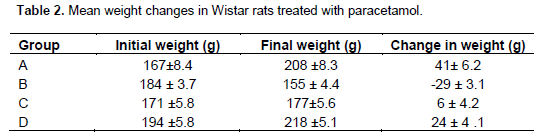
Table 3 shows that there was a significant increase in the plasma levels of the liver enzymes AST and ALT in group B as compared to those of group A (p>0.05). The plasma levels of AST and ALT in group C Wistar rats significantly increased when compared with rats in group A (ρ<0.5). However, these enzymes were significantly decreased when compared with rats in group B (p>0.05). There was no significant variation in plasma ALP level of group C from those of groups A and B rats (p>0.05).
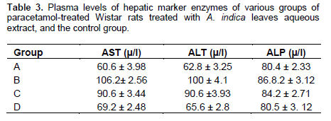
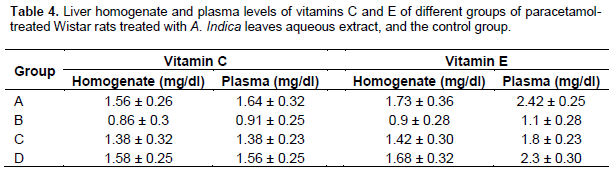
Table 4 shows that the homogenate and plasma levels of vitamins C and E in group B were significantly decreased when compared with control group A Wistar rats (p>0.05). But there was no significant difference in these vitamins in groups C and D when compared with group A (p>0.05). The homogenate and plasma vitamins C and E levels in groups C and D were observed to be significantly increased when compared with group B (p<0.05). Analysis shows that the differences between the homogenate/plasma levels of both vitamins C and E in groups C and D were significant (p<0.05).
DISCUSSION
In this study, it appears that the overall metabolic effects of various treatments of the different groups of rats were summarized in their respective weight changes. The finding of this study shows a significant increase in the body weights of group A wistar rats, which is consistent with the normal physiological features as a result of normal metabolic processes in normally fed and conditioned animal. On the contrary, rats in group B show a significant decrease in the body weight, which may be attributed to the negative biochemical effect engendered by the paracetamol induced oxidative stress (Bhanwra et al., 2000). For groups C and D which were treated with paracetamol and varied doses of A. indica leaves aqueous extract, there was no significant weight increase in group C but significant increase in group D. This is reminiscent of the antioxidant effect of the extract Nwanjo and Orjiako, 2006), thus obliterating the effects of the oxidative stress, which could have been induced by paracetamol (Hazai et al., 2002).
The observed significant increase in the hepatic marker enzymes (AST and ALT) in paracetamol treated Wistar rats (group B) as compared to group A may implicate stress on the liver enzymes by paracetamol. This is further supported by the plasma AST/ALT ratio of less than one (AST/ALT <1) which may indicate ensuing acute or chronic liver injury (Essani et al., 1995). This could be due the fact that cellular enzymes extrude into the extracellular fluid, thus raising their concentrations in plasma (Adeyemi and Bukola, 2014) due to hepatocellular damage, hence the rise in their plasma levels. The significant decrease in AST and ALT observed in groups C and D when compared with group B may be attributed to the antioxidative or the hepatoprotective effect of leaves aqueous extract of A. indica which is similar to documented reports on the hepatoprotective activity of A. indica (Ha et al., 2010). The extract may have an effect on the Nrf2 regulation, thereby conferring the preservation of hepatocellular integrity against paracetamol-induced hepatotoxicity, which is in tandem with the work of Thomson (2013). The non-significant increase in plasma ALP in group B may mean that though paracetamol intoxication causes hepatocellular damage (Bhanwra et al., 2000), cholestasis may not be primarily involved.
Also, in this study, the result indicates that homogenate and plasma levels of vitamins C and E in group B are significantly decreased when compared with group A Wistar rats. But there was no significant difference in these vitamins in groups C and D when compared with group A. The depletion of vitamins C and E observed in the paracetamol intoxicated Wistar rats (group B) could be correlated with the excessive utilization of non-enzymic antioxidants in scavenging enormous free radicals produced and this corroborates with the widely studied role of antioxidants like vitamins C and E in xenobiotics-induced oxidative stress and hepatoprotection (Sharma et al., 2010). On treatment with both paracetamol and A. indica leaves aqueous extract, animals in groups C and D showed minimal or no change in levels of vitamins C and E when compared with the control group. The extract may have impacted positively on the Nrf2 which in turn increased the production of antioxidant proteins which mopped up the oxidants, thereby conserving vitamins C and E. However, there is a significant increase when compared with group B. A. indica as an antioxidant prevents lipid peroxidation thereby reducing
CONCLUSION
In conclusion, based on these findings, it could be inferred that A. indica leaves aqueous extract enhances vitamins C and E levels in paracetamol induced hepatocellular damage in Wistar rats. This could be due to its antioxidant activity and its effect on Nrf2 regulation. It, therefore, becomes pertinent to develop ways to modulate cell specific Nrf2 activity to facilitate the development of novel strategies for the treatment of oxidative stress-induced diseases.
CONFLICT OF INTERESTS
The authors have not declared any conflict of interests.
REFERENCES
|
Adeyemi OS, Bukola TO (2014). Lipid profile and oxidative stress markers in Wistar rats following oral and repeated exposure to fijk herbal mixture. J. Toxicol. 2014:876035. |
|
|
Alimba CG, Bakare AA, Aina OO (2012). Liver and kidney dysfunction in Wistar rats exposed to munincipal landfill leachate. Resour. Environ. 2(4):150-163. |
|
|
Bhanwra S, Singh P, Koshla P (2000). The effect of Azadirachta indica (Neem) leaf aqueous extract on paracetamol induced liver damage in rats. Indian J. Physiol Pharmacol. 44:4-69. |
|
|
Biswas K, Chattopadhyay I, Banerjee RK, Bandyopadhyay U (2002). Biological activities and Medicinal properties of neem (Azadirachta indica). Curr. Sci. 82(II):1336-1345. |
|
|
Boeke SJ, Boersma MG, Alink GM, van Loon JJ, van Huis A, Dicke M, Rietjens IM (2004). Safety evaluation of neem (Azadirachta indica) derived pesticides. J. Ethnopharmacol. 94(1):25-41. |
|
|
Challopadhyay RR, Bandyopadhyay M (2005). Possible mechanism of hepatoprotective activity of Azadirachta indica leaf extract against paracetamol induced hepatic damage in rats. Part.III. Indian J. Pharmacol. 37(3):184-185. |
|
|
Essani NA, Fisher MA, Farhood A, Manning AM, Smith CW, Jaeschke H (1995). Cytokine-induced hepatic intercellular adhesion molecule-1 (ICAM-1) messenger RNA expression and its role in the pathology of murine endotoxin shock and acute liver failure. Hepatology 21:1632-1639. |
|
|
Frie B, Stocker R, Ames BN (1988). Antioxidant defences and lipid peroxidation in human blood plasma. Proc. Nat. Acad. Sci. USA 85:9748-9752. |
|
|
Halliwell B, Glutteridge JMC (1989). (Eds.). Free Radicals in Biology and Medicine. 2ndedition. Oxford, UK: Clarendon Press. |
|
|
Hazai E, Vereczkey L, Monostory K (2002). Reduction of toxic metabolites formation of acetaminophen. Broch. Biophys. Res. Ommun. 291(4):1089-1094. |
|
|
Ha HL, Shin HJ, Feitelson MA, Yu DY (2010). Oxidative stress and antioxidants in hepatic pathogenesis. World J. Gastroenterol. 16(48): 6035-6043. |
|
|
Hossain MA, Al-Toubi WAS, Weli AM, Al-Riyami QA, Al-Sabahi JN (2013). Identification and characterization of chemical compounds in different crude extracts from leaves of Omani neem. J. Taibah Univ. Sci. 7(4):181-188. |
|
|
Ihim AC, Nwosu DC, Nwanjo HU, Onyenekwe CC, Nnodim JK, Manafa PO, Okeke AC, Oluboye AO, Nwobodo EI, Nwadike C, Oze G (2013). Antilipid peroxidative and hypolipidemic potentials of Nauclea latifolia leaves extract on ciprofloxacin induced oxidative stress in rats. Asian J. Pharm. Biol. Res. 3(1):18-23. |
|
|
King EJ, King PR (1954). Estimation of plasma phosphates by determination of hydrolyzed phenol with amino antipyrene. J. Chem. Pathol. 7:322-326. |
|
|
Kutlu M, NaziroÄŸhu M, SimÅŸek, H, Yilmaz T, Sahap Kükner A (2005). Moderate exercise combined with dietary Vitamin C and E counteract oxidative stress in kidney and lens of streptozotocin - induced diabetic rats. Int. J. Vit. Nutr. Res. 75:71-80. |
|
|
Litch JT, Wilcoxon F (1959). Determination of acute toxicity tests. J. Pharmacol. Exp. Ther. 96:99-113. |
|
|
Masalkar PD, Abhang SA (2005). Oxidative stress and antioxidant status in patients with alcoholic liver disease. Clin. Chim. Acta 355(1-2):61-65. |
|
|
Molavi B, Mehta JL (2004). Oxidative stress in cardiovascular disease: Molecular basis of its deleterious effects, its detection and therapeutic considerations. Curr. Opin. Cardiol. 19(5):488-493. |
|
|
Niki E (1991). Actions of ascorbic acid as a scavenger of active and stable oxygen species. Am. J. Clin. Nutr. 54:1119S-24S. |
|
|
Nunomura A, Castellani R, Zhu X, Moreira PI, Perry G, Smith MA (2006). Involvement of oxidative stress in Alzheimer disease. J. Neuropathol. Exp. Neurol. 65(7):631-641. |
|
|
Nwanjo HU, Orjiako AO (2006). Effects of vitamins E and C on exercise-induced oxidative stress. Glob. J. Pure Appl. Sci. 12(2):199-202. |
|
|
Omaye ST, Turnbull JD, Sauberlich HE (1979). Selected methods for the determination of Ascorbic acid in animals cells, tissues and fluids. Methods Enzymol. 62:3-11. |
|
|
Pennington TD, Styles BT (1975). A generic monograph of the melicicea. Blumea 22:419-540. |
|
|
Quaife ML, Scrimshaw NS, Lowry OH (1949). A micromethod for assay of totaltocopherols in blood serum. J. Biol. Chem. 180(3):1229-1235. |
|
|
Rao GM, Sumita P, Roshni M, Ashtagimatt MN (2005). Plasma antioxidant vitamins and lipid peroxidation products in pregnancy induced hypertension. Indian J. Clin. Biochem. 20(1):198-200. |
|
|
Reitman S, Frankel S (1957). Colorimetric method for the determination of serum glutamate oxaloacetic transaminase and glutamate pyruvate transaminases. Am. J. Clin. Pathol. 28(1):56-63. |
|
|
Sen P, Medinata PK, Ray A (1992). Immunostimulant activities of A. indica. Indian J. Exp. Biol. 12:1170-1175. |
|
|
Sharma P, Shankar S, Agarwal A, Singh R (2010). Variations in serum lipids and liver function markers in lindane exposed female Wistar rats: Attenuating effect of circumin, vitamin C and vitamin E. Asian J. Exp. Biol. Sci. 1(2):440-444. |
|
|
Thomson G (2013). Review: Nrf2, a multi organ protector. Available at: |
|
|
Valko M, Leibfritz D, Moncol J, Cronin MT, Mazur M, Telser J (2007). Free radicals and antioxidants in normal physiological functions and human diseases. Int. J. Biochem. Cell Biol. 39(1):44-84. |
|
|
Veinbergs I, Mallory M, Sagara Y, Masliah E (2000). Vitamin E supplementation prevents spatial learning deficit and dendritic alteration in aged apolipoprotein E deficient mice. Eur. J. Neurosci. 12(12):4541-4546. |
|
|
Willcox JK, Ash SL, Catignani GL (2004). Antioxidants and prevention of chronic diseases. Crit. Rev. Food Sci. Nutr. 44(4):275-295. |
|
|
Zaidi SM, Al-Qirim TM, Banu N (2005). Effects of antioxidant vitamins on glutathione depletion and lipid peroxidation induced by restraint stress in the rat liver. Drugs R D. 6(3):157-65. |
|
Copyright © 2024 Author(s) retain the copyright of this article.
This article is published under the terms of the Creative Commons Attribution License 4.0


