Full Length Research Paper

ABSTRACT
This research evaluates the value of loading Nigella sativa on chitosan nanoparticles (ChNPs) in the treatment of Schistosoma mansoni infection compared to praziquantel. Chitosan has efficient oral administrative capability to penetrate mucosal surface being mucoadhesive and non-toxic, as well as combination with NPs add characters of controlled release and safe serum levels. The study was done on 40 S. mansoni infected male mice which were divided into 4 groups: positive control group (infected, non treated), group treated with N. sativa only (NS), group treated with N. sativa loaded chitosan NPs (NC) and group treated with combined praziquantel and N. sativa loaded chitosan nanoparticles (NCP). Results showed decreased worm burden in NC and NCP groups concerning the control and NS groups while significant decrease occurred in oogram pattern and tissue egg loads reached reduction percentages more than 90% in NC and NCP groups. On the level of granuloma diameter reduction, it was 46.3% in NC and 41.3% in NCP group while granuloma number was reduced by 50.9% in NC and reduced by 32% in NCP group revealing the apparent role of ChNPs in improvement of bilharzial hepatic changes. Thus, ChNPs improved the effects of N. sativa in therapy of murine schistosomal infection by enhancing the effects of praziquantel and reducing the resistance to it. This can be considered as a new strategy using ChNPs as anti-schistosomal drug carrier in mice models.
Key words: Nigella sativa, chitosan nanoparticles, Schistosoma mansoni, praziquantel
INTRODUCTION
Regardless of the many efforts to control schistosomiasis infection in Egypt, it is still one of the most common endemic diseases achieving high prevalence in Egypt like many other developing countries. Schistosomiasis represents one of the most common parasitic diseases worldwide and the second most harmful parasitic infection after malaria in the aspect of economic impact and disabling infection (Ali et al., 2016).
Drug-resistant Schistosoma strains were shown in endemic areas due to the repeated use of praziquantel and oxamniquine (Zhang and Coultas, 2013), hence in the last few years, there was an increased request for the use of anti-parasitic drugs of natural plant sources (Magalhães et al., 2009). Accordingly, the importance of exploring new effective natural compounds to be used in treatment of schistosomiasis is a must (John et al., 2007).
Nigella sativa oil is considered one of the plant derivatives which have been screened as active being active agent for adult Schistosoma mansoni and is a promising drug of a plant origin that has anti-schistosomal efficacy (Mohamed et al., 2005).
Seeds of N. sativa which are known as black seeds were a fertile source for treatment of abdominal pain, diarrhea, cough, asthma, and rheumatism and other disease as a part of traditional medicine in Asia, Middle and Far Eastern countries. Many phytochemicals and pharmacological studies proved the antioxidant, anti-inflammatory, anticancer and antimicrobial activities of the aqueous and oil extracts of these seeds. The incorporation of N. sativa into solid nanoparticles as a new delivery system revealed them as a suitable carrier in the pharmaceutical fields with high physical stability (Al-Haj et al., 2010).
Chitosan is produced by removal of acetate moiety of chitin, a powerful source of chitosan is the crustacean cells of crabs in addition to fungal cell walls, and chitosan is a natural biocompatible polymer, which is characterized by being highly basic, mucoadhesive, and cationic polysaccharide. Being a penetration enhancer, chitosan promotes both paracellular and intercellular transport of drugs by opening the tight junctions of the epithelium (Mohammed et al., 2017).
On testing resistance of chitosan NPs to the effect of pH and temperature in media that simulate the stomach and intestine, it was found very stable at 37°C in different buffers (Oliveira et al., 2012). Being a polysaccharide source for nanoparticles, chitosan was successfully applied as drug delivery system because polysaccharides are stable, safe, non-toxic, hydrophilic and biodegradable; also, the electrostatic interaction between positively charged polymers as chitosan and negatively charged mucin makes chitosan more mucoadhesive, as well as its initial burst effect and slow drug release which were proven in vitro contribute to their urgent necessity to be used in vivo ChNPs (Bilia et al., 2014).
Nanoparticle technology became one method offering a valuable tool to novel drug delivery strategies, having the characters of controlled release, safe serum levels of active components from enzymatic or environmental degradation and internal retention. Nanoparticle manufacturing methods apply to a wide range of drugs (Nagpal et al., 2010). Chitosan-based nanoparticles are also used in the treatment of cancer, gastrointestinal diseases, pulmonary diseases, drug delivery to the brain and ocular infections (Mohammed et al., 2017). The aim of this study is to evaluate the value of loading N. sativa on chitosan nanoparticles (ChNPs) in treatment of S. mansoni adult worms infection compared to praziquantel.
MATERIALS AND METHODS
This experimental study was conducted in the Schistosoma Biological Supply Centre (SBSC), Theodore Bilharz Research Institute of Giza in Egypt during the period from July 2018 to October 2018. No specific grant from any funding agency was provided towards this research.
Materials and drugs
Chitosan (deacetylation degree of 93%) and sodium tripolyphosphate (Na TPP) were purchased from Sigma-Aldrich, USA. PBS and acetic acid were obtained from Sigma–Aldrich, USA. N. sativa oil (Baraka) in the form of gelatinous capsules 450 mg was obtained from Pharco, Egypt. The inotropic gelation of chitosan with Na TPP anions helps in the synthesis of ChNPs (Kawashima et al., 1985; Werle et al., 2009). This interaction was controlled by the charge density of Na TPP and chitosan under the effect of the solution pH. Various concentrations of acetic aqueous solutions 1, 2 and 3 mg/ml were used to dissolve chitosan. Na TPP solution (1 mg/ml) was prepared by double-distilled water. ChNPs were produced by dropwise addition of 5 ml of the chitosan solution on 2 ml of Na TPP solution by the effect of 1000 rpm magnetic stirring for 1 h at room temperature. By centrifugation at 20000 g at 14°C for 30 min, we did separations of the nanoparticles and then they were freeze-dried and stored at 4°C.
Loading of N. sativa to ChNPs was made by adding chitosan solution to Na TPP solution (containing N. sativa at a concentration of 500 mg/2 ml). N. sativa loaded on ChNPs (NS/ChNPs) was separated from the suspension by centrifugation (20000 g at 14°C) for 30 min. Thereafter, sediment was collected and weighed. The particle size was determined by dynamic light scattering (DLS) using Zetasizer Nano Instrument (Malvern Instruments, UK) according to (Koukaras et al., 2012) to record that at chitosan/Na TPP w/w ratio of 2.5/1, the nanoparticle diameter was 200-396 nm and physical appearance was opalescent suspension. The total protein content/mg of chitosan encapsulating powder was calculated by dividing the protein concentration of the loaded N. sativa/the nanoparticles’ weight (Danesh-Bahreini et al., 2011). The loading efficiency of the nanoparticles was determined by:
%LC= [(A-B)/C] × 100
Where A letter points to the total amount of N. sativa, B letter points to the free amount of N. sativa and C letter points to the weight of nanoparticles.
N. sativa (Baraka) gelatinous capsules (450 mg) were obtained from Pharco Pharmaceuticals, Egypt, dissolved in corn oil and adjusted to be given at dose 1140 mg/kg. Praziquantel tablets (Distocide, EIPICO, El-Asher Men Ramadan, Egypt) were crushed, administered orally as a suspension in 2% Cremophore-E1 (Sigma-Aldrich Chemical Co., St. Louis, MO) (Fallon and Doenhoff, 1994). Praziquantel was given to mice in a dose of 500 mg/kg divided in half and given on two consecutive days (half full dose).
Experimental design
40 male mice, aged 6-8 weeks and weighed 20–25 g, were included in our study. Mice were housed in well-ventilated cages and fed standard pellet food with free access to water (El Fakhry et al., 1998). Mice were divided into four groups (10 mice/group):
Control group: control positive, infected mice and not treated.
NS group: treated with N. sativa only.
NC group: treated with NS/ChNPs.
NCP group: treated with combined praziquantel and NS/ChNPs.
Mice infection and treatment
Cercariae of S. mansoni were obtained from infected Biomphalaria Alexandrina snails, reared and maintained at Schistosome Biological Supply Program (SBSP), Theodore Bilharz Research Institute, in the governorate of Giza, Egypt. Mice were infected subcutaneously with freshly shed 60±10 cercariae/mouse (Liang et al., 1987). The treatment schedule included that N. sativa only was given by a dose of 1140 mg/kg day after day. NS/ChNPs was given in a dose of 1140 mg/kg daily. Combined praziquantel and NS/ChNPs were given at a dose of 500 mg/kg praziquantel divided in half and given on two consecutive days + 570 mg/kg NS/ChNPs daily. Treatment started from the 7th week post-infection and continued for two weeks. All mice were sacrificed by cervical dislocation at 9 weeks post-infection.
Worm recovery
The worm burden of S. mansoni recovered from the hepatic portal system and mesenteric veins of sacrificed mice was done by the perfusion technique described by (Smithers and Terry, 1965).
Egg count examination
a) Oogram pattern: small intestine of sacrificed mice was divided into 3 parts (each 1 cm in length), then squeezed between a glass slide and coverslip to show and count immature, mature and dead egg stages of S. mansoni in each part and the mean number of each stage was calculated (Pellegrino et al., 1962).
b) Tissue egg load: the number of ova per gram the liver or intestinal tissue was determined by digesting a part of liver or middle intestine in 5% potassium hydroxide overnight (Cheever, 1968).
Histopathological examination
It is done by fixing liver samples from all groups of the experiment in a 10% formalin solution and then embedding these samples in paraffin wax and staining of five sections with hematoxylin and eosin (Bancroft and Stevens, 1975) and Masson Trichrome stain (Masson, 1929). The number of liver egg granulomas was assessed by microscopic examination of the prepared slides through five successive low power fields (10X) while the granuloma mean diameter was calculated by dividing the sum of vertical and transverse diameters/2 (Mahmoud and Warren, 1974).
Ethical considerations
The Ethical Committee of the Faculty of Medicine, Benha University, Egypt and that of Theodore Bilharz Research Institute approved the study according to the international guiding principles for biomedical research involving animals as issued by the international organizations of medical sciences.
Statistical analysis
Results were collected, tabulated and statistically analyzed using the statistical package SPSS version 12. Data were tabulated as mean and standard deviation (SD) for quantitative variables and percent for qualitative variables. ANOVA was used to detect significance in the quantitative variables, and P values <0.05 were considered as statistically significant. Post-hoc Bonferroni test was used.
RESULTS
Parasitological study
Worm burden
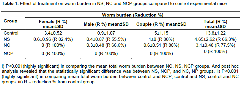
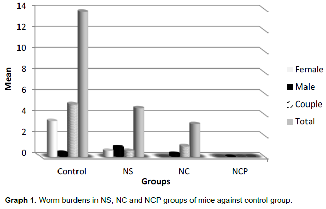
The mean female worm burden of control mice and NS group was 3.4±0.51 and 0.6±0.96 respectively with no female worms found in NC and NCP groups achieving reduction percentage of 100% while the mean male worm burden in control, NS and NC groups was 0.9±0.87, 0.4±1.07 and 0.3±0.48 respectively with no male worms in NCP group. Also, mean couple worm burden in control, NS and NC groups was 5±1.15, 1±0 and 0.6±0.51 respectively showing high reduction percentage of 88% in NC group and no couple worms in NCP group while total worm burden in the same group was 13.8±1.22, 4.65±2.62 and 3.1±0.48 and 0 respectively. These results revealed achievement of a reduction in total worm burdens from the control group by 66.3, 77.5 and 100% in NS, NC and NCP groups respectively (Table 1, Graph 1).
Oogram pattern
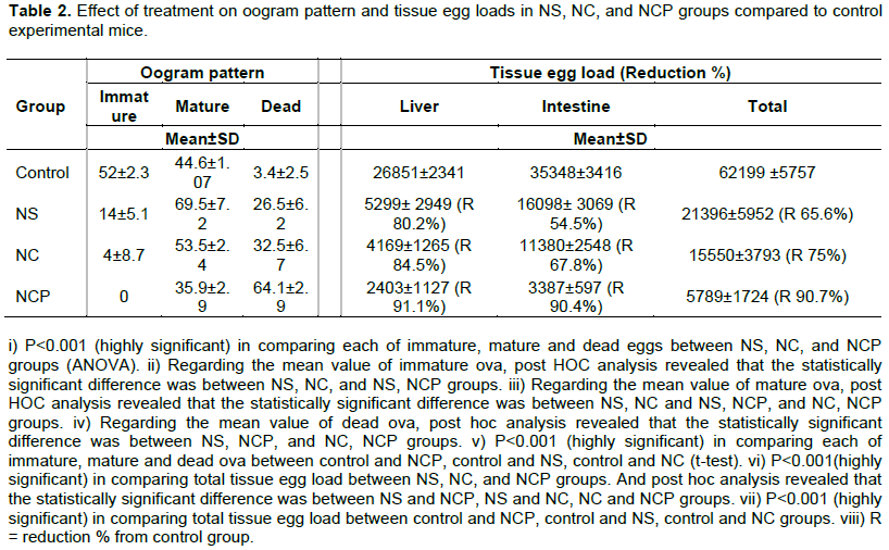
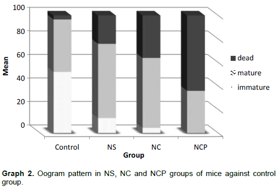
Per every 100 eggs in control group mice we found that mean number of immature, mature and dead eggs was 52±2.3, 44.6±1.07 and 3.4±2.5 respectively while in NS group mice the mean number of immature, mature and dead eggs was 14±5.1, 69.5±7.2 and 26.5±6.2 respectively. Regarding the NC group mice the mean number of immature, mature and dead eggs was 4±8.7, 53.5±2.4 and 32.5±6.7 respectively while in NCP group mice, there was no immature eggs; also, mean number of mature and dead eggs was 35.9±2.9 and 64.1±2.9 (Table 2, Graph 2) regarding the mean number of immature eggs.
Tissue egg load (No. of eggs per gram)
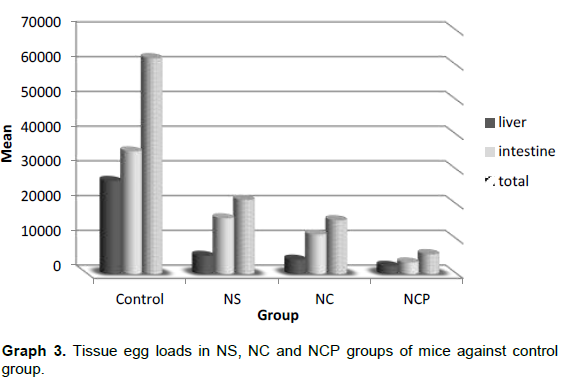
The mean liver egg count of control, NS, NC and NCP groups of mice was 26851±2341, 5298.55±2949, 4169.38±1264.6 and 2402.54±1127 respectively with highest reduction percentages from control in NC and NCP groups by 84.5 and 91.1% respectively, while the mean intestine egg count of control, NS, NC, NCP groups of mice was 35348±3416, 16098± 3069, 11380±2548 and 3387±597 respectively with highest reduction percentages in NC and NCP groups as 67.8 and 90.4% respectively; also, the mean total tissue egg load for the same groups respectively was 62199±5757, 21396±5952, 15550±3793 and 5789±1724 (Table 2, Graph 3).
Histopathological study
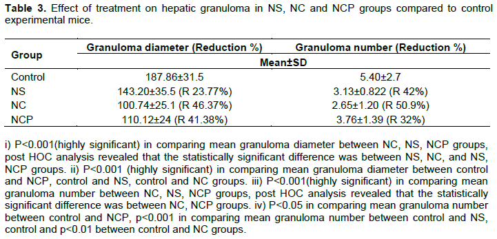
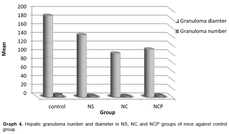
The mean diameter of liver granuloma of control, NS, NC, and NCP groups was 187.86±31.5, 143.20±35.5, 100.74±25.1 and 110.12±24 with reduction of the NS, NC and NCP groups from control by 23.77, 46.37 and 41.38 % respectively showing highest reduction percentage in NC group as 46.37%. The mean number of liver granulomas in the same groups was 5.40±2.7, 3.13±0.822, 2.65±1.20 and 3.76±1.39 respectively with the highest reduction percentage in NC group as 50.9% (Table 3, Graph 4).
DISCUSSION
N. sativa has a remarkable anti-oxidant and anti-helminthic activity with a considerable reduction in the total number of recovered worms and the possible explanation of its lethal effect on worms is due to its content of alkaloid nigellicine (El Shenawy et al., 2008). The importance of chitosan as a drug carrier seems obvious when the successful administration of many drugs and macromolecules was enhanced by the availability of drug carriers to concentrate it at target site, thus decreasing therapeutically unused drug levels and their side effects (Grenha et al., 2010). Chitosan was synthesized by adding Na TPP which is nontoxic, multivalent and can form gels via ionic interactions so it has been used to prepare ChNPs (Kawashima et al., 1985; Werle et al., 2009).
In our study, we used N. sativa alone and NS/ChNPs in combination with praziquantel to highlight the importance of treatment of S. mansoni infections in experimental mice with NS/ChNPs which is a novel issue. The total worm burden was reduced by 66.3% in the NS group, by 77.5% in the NC group while all worms were killed in the NCP group. Our results were near to those of Ali et al. (2016) who revealed reduction in total worm burden in mice infected by S. mansoni by 57% when treated by N. sativa oil alone and by 47% when treated by N. sativa oil and Chroococcus turgidus algae. Regarding the female and male worm burdens, in the present work there are reductions in both by 82 and 55% respectively in NS group and by 100 and 66% respectively in NC group coinciding with the results of Ali et al. (2016), who revealed reduction of both female and male worm burdens by 41 and 64% respectively in N. sativa oil treatment while in contrast to combined treatment which showed reduction by 21 and 49% respectively, this highlights the importance of loading N. sativa on ChNPs in our study in addition to the enormous results achieved by combination with praziquantel which caused depletion of all worms. Our results were higher than Abououf et al. (2018) who used N. sativa only and combined with praziquantel against mature S. mansoni in mice revealing reduction percentage in female, male, couple and total worm burdens as 65.9, 50, 68.6 and 57.5% respectively in their group which was treated by N. sativa only and reductions were 89.2, 97.5, 91.6 and 93.7% respectively for the same worm burdens in their group which was given combined treatment of praziquantel and N. sativa oil; proving highly effective NS/ChNPs in our study which showed 100% reduction in all worm burdens when combined with praziquantel. On comparing efficacy of black seeds (N. sativa) oil on other parasitic infections like Hymenolepis nana in infected mice, results revealed there was a significant decrease in the mean number of eggs per gram of feces by 88.85% at 7th day after treatment with 5 ml/kg and by 100% at 14th day of treatment. This was proof of the wide scaled efficacy of N. sativa in parasitic infections (Al-Megrin, 2016).
In the current study, NS/ChNPs affects greatly the oviposition of female worms with decreased immature eggs in oogram pattern from 52% in control group mice to 4% in NC group while there were increased dead eggs from 3.4% in control group mice to 32.5% in NC group indicating affection of fecundity of female worms by NS/ChNPs. These observations were also reported by Utzinger et al. (2002), Suleiman et al. (2004) and Mati et al. (2010), as they found that reduction in the worm burden and egg density in treated mice was considered as a strong indication of the effectiveness of anti-schistosomal agents. In the study of Abououf et al. (2018) by using N. sativa oil alone against mature worms, the dead eggs increased from 5.25 to 21.4% of oogram pattern and immature eggs decreased from 62.5 to 31% of oogram in the untreated mice group while our results relying on NS/ChNPs achieved more treating effects as mentioned above, thus proving the highly effective ChNPs as drug delivery system in treatment of S. mansoni mature worms. In other study, dead eggs increased from 9 to 16% of oogram in S. mansoni infected mice when treated with a mixture of N. sativa oil and aqueous garlic extract and immature eggs decreased from 54 to 20% of oogram pattern in the same group of mice (El Shenawy et al., 2008). On comparison with the study of Metwally et al. (2018) by using other treating substances like garlic and allicin, dead eggs increased from 6.1 to 7.5% for garlic and 8% for allicin in oogram pattern while immature eggs decreased from 54.5 to 47.9% for garlic and 46.4% for allicin in oogram. In vivo activity experiments of Epiisopiloturine against adult S. mansoni revealed increase in dead eggs from 2 to 36% in oogram pattern while immature eggs decreased from 79 to 24% when a dose of 40 mg/kg was given 45 days post-infection (Guimarães et al., 2015).
In the group treated with combined therapy of N. sativa with praziquantel, dead ova increased from 3.4 to 64.1% in oogram pattern while immature ova were completely disappeared and this coincided with the study of Abououf et al. (2018).
On using praziquantel alone in treatment of mice infected with S. mansoni, the dead eggs increased from 12 to 81.1%, using N. sativa only dead eggs increased from 12 to 17.8% and using praziquantel combined with N. sativa, the dead eggs increased from 12 to 95.8% in oogram pattern (Mahmoud et al., 2002). These results were higher than our study revealing that praziquantel in combinations improved the treating abilities of N. sativa oil while NS/ChNPs without being combined with praziquantel have the second higher treatable effects.
In our study, NS/ChNPs showed a reduction in liver egg load by 84.5% and intestine egg load by 67.8% from the control mice. This significant reduction was higher than that of Abououf et al. (2018) who recorded reduced liver egg load by 57.8% and intestine egg load by 81.4% when N. sativa oil was used alone indicating shift of NS/ChNPs to liver tissue. Also, these results coincided with those of Ali et al. (2016) who showed significant reduction in number of eggs per gram liver tissue when treated with algal extract, N. sativa oil and both by percentages of 56, 65 and 74% respectively, while the reduction percentages in intestine egg loads were 47, 44 and 62% of the same treated groups respectively. At the same time, our results achieved higher treating results than Mahmoud et al. (2002) who showed reduction in liver and intestine egg loads in mice treated by N. sativa alone by 33.7 and 33.2% respectively, while when combined with praziquantel of different doses, reductions were by 77.1 - 80.7% and 93.8 - 92.9% in liver and intestine egg loads respectively. Also, Mahmoud et al. (2002) results proved that away from combinations with praziquantel, our NS/ChNPs have the upper hand in treating mice infected with S. mansoni.
Regarding the hepatic granuloma formed by S. mansoni, the present study revealed high effective treating abilities of NS/ChNPs on reducing the granuloma diameter by 46.3% and granuloma number by 50.9% from the control group while when combined with praziquantel, it showed lower results by reduction percentages of 41.3 and 32% respectively from the control indicating the high efficacy of NS/ChNPs in improvement of bilharzial hepatic changes. These results were different from Mahmoud et al. (2002) who revealed that the combination of praziquantel with N. sativa did not improve the hepatic granuloma diameter. Abououf et al., (2018) showed a reduction in granuloma diameter by 26.7% when treated with N. sativa oil, thus proving the high effect of NS/ChNPs but the reduction in granuloma number had nearly the same effect (51%). Comparing our results with those of Sheir et al. (2015) who showed reduction in granuloma diameter when mice treated by combined N. sativa with artemether and/or praziquantel to 35.4 and 32.2% respectively, we found that effective action was in the use of NS/ChNPs in our study.
Due to the ability of nanostructures to cross the cell and tissue barriers as a result of their very small size, they are considered now as a wide base in the biomedical sciences (Dou et al., 2016). Characters of nanostructures that make them more distinctive than the ordinary drug delivery systems include high specificity, high drug-carrying capacity, high stability, possibility to use different routes of administration and ability for controlled release (Pal et al., 2011). Chitosan has anti-bacterial and anti-fungal properties being soluble in diverse acids and able to form gels and complexes, and is also safe and non-toxic (Kim and Rajapakse, 2005). Moreover, ChNPs when used in mice infected with S. mansoni, produced 47% protection suggesting an important role in inducing immune response protecting against schistosomiasis (Oliveira et al., 2012).
Regarding the toxicity and safety of chitosan, there was no published data inquiring about safety of chitosan for human use or providing data about human toxicity of chitosan-based formulations. However, good safety was proved by several animal toxicity studies (Mohammed et al., 2017). Degradation of chitosan depends on the degree of its deacetylation and it is approved safe to be used in dietary consumption and for wound dressing (Wang et al., 2011).
CONCLUSION
This study thus revealed that NS/ChNPs is a novel anti-schistosomal combination that gives unique results in the elimination of adult schistosomal worms and hepatic granuloma size-reduction whether alone or combined with praziquantel.
CONFLICT OF INTERESTS
The authors have not declared any conflict of interests.
REFERENCES
|
Abououf EA, Elhamshary AMS, Nagati IM, Eraky MA, Elkholy AA, Ibrahim AN, Omar GH (2018). Effect of Nigella sativa oil on Schistosoma mansoni mature worms in experimentally infected mice. Journal of Egyptian Society of Parasitology 48(1):55-66. |
|
|
Al-Haj NA, Shamsudin M, Alipiah N, Zamri H, Ahmad B, Siddig I, Rasedee A (2010). Characterization of Nigella sativa L. essential oil-loaded solid lipid nanoparticles. American Journal of Pharmacology and Toxicology 5(1):52-57. |
|
|
Al-Megrin W (2016). Efficacy of black seeds oil (Nigella sativa) against Hymenolepis nana in infected mice. European Journal of Medicinal Plants 13(4):1-7. |
|
|
Ali M, Abou-Eldahab MA, Mansour HA, Nigm A (2016). Schistosoma mansoni: Antiparasitic effects of orally administered Nigella sativa oil and/or Chroococcus turgidus extract. Acta Biologica Hungaria 67(3):247-260. |
|
|
Bancroft JD, Stevens A (1975). Histopathological stains and their diagnostic uses. UK: Edinburgh & London: Churchill Livingstone. P 149. |
|
|
Bilia AR, Guccione C, Isacchi B, Righeschi C, Firenzuoli F, Bergonzi MC (2014). Essential oils loaded in nanosystems: a developing strategy for a successful therapeutic approach. Evidence-Based Complementary and Alternative Medicine. Volume 2014, Article ID 651593, 14 pages |
|
|
Cheever AW (1968). Conditions affecting the accuracy of potassium hydroxide digestion techniques for counting Schistosoma mansoni eggs in tissues. Bulletin of the World Health Organization 39(2):328-331. |
|
|
Danesh-Bahreini MA, Shokri J, Samiei A, Kamali-Sarvestani E, Barzegar-Jalali M, Mohammadi-Samani S (2011). Nanovaccine for leishmaniasis: preparation of chitosan nanoparticles containing Leishmania superoxide dismutase and evaluation of its immunogenicity in BALB/c mice. International Journal of nanomedicine 6:835-842. |
|
|
Dou Z, Wang G, Zhang E, Ning F, Zhu Q, Jiang J, Zhang T (2016). Effect of Al2O3 Nanoparticles Doping on the Microwave Dielectric Properties of CTLA Ceramics. Journal of Material Sciences and Engineering 5:256. |
|
|
El Fakhry Y, Achbarou A, Desportes-Livage I, Mazier D (1998). Encephalitozoon intestinalis: humoral responses in interferon-gamma receptor knockout mice infected with a microsporidium pathogenic in AIDS patients. Experimental parasitology 89(1):113-121. |
|
|
El Shenawy NS, Soliman MF, Reyad SI (2008). The effect of antioxidant properties of aqueous garlic extract and Nigella sativa as anti-schistosomiasis agents in mice. Revista do Instituto de Medicina Tropical de Sao Paulo 50(1):29-36. |
|
|
Fallon PG, Doenhoff MJ (1994). Drug-resistant schistosomiasis: resistance to praziquantel and oxamniquine induced in Schistosoma mansoni in mice is drug-specific. The American Journal of Tropical Medicine and Hygiene 51(1):83-88. |
|
|
Grenha A, Al-Qadi S, Seijo B, Remuñán-López C (2010). The potential of chitosan for pulmonary drug delivery. Journal of Drug Delivery Science and Technology 20(1):33-43. |
|
|
Guimarães MA, de Oliveira RN, Véras LM, Lima DF, Campelo YD, Campos SA, Kuckelhaus SA, Pinto PL, Eaton P, Mafud AC (2015). Anthelmintic activity in vivo of epiisopiloturine against juvenile and adult worms of Schistosoma mansoni. PLOS Neglected Tropical Diseases 9:e0003656. |
|
|
John AOO, Tinuade AO, Oyeku AO, Lateef AA (2007). Effectiveness of dried Carica papaya seeds against human intestinal parasitosis: a pilot study. Journal of Medicinal Food 10(1):194-196. |
|
|
Kawashima Y, Handa T, Kasai A, Takenaka H, Lin SY, Ando Y (1985). Novel method for the preparation of controlled-release theophylline granules coated with a polyelectrolyte complex of sodium polyphosphate-chitosan. Journal of pharmaceutical sciences 74(3):264-268. |
|
|
Kim SK, Rajapakse N (2005). Enzymatic production and biological activities of chitosan oligosaccharides (COS): A review. Carbohydrate Polymers 62(4):357-368. |
|
|
Koukaras EN, Papadimitriou SA, Bikiaris DN, Froudakis GE (2012). Insight on the formation of chitosan nanoparticles through ionotropic gelation with tripolyphosphate. Molecular Pharmaceutics 9(10):2856-2862. |
|
|
Liang Y, Bruce J, Boyd D (1987). Laboratory cultivation of schistosome vector snails and maintenance of schistosome life cycles. In: Proc First Sino-American Symposium pp. 34-48. |
|
|
Magalhães LG, Machado CB, Morais ER, Moreira ÉB, Soares CS, da Silva SH, Da Silva FAA, Rodrigues V (2009). In vitro schistosomicidal activity of curcumin against Schistosoma mansoni adult worms. Parasitology research 104(5):1197-1201. |
|
|
Mahmoud AA, Warren KS (1974). Anti-inflammatory effects of tartar emetic and niridazole: suppression of schistosome egg granuloma. The Journal of Immunology 112(1):222-228. |
|
|
Mahmoud M, El-Abhar H, Saleh S (2002). The effect of Nigella sativa oil against the liver damage induced by Schistosoma mansoni infection in mice. Journal of Ethnopharmacology 79(1):1-11. |
|
|
Masson P (1929). Some histological methods: trichrome staining and their preliminary technique. Journal of Technical Methods 12:75-90. |
|
|
Mati V, Freitas R, Melo A (2010). Effects of pentoxifylline during Schistosoma mansoni infection in Swiss mice: an analysis of worm burden, fecundity, and liver histopathology. Journal of Helminthology 84(4):348-354. |
|
|
Metwally DM, Al-Olayan EM, Alanazi M, Alzahrany SB, Semlali A (2018). Antischistosomal and anti-inflammatory activity of garlic and allicin compared with that of praziquantel in vivo. BMC complementary and alternative medicine 18(1):135. |
|
|
Mohamed AM, Metwally NM, Mahmoud SS (2005). Sativa seeds against Schistosoma mansoni different stages. Memórias do Instituto Oswaldo Cruz 100(2):205-211. |
|
|
Mohammed MA, Syeda JTM, Wasan KM, Wasan EK (2017). An Overview of Chitosan Nanoparticles and Its Application in Non-Parenteral Drug Delivery. Pharmaceutics 9(4). |
|
|
Nagpal K, Singh SK, Mishra DN (2010). Chitosan nanoparticles: a promising system in novel drug delivery. Chemical and Pharmaceutical Bulletin 58(11):1423-1430. |
|
|
Oliveira CR, Rezende CM, Silva MR, Borges OM, Pego AP, Goes AM (2012). Oral vaccination based on DNA-chitosan nanoparticles against Schistosoma mansoni infection. The Scientific World Journal 2012:938457. |
|
|
Pal SL, Jana U, Manna PK, Mohanta GP, Manavalan R (2011). Nanoparticle: An overview of preparation and characterization. Journal of Applied Pharmaceutical Science 1(6):228-234. |
|
|
Pellegrino J, Oliveira CA, Faria J, Cunha AS (1962). New approach to the screening of drugs in experimental schistosomiasis mansoni in mice. The American journal of tropical medicine and hygiene 11:201-215. |
|
|
Sheir SK, Maghraby A, Mohamed AH, Osman GY, Al-Qormuti SA (2015). Immunomodulatory and ameliorative role of Nigella sativa oil on Schistosoma mansoni infected mice. Canadian Journal of Pure and Applied Sciences 9:3345-3355. |
|
|
Smithers SR, Terry RJ (1965). The infection of laboratory hosts with cercariae of Schistosoma mansoni and the recovery of the adult worms. Parasitology 55(4):695-700. |
|
|
Suleiman M, Akarim E, Ibrahim K, Saad A, Mohammed A, Ahmed B, Sulaiman S (2004). Antischistosomal effects of praziquantel, its alkaline hydrolysis and sun decomposed products on experimentally S. mansoni infected albino mice.(A) Efficacy assessment based on clinicopathological findings. Journal of the Egyptian Society of Parasitology 34(1):131-142. |
|
|
Utzinger J, Chollet J, Tu Z, Shuhua X, Tanner M (2002). Comparative study of the effects of artemether and artesunate on juvenile and adult Schistosoma mansoni in experimentally infected mice. Transactions of The Royal Society of Tropical Medicine and Hygiene 96(3):318-323. |
|
|
Wang JJ, Zeng ZW, Xiao RZ, Xie T, Zhou GL, Zhan XR, and Wang SL (2011). Recent advances of chitosan nanoparticles as drug carriers. International Journal of Nanomedicine 6:765-774. |
|
|
Werle M, Takeuchi H, Bernkop-Schnurch A (2009). Modified chitosans for oral drug delivery. Journal of pharmaceutical sciences 98(5):1643-1656. |
|
|
Zhang SM, Coultas KA (2013). Identification of plumbagin and sanguinarine as effective chemotherapeutic agents for the treatment of schistosomiasis. International Journal for Parasitology: Drugs and Drug Resistance 3:28-34. |
|
Copyright © 2024 Author(s) retain the copyright of this article.
This article is published under the terms of the Creative Commons Attribution License 4.0


