Full Length Research Paper

ABSTRACT
Sargentodoxa cuneata is widely used in the treatment of rheumatic arthritis in China. This study was conducted to investigate the anti-inflammatory and immunoregulatory activities of the ethanol extracts from S. cuneata and analyze the bioactive constituents. The ethyl acetate (ESCe), dichloromethane (ESCd) and aqueous (ESCa) fractions at a dose of 200 mg/kg significantly inhibited mice ear edema. Furthermore, the ethyl acetate (ESCe) and aqueous fractions (ESCa) inhibited rat paw edema, decreased the levels of Malondialdehyde (MDA) and biosynthesis of the prostaglandin (PGE2), increased superoxide dismutase (SOD) activity, inhibited adjuvant arthritis (AA) rat paw edema, enhanced splenocyte proliferation and macrophage phagocytosis activity. ESCa at a dose of 50 mg/kg significantly suppressed IL-1β and TNF-α levels to 0.101 ng and 15.45 pg in each million macrophages. S. cuneata possessed anti-inflammatory and immunoregulatory activities, and phenolic compounds are the important bioactive constituents of this plant.
Key words: Sargentodoxa cuneata, anti-inflammatory, edema, immunoregulatory, total phenolics.
INTRODUCTION
Some bioactive constituents from anti-inflammatory and anti-rheumatic Traditional Chinese Medicine (TCM), such as triptolide from Tripterygium wilfordii, and sinomenine from Sinomenium acutum, exhibited excellent immunosuppressive and anti-neurodegenerative effects based on inhibition of neuro-inflammation (Jiao et al., 2008; Shukla et al., 2011).
Recently, several anti-rheumatic herbs have become of great interest due to their potentials as possible anti-inflammatory and anti-neuroinflammative agents (Suk, 2012). S. cuneata (Family: Sargentodoxaceae, previously attributed to Lardizabalaceae), a plant endemic to China, has been widely used in TCM or ethnic medicine (called as Hongteng) for the treatment of rheumatic arthritis, acute appendicitis, amenorrhea and menstrual pains. The ethanol and water extracts could relieve inflammation, exhibit immunoregulatory and antioxidant effects, referred to as ZhongHua BenCao (1998). Previous phytochemical studies on S. cuneata revealed the presences of various kinds of constituents such as anthraquinones, lignans, flavonoids and phenolic compounds (Damu et al., 2003; Tang et al., 2012). But the systematic investigation on the bioactive components present in the plant have rarely been reported, therefore this present study was carried out to investigate the anti-inflammatory and immunoregulatory constituents from S. cuneata.
MATERIALS AND METHODS
Materials
Carrageenan was obtained from Sigma Chemical Co. (St. Louis, MO, USA). Folin-Ciocalteu reagent was purchased from Merck Co. (Santa Ana, CA, USA). SOD, MDA and PGE2 kits were obtained from Jiancheng Biotech (Nanjing, China). The plant was collected in Qin Mountain, Shanxi Province, China in September, 2009 and authenticated as Sargentodoxa cuneata Rehd. Et Wils. A voucher specimen (DXT0909) was deposited at the Herbarium of Pharmacognosy laboratory, School of Pharmacy, Jiangsu University.
Extraction
S. cuneata stem powder (80 g) was extracted with 80% ethanol under reflux for 2 h (800 mL ×2). The extract was evaporated, and further portioned successively with petroleum ether, dichloro-methane and ethyl acetate. The residual aqueous fraction was subjected to a macro resin column (4.0 cm×35 cm), eluting with distilled water (5-fold) and 95% ethanol (5-fold). The solvents were removed on a rotary evaporator or a lyophilizer to obtain petroleum ether fraction (ESCp, 0.11 g), dichloromethane fraction (ESCd, 0.77 g), ethyl acetate fraction (ESCe, 2.09 g) and aqueous fraction (ESCa, 4.77 g). All samples were stored at -20°C until further use.
Animals
Healthy adult male Sprague-Dawley (SD) rats (180 to 200 g) and Kunming mice (18 to 22 g) of either sex were used for the experiment. The animals were obtained from the animal center of Jiangsu University.
Acute toxicity in mice
Groups of 6 mice received orally doses of 0.5, 1.0 and 2.0 g/kg of ESCp, ESCd, ESCe and ESCa, respectively, and 1.0, 2.0 and 4.0 g/kg of ESC, while the control group received only 1% Tween 80 (25 mL/kg). The groups were observed for 48 h and at the end of this period LD50 values were calculated for each group (Franzotti et al., 2000).
Xylene-induced ear edema in mice
Kunming mice of either sex were randomly divided into 12 groups of eight animals per group. Groups 1 to 5 were treated orally with ESC, ESCp, ESCd, ESCe, ESCa at a dose of 100 mg/kg respectively, while groups 6 to 10 were treated with a dose of 200 mg/kg of ESC, ESCp, ESCd, ESCe, ESCa, respectively. Group 11 received the vehicle control (1% Tween 80, 25 mL/kg), while group 12 was treated with the standard drug indomethacin at 25 mg/kg orally. One hour after administration, edema was induced by applying 50 μL of xylene to the inner surface of the right ear. The mice were sacrificed 30 min after induction of edema by cervical luxation and yield discs (ID 6 mm) was taken from each ear. The anti-inflammatory activity was tested and expressed as percentage inhibition of ear edema of treated mice compared with controls (Yang et al., 2008).
Carrageenan-induced paw edema in rats
Male SD rats were randomly divided into six groups (six rats per group), group 1 was treated with the vehicle control (1% Tween 80, 10 mL/kg), group 2 positive control (indomethacin, 10 mg/kg), while groups 3-6 were treated orally with ESCe and ESCa at doses of 50 and 100 mg/kg, respectively (Yang et al., 2008). One hour after the oral administration of the extracts, 100 μL of 1% carrageenan was injected subcutaneously into the planar tissue of the right hind paws. Paw volumes were measured using a plethysmometer at 0, 1, 2, 3 and 4 h after administration. After 4 h, the animals were sacrificed and the right hind paws were dissected rinsed in ice-cold normal saline, and placed in 0.5% trichloroacetic acid at 4°C. The homogenates were centrifuged at 3000 rmp for 5 min. The supernatant was obtained and stored at -80°C for the MDA and PGE2 assays. The blood and liver were likewise obtained from the animals; the serum was separated by centrifugation at 3000 rpm for 10 min and utilized for the estimation of MDA and SOD assays.
Determination of MDA and PGE2 in paw
The lipid peroxidation product malondialdehyde (MDA) in the hind paw was estimated by determination of the level of thiobarbituric acid reactive substances (TBARS) at 532 nm, according to the reported method of Ohishi et al. (1985). MDA content was expressed as absorbance/g of tissue. PGE2 was determined by ELISA according to the instructions in the manuals of the kit.
Determination of SOD and MDA in serum and liver
MDA level and SOD activity in serum and liver tissues were assayed according to the manufacturer’s instructions in the assay kit.
Adjuvant arthritis induction
AA was induced by intradermal injection into the right hind paws with complete Freund’s Adjuvant as previously reported by Nair et al. (2010). Male SD rats were divided into 7 groups (eight rats per group) as follows: group 1; normal control (1% Tween 80, 10 mg/kg), group 2; arthritis control, group 3; positive control (tripterygium glycosides, TG, 10 mg/kg), groups 4 to 7 were treated with ESCe and ESCa at doses of 25 and 50 mg/kg by gastric probe to AA rats from thirteenth day to twenty forth day. Paw volumes were measured with a plethysmometer at 0, 4, 8, 12 and 16 days after the administration.
Splenocyte viability assay by MTT method
Splenocytes were isolated aseptically from rats and suspended in RPMI 1640 medium supplemented with 10% fetal bovine serum (FBS). Splenocytes (1×10⁵cells/well) was incubated with lipopolysaccharide (LPS) (6 mg/L) for 44 h at 37°C, 5% CO2. After incubation, 50 μg/well of methylthiazol-2-yl-2, 5-diphenyl tetrazolium bromide (MTT) was added to the cell suspension for 4 h, and the resulting solution was centrifuged to remove the supernatant. The insoluble formazan was dissolved in 100 μl of Dimethylsulfoxide (DMSO) for 30 min. The optical density of the cultured wells was then measured using an ELISA reader at 570 nm. Results were expressed as ratio of optical density values between treatment and blank control cells.
Macrophage pinocytosis assay
Peritoneal macrophages was collected from rats peritoneal cavity, centrifuged, washed with D-Hanks (5% FBS), and suspended in RPMI 1640 with 10% FBS at 5×10⁶ cells/ml. After attachment, the cells were seeded with RPMI 1640 (100 μl/well) and further incubated for 24 h at 37°C in a 5% CO2 incubator. After incubation, 0.075% of neutral red dye was added (100 ml/well) and further incubated for another 30 min. The cells were washed with D-Hanks and lysed, and the optical density was read at 570 nm on a Bio-Rad microplate reader.
Determination of IL-1β and TNF-α in peritoneal macrophages
The macrophages (1 ml/well) with RPMI 1640 were incubated for 2 h at 37°C in a 5% CO2 incubator. The cells were washed with D-Hanks solution to gain monolayer macrophages. The cells were incubated for another 48 h in 1 ml of RPMI 1640 including LPS (5 mg/L). The supernatant after centrifugation was evaluated for IL- 1β and TNF-α levels according to the ELISA manufacturer's instructions.
Determination of total phenolic content
The total phenolic contents were determined by the Folin-Ciocalteu colorimetric method (Beyhan et al., 2010). Folin-Ciocalteu reagent was used and a standard calibration curve: Y= 0.1174X+0.0139 (R2=0.9996) was prepared using different concentrations of gallic acid. The measurement was carried out in triplicate and the results were expressed as mg gallic acid equivalents (GAE) per gram of extract (GAE/g).
Statistical analysis
Results were expressed as mean ± SD. Statistical differences between control and treated group were tested using a one-way analysis of variance (ANOVA). Following ANOVA analyses, LSD post hoc tests were used.
RESULTS
Acute toxicity evaluation and dosage determination
LD50 of ESC was above 4.0 g/kg in mice, compared to 12 g/kg of water extract injected intraperitoneally. LD50 values of ESCp, ESCd, ESCe, ESCa were 0.8, 1.0, 1.0 and 1.3 g/kg, respectively. A series of dosages under 200 mg/kg were chosen in the further activity evaluation.?
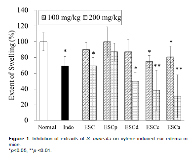
Inhibition of xylene-induced ear edema in mice
As shown in Figure 1, all tested samples inhibited xylene induced ear edema in a dose dependent manner. ESCd, ESCe and ESCa significantly (p < 0.05, p < 0.01) inhibited the ear edema at a dosage of 200 mg/kg. The inhibition rate was found to be 50.3, 61.8 and 69.2% in ESCd, ESCe, ESCa, respectively, while the standard drug indomethacin showed 39% inhibition.
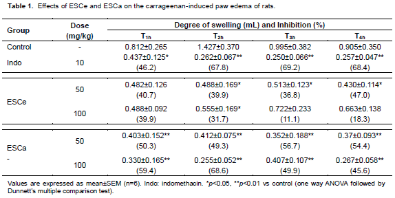
Inhibition of carrageenan-induced rat paw edema
Table 1 shows the effect of various extracts on carrageenan-induced rat paw edema. ESCe and ESCa signi?cantly inhibited rat paw edema within the first 4 h of administration (p < 0.05, p < 0.01). However, ESCa had a higher suppression on edema than ESCe, with ESCa having the highest peak of 68.6% (100 mg/kg) observed at 2 h, similar to the maximum inhibitory effect displayed by indomethacin (69.2%) at 3 h.
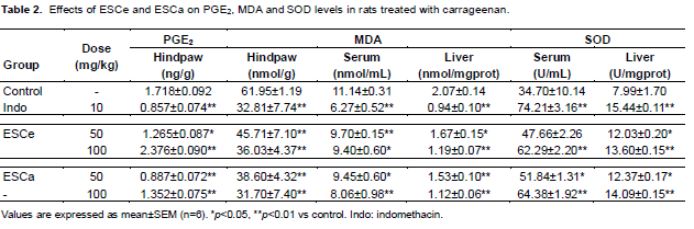
Determination of PGE2, MDA and SOD levels
As shown in Table 2, the level of PGE2 in the hind paw was found to be significantly reduced by 26.4% in ESCe and 48.4% in ESCa treated models at the dosage of 50 mg/kg. MDA levels in hind paw, serum and liver, decreased dose-dependently and significantly (p < 0.05, p < 0.01) in ESCe and ESCa treated groups (50 and 100 mg/kg), while SOD levels were signi?cantly increased in the serum and liver tissue of ESCe and ESCa treated groups.
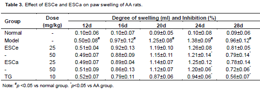
Inhibition on AA rat paw edema
The edema of untreated AA model group as observed in Table 3 was increased from day 12 to day 24, and gra-dually decreased from day 24 to day 28. A similar pattern was observed in animals treated with ESCe and ESCa, however, there was a signi?cant inhibition on paw edema of the treated groups when compared to that of the untreated model group (Table 3).
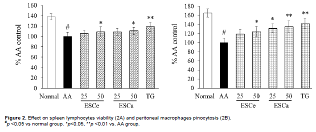
Splenocyte viability and macrophages pinocytosis activity
There was a significant increase in living splenocyte and the pinocytosis activity of macrophages in ESCe and ESCa treated group. The activity was found to be displayed in a dose-dependent manner, with the highest dose stimulating the maximum increase of living spleno-cyte and pinocytosis activity of macrophages (Figure 2). In general, ESCe and ESCa were more sensitive to the macrophage phagocytosis activity than splenocyte growth. ESCa (50 mg/kg) exhibited a 34.8% growth in en-hancing macrophages pinocytosis ability, which is almost similar to the effect observed in the positive control treated group (41.7%).
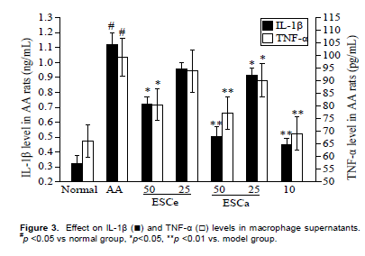
IL-1β, TNF-α level of peritoneal macrophages
From the result (Figure 3), it was observed that ESCa at a dose of 50 mg/kg and the positive control (TG) exhibi-ted significant inhibition of the IL-1β and TNF-α expression in macrophages (p < 0.05, p < 0.01). ESCa inhibited the IL-1β and TNF-α levels to 0.101 ng and 15.45 pg in each million macrophages when compared to higher levels of 0.225 ng/million macrophages (IL-1β) and 19.87 pg/million macrophages (TNF-α) observed in the untreated model group.
Analysis of bioactive constituents of S. cuneata extracts
In order to find the relationship between the activity and total phenolic content of the fractions, Folin-Ciocalteu colorimetric method was used to determine total phenolic content. ESCa was found to contain the highest amount of total phenols (449.83 mg/kg), while ESCe, ESCd, ESC, and ESCp contain 346.76, 238.59, 242.84, and 63.69 mg /kg of total phenolic contents, respectively.
DISCUSSION
The results obtained in this study provided scientific basis for the traditional usage of S. cuneata as an anti- in?ammatory agent, with ESCa and ESCe having high contents of phenols and showing markedly anti-inflammatory activity. This study preliminarily clarifies possible mechanisms mediating the anti-in?ammatory action of S. cuneata, including the suppression of MDA and PGE2 production, and SOD activity reinforcement in different tissues. Interestingly, after 3 h of administration, the edema observed in 100 mg/kg treated group was found to be more visible than that of the 50 mg/kg treated group, which correlated to the levels PGE2 contents in hind paws (Table 1 and 2).
This implied that PGE2 could be an important inflame-matory biomarker induced during swelling. ESCa and ESCe exhibited significant suppression on MDA levels, which was probably due to the increase in SOD activities. These were probably the mechanisms responsible for anti-inflammatory activity of S. cuneata. The immunoregulatory activities were also evaluated for a comprehensive explanation of its traditional use. Splenocyte proliferation is an important indicator reflec-ting the organism’s immune function, and macrophage cells could affect immune response and pro-inflammatory factor’s release. ESCe and ESCa could enhance splenocyte proliferation and macrophage phagocytosis, yet inhibit the IL-1β and TNF-α expression, thus implying that ESCe and ESCa possesses good immunoregulatory activity. This partly confirms the basis for the traditional usage of S. cuneata in the treatment of rheumatic arthritis. The immunosuppressive positive control TG was found to enhance the splenocyte proliferation and macrophage phagocytosis, which was contrary to the previous report (Li et al., 2000). This disparities might owe to the lower dosage of 10 mg/kg used.
In other to ascertain the bioactive constituents res-ponsible for the observed activity in S. cuneata, we analyzed the total phenolic content in each fraction. Our findings suggested that total phenolic content was positively correlated to the anti-inflammatory activity of each fraction, suggesting that the phenolic compounds could be the bioactive constituents present in the plant.
CONCLUSION
Further investigation will be carried out to isolate and elucidate the bioactive constituents of S. cuneata contributing to its remarkable anti-inflammatory and immunoregulatory activities, and to evaluate its potential as a possible anti-neurodegenerative agent based on inhibition of neuro-inflammation.
ACKNOWLEDGEMENTS
This work was supported by the National Natural Science Foundation of China (31270947, 81370084), Postdoctoral Foundation (1201027B, 2012M521023) and Jiangsu University Foundation (12A481).
CONFLICT OF INTEREST
Authors have no conflict of interest.
REFERENCES
| Beyhan Ö, ElmastaÅŸ M, Gedikli F (2010).Total phenolic compounds and antioxidant capacity of leaf, dry fruit and fresh fruit of feijoa (Acca sellowiana, Myrtaceae). J. Med. Plants Res. 4:1065-1072. | ||||
|
Damu AG, Kuo PC, Shi LS, Hu CQ, Wu TS (2003). Chemical constituents of the stem of Sargentodoxa cuneata. Heterocycles 60: 1645-1652. Crossref |
||||
| Editorial Board of ZhongHua BenCao of State Administration of Traditional Chinese Medicine (1998). ZhongHua BenCao 1st edn. Shanghai: Shanghai Science and Technology Press. P 434. | ||||
|
Franzotti EM, Santos CVF, Rodrigues HMSL, Mourão RHV, Andrade MR, Antoniolli AR (2000). Antiinflammatory, analgesic activity and acute toxicity of Sida cordifolia L. (Malva-branca). J. Ethnopharmacol. 72:273–277. Crossref |
||||
|
Jiao J, Xue B, Zhang L, Gong YT, Li KR, Wang HM, Jing LM, Xie JX, Wang XM (2008). Triptolide inhibits amyloid-β1-42-induced TNF-α and IL-1β production in cultured rat microglia. J. Neuroimmunol. 205:32-36. Crossref |
||||
| Li H, Jia YF, Li D (2000). Effects of polyglycosides of Tripterygium wilfordii hook on inflammation and immune suppression. J. Shanghai Med. Univ. 27:502-505. | ||||
|
Nair V, Singh S, Gupta Y (2010). Anti-arthritic and disease modifying activity of Terminalia chebula Retz. in experimental models. J. Pharm. Pharmacol. 62:1801–1806. Crossref |
||||
|
Ohishi N, Ohkawa H, Miike A, Tatano T, Yagi K (1985). A new assay method for lipid peroxides using a methylene blue derivative. Biochem. Int. 10:205-211. Pubmed |
||||
|
Shukla SM, Sharma SK (2011). Sinomenine inhibits microglial activation by Aβ and confers neuroprotection. J. Neuroinflammation 8:117. Crossref |
||||
|
Suk K, Ock J (2012). Chemical genetics of neuroinflammation:natural and synthetic compounds as microglial inhibitors. Inflammopharmacology 20:151-158. Crossref |
||||
|
Tang J, Ma RL, Ouyang Z, Chen HS (2012). Chemical constituents from the water-soluble fraction of wild Sargentodoxa cuneata. Chin. J. Nat. Med.10:115-118. Crossref |
||||
|
Yang XW, Zeng HW, Liu XH, Li SM, Xu W, Shen YH, Zhang C, Zhang WD (2008). Anti-inflammatory and anti-tumour effects of Abies georgei extracts. J. Pharm. Pharmacol. 60:937-941. Crossref |
||||
Copyright © 2024 Author(s) retain the copyright of this article.
This article is published under the terms of the Creative Commons Attribution License 4.0


