Full Length Research Paper
ABSTRACT
Study on endoparasites in wild pigs (Sus scrofa) interfering with agriculture, was carried out in areas adjoining the Western Ghats (Mudumalai tiger reserve, Anamalai tiger reserve) and Eastern Ghats (Sathyamangalam region) of Tamil Nadu state in India during November, 2013 to May, 2014. Ninety faecal samples in total (n=30 of Wild pigs, n=30 of Desi pigs and n=30 Cross bred pigs each of the study areas) were subjected to the parasitological examination using standard methods in wild pigs as well as in domestic pigs. Domestic pigs were involved to find the difference in the prevalence level between wild pigs and domestic pigs as there is a wide variation in their habitat. Prevalence of endoparasitic infections revealed the evidences of Ascaris suum, Trichuris suis, Strongyles, Strongyloides sp. and mixed parasitic infections comprising of Ascaris sp. with Trichuris sp., in addition to Strongyles with Strongyloides sp. The overall positivity of internal parasitic prevalence in wild pigs, desi pigs and cross bred pigs pertaining to Mudumalai, Anamalai and Sathyamangalam were documented, and differences among their prevalence data indicated an over-dispersed helminth distribution. These results indicate that populations of wild pigs although living under optimal conditions, are heavily affected by a burden of parasitic disease and some parasites are likely to limit population growth via a high mortality of piglets and infections throughout the lifespan of adults.
Key words: Wild pigs, domestic pig, endoparasites, prevalence.
INTRODUCTION
Although, the helminth parasites of domestic pigs are well documented there is paucity of information with regard to wild pigs. In wild populations there is usually a balanced host-parasite relationship, but pathogenicity. However, anthropogenic changes of the environment, the increase of human populations and the introduction of other animal species, may introduce unknown factors that can disrupt the natural balance and induce pathological conditions. This study is a contribution to the knowledge of wild pig helminthic fauna as these animals have been co-inhabiting with human beings and domestic pigs sharing the same resources such as land, water and also air. The epidemiology of parasitic diseases is very important as they have a zoonotic potential that can lead to various deleterious effects. In this study, a comparison of the prevalence rate of various parasitic infections in wild pigs, desi pigs and cross bred pigs have been recorded by standard techniques like floatation and centrifugation and their Egg Per Gram (EPG) was constituted to know the intensity of the infections.
MATERIALS AND METHODS
Study area
The study on endoparasites in wild pigs (Sus scrofa) interfering with agriculture was carried out in areas adjoining the Western Ghats (Mudumalai tiger reserve, Anamalai tiger reserve) and Eastern Ghats (Sathyamangalam region) of Tamil Nadu state in India during November, 2013 to May, 2014.
Collection and preservation of coprological samples for endoparasitic examination
Throughout this study programme, 30 fresh faecal samples of wild pigs from described sampling areas were collected in small containers with 10% formalin for parasitic examination, and were properly labelled and sealed with parafilm subsequently. The wild pigs were tracked by foot and their locations and resting nests were identified by their field signs (Boon et al., 2015). Similarly, 30 faecal samples from desi-pigs that are semi-free ranging and 30 faecal samples from cross-bred pigs that were maintained in organised farms were collected and processed.
Examination of samples
The faecal samples were processed by both centrifugal sedimentation technique and floatation technique as described by Soulsby (1982).
Centrifugal sedimentation technique
Approximately 2 g of faeces was taken in a 100 ml beaker and was thoroughly mixed with about 10 to 15 ml of tap water. The mixture was strained through a tea strainer into a cup and then, it was transferred into a centrifuge tube. The centrifuge tubes were placed in a balanced state and were subsequently centrifuged for 2 to 4 min, at 1500 rpm. Then, the supernatant was discarded leaving 1 to 2 ml of supernatant, without disturbing the sediment at the bottom and finally, small drop from thoroughly homogenized sediment was taken on clean glass slide and was observed under both low and high power objectives of microscope (Soulsby, 1982).
Floatation technique
Faecal samples were taken in a 100bml beaker and were thoroughly emulsified with about 10 to 15 ml of saturated solution of sodium chloride with a specific gravity of 1.18 to 1.20. The mixture was strained into a cup and then, it was transferred into a floatation tube till the mixture reaches the brim of the tube and forms a positive meniscus and was left undisturbed for 15 to 20 minutes. The tip of the positive meniscus was gently touched with a clean cover slip and then the cover slip was placed on a slide, and was examined microscopically under both low and high power objectives (Soulsby, 1982).
Quantitative analysis
One gram of faecal sample was mixed with 4 to 5 ml of saturated solution of sodium chloride and was strained trough tea-strainer into floatation tube and volume was adjusted up to 12 ml with saturated salt solution and kept for 20 min undisturbed. From this, 0.3 ml of suspension was added to McMaster slide chamber and the eggs were counted and multiplied by 40 and thus, the number of eggs per gram of faeces was calculated.
STATISTICAL ANALYSIS
The statistical analysis of the data was carried out as per the guidelines, using one way ANOVA, wherever applicable.
RESULTS
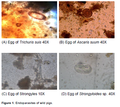
Evidence of endoparasitic fauna was recorded in free ranging wild pigs as well as domestic pigs, comprising of desi pigs and cross bred pigs. Ascaris suum, Trichuris suis, Strongyles, Strongyloides sp. and mixed parasitic prevalence comprising of Ascaris sp. with Trichuris sp. in addition to Strongyles with Strongyloides sp. were documented during the study programme with all these pigs with a varying level of intensity that was high in wild pigs, moderate in desi pigs and negligible in cross bred pigs (Figure 1).
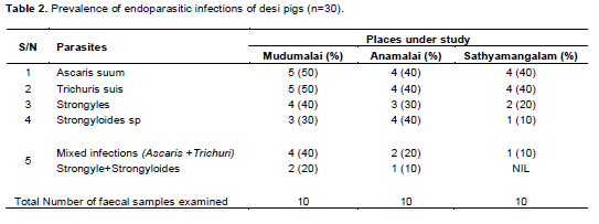
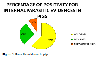
Prevalence of endoparasites with regard to different internal helminthic fauna of wild pigs, desi pigs and cross bred pigs pertaining to adjoining areas of Mudumalai, Anamalai, and Sathyamangalam wildlife regions were presented in Table 1 to 3. Positivity for internal parasitic prevalence with regard to wild pigs, desi pigs and cross bred pigs were 62, 29 and 9% respectively (Figure 2).
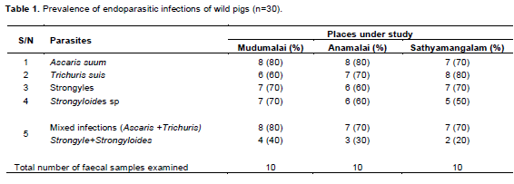

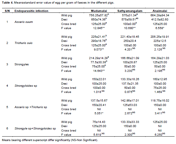
The EPG values pertaining to the parasitic prevalence comprising of A. suum, T. suis, Strongyles, Strongyloides sp. and mixed parasitic prevalence comprising of Ascaris sp. with Trichuris sp. in addition to Strongyles with Strongyloides sp. in wild pigs, desi pigs and cross bred pigs were presented in Table 4. The mean ±S.E. values of EPG among wild pigs, desi pigs and cross bred pigs with regard to individual endoparasitic species during this study in different adjoining areas of different wildlife regions were documented in Table 4. Highly significant variations (P≤0.01) were encountered with regard to A. suum, T. suis, Strongyles in Mudumalai regions, and with regard to A. suum in Sathyamangalam region. Similarly, significant variations (P≤0.05) were encountered among these pigs with A. suum in Anamalai region, and also with regard to mixed parasitic infection comprising of Ascaris sp. with Trichuris sp. among the pigs in Mudumalai region.
DISCUSSION
Parasitic prevalence
Overall parasitism in wild pigs and other pigs
The overall parasitic prevalence pertaining to the internal parasites in wild pigs (Figure 2) was found to be 62% wild pigs, 29% in desi pigs and 9% in cross bred pigs. Even though literatures pertaining to the occurrence of internal parasites in domestic swine are more, there is paucity of information in helminthic fauna of wild pigs in general. The parasitic eggs in this study were identified based on the morphological keys furnished by Encountering the increased percentage of parasitic prevalence in the samples from wild pigs which was supported by the report furnished by Jarvis et al. (2007) who quoted that none of the 25 examined carcasses of wild boars from Central Spain and those imported from France was free of helminths. Similarly, Eslami and Hamdi (1992) opined that the majority of wild boars examined (74%) had at least one species of helminth in the internal organs, and parasitic infections with several species were common in the wild boars. Further, encountering the increased overall positivity of parasitic prevalence in the wild pigs under study was in argument with the reports furnished by Bhat and Manickam (1998) who opined that high faecal egg counts were common in free ranging animals’ state than those living in captivity.
Similarly, when compared to the overall parasitic prevalence between desi pigs and the cross bred pigs of the adjoining areas studied; the percent prevalence of helminthic fauna was more in the case of desi pigs. The reasons for such an intensive prevalence of helminthic fauna in case of desi pigs might be assigned to the lesser veterinary care, straying of desi pigs outside consuming different kinds of intermediate hosts like earthworms, small sized reptiles or other creatures in or around the drainage areas noticed in the adjoining areas of wildlife regions. Comparatively, due to a better management system that includes provision of required health care measures, maintenance of good feeding regime, feeding based enhanced immune status of the animal etc. while the overall prevalence in the case of cross bred pigs was found to be less than 9%.
The reasons for encountering higher percentage of overall positivity of parasitism in wild pigs might be assigned to the reasons like diversified feeding activities of the animals, straying into the peripheral-areas of the wild regions, consumption of different types of intermediate host, absolute lack of health care related measures, consumption of feed materials contaminated by excreta of the co-existing wild animals like gaur, nilgiri tahr, spotted deer and sambar deer that co-exist in the wild environment etc. Different types of management patterns, variations in feed offered, extent of health care measures like deworming activities, varying immune status of the swines might be the reasons that could be attributed to the variation in the positivity of parasitism in desi as well as the cross bred pig of the adjoining areas studied. However, since wild pigs entering the agricultural fields adjoining those regions in the study are basically wild in nature with more or less similar type of feeding activities, wild pigs of all these regions were found to have higher percentage of overall parasite prevalence with values of 90% and above, and this could be a potential source of transmitting infection to the domestic pigs. Further, the encountering of increased overall parasitic prevalence in wild pigs under study was in agreement with the report given by Jarvis et al. (2007) who opined that the natural peculiarities of the area, including the sufficient availability of intermediate hosts of helminths were the important factors that affected wild pigs with helminths.
Parasitic prevalence in different adjoining regions and the intensity of infection
The percentage of positivity pertaining to different parasitic fauna encountered in wild pigs,desi pigs and cross bred pigs are presented in Tables 1 to 3. A. suum and T. suis were the frequently encountered parasitic fauna, with wild pigs in general. The EPG values of A. suum, in case of wild pigs as well as desi pigs differed highly significantly when compared to the value encountered in case of cross bred pigs in Mudumalai as well as Sathyamangalam area. Similarities in certain feeding related activities, varying immune status of pigs etc. might be assigned as the etiological factors pertaining to such highly significant variations in them.
In this regard, it was noteworthy to mention the report furnished by Tiwari et al. (2009) who quoted that in Botswana, A. suum was the most prevalent helminth encountered in the case of pigs. Further variations in the number of wild pigs affected with A. suum was encountered in this study was supported by Foata et al. (2005) who opined that among the numerous most often cited in the literature, A. suum was found to be one of the only three parasitic species that were index at the time of their study in case of wild boars. Variations in EPG values pertaining to the A. suum as encountered in the wild pigs as well as in others was further in agreement with the findings revealed by Popiolek et al. (2010) who further stated that though the A. suum was common in pigs and wild boars worldwide, the level of wild boar infection was not very high. In this regard, Tomass et al. (2013) quoted that A. suum was the most common helminth in all age categories of pigs and Ascaris suum was a natural parasite of pigs as it could also infect human, and the potential of the A. suum to infect human might be due to the fact that it stored similar protein molecular with A. lumbricoides for which man is the natural host. Urquart et al. (1996), also quoted that the eggs pertaining to A. suum were very resistant to extreme temperature and the eggs of A. suum were found to be viable for more than four years.
Encountering A. suum in domestic pigs as noticed with desi as well as cross bred pigs under study, was in agreement with the findings presented by Radostitis et al. (2007) and Urquart et al. (1996). Encountering T. suis in the wild pigs in this study was in agreement with the report furnished by Jarvis et al. (2007) who revealed that though, T. suis was noticed as one of the seven helminth species in wild boars. During this study, the EPG value pertaining to wild pigs as well as the desi pigs were found to be significantly different, when compared with the values encountered with cross bred pigs in Mudumalai region alone. The different types of management systems that are maintained with the cross bred pigs including the reasonably intensive health care related measures lack of opportunities for the consumption of diversified types of feeds including different types of intermediate hosts etc. might be assigned as the etiological factors for the encountering of highly significant variations in the EPG values of T. suis in the case of cross bred pigs when compared to that of wild pigs as well as desi pigs. Encountering the T. suis as noticed in the studyis also in agreement with the report presented by Tomass et al. (2013). It becomes noteworthy to mention the report presented by Nansen and Roepstorff (1999) who opined that often moderate numbers of adult T. suis were present in the caecum and colon, and if there was high prevalence of T. suis infections in the swines, it might lead to unthriftiness and death, and it was demonstrated that severe clinical disease might be associated with T. suis induced suppression of mucosal immunity to resident bacteria.
The EPG value of strongyles in wild pigs was found to be 214.29±14.29, where it was 77.50±30.38 in desi pigs and 75.00±25.00 in cross bred pigs. Findings on the prevalence of strongyles in wildlife under study was in agreement with the report presented by Souse et al. (2004) who encountered presence of gastrointestinal strongyles eggs in all the faecal samples of wild boars examined and further quoted about the average EPG of 2142 in the case of gastrointestinal strongyles in wild pigs when compared to the EPG values of desi pigs, in addition to the cross bred pigs which might be attributed to the existing different kinds of biotic as well as the abiotic environmental factors. The encountering of prevalence of strongyles was further in agreement with the report furnished by Magi et al. (2005).
The EPG values of Strongyloides sp. were presented in Table 4 and however, there was no statistical significance among the pigs comprising of wild pigs, desi pigs and cross bred pigs. Encountering the Stronglyloides sp. with pigs under this study was in agreement with the findings reported by Varadhrajan and Pythal who however, encountered mixed infection associating with Fasciola sp., Strongyles and Strongyloides sp. in wild boars. In this regard, Tiwari et al. (2009) quoted that parasitic fauna like Strongyloides sp. were found to be related to the occurrence of clinical signs like diarrhoea as well as emaciation in pigs. With regard to encountering the prevalence of Strongyloides sp. in this study, Coombs and Springer (1974) opined that since wild pigs were associated with feeding of earthworms, beetles, bugs and numerous larvae which functioned intermediate or paratenic hosts for various helminthic fauna, more helminth fauna were encountered including the Strongyloides ransomi. Urquart et al. (1996), reported the experimental demonstration of prenatal infection associated with Stronglyloides sp. in pigs.
CONCLUSION
Even though increased parasitic prevalence was encountered in the faecal samples of wild pigs entering the agriculture fields near wildlife regions, immediate conclusion about the existence of a clinical disease condition could not be drawn. Many factors may be involved and lack of extensive studies pertaining to the parasitic fauna in both core and buffer zone areas of wildlife regions. However, it might be understood that wild pigs revealed existence of different helminthic fauna comprising of A. suum, T. suis, Strongyles, Strongyloides sp. and mixed parasitic infections associated with Ascaris sp. with Trichuris sp., in addition to Stongyles with Strongyloides sp. Availability and feeding of diversified feed materials which comprises of earthworms, beetle larvae, insects, small rodents, egg and chicks of birds nesting on the ground as well as in short grass, including the feeding of carrion sometimes might be the significant factors that lead to the encountering of different parasitic fauna in the wild pigs studied. The encountering of significant variations in the mixed parasitic prevalence associated with Ascaris sp. and Trichuris sp. might be attributed to differences in feed materials consumed, variations in the immune level, management practices etc. The finding of the presence of endoparasites in wild pigs will be helpful in designing strategy management practices and curbing the disease at the initial levels and this can be done with ease.
CONFLICT OF INTEREST
The authors declare that they have no conflicts of interest.
REFERENCES
| Boon A, Jayathangaraj MG, Palanivelrajan M, Raman M (2015). Wild pig field signs - identity and confirmation. Int. J. Life Sci. Res. 3(1):205-213. | ||||
| Bhat MN, Manickam (1998). Coproculture and Study of seasonal variation in parasitic burden in spotted deer (Axis axis) in different sanctuaries in Tamil Nadu. Indian Vet. J. 75:469-471. | ||||
|
Coombs DW, Springer DM (1974). Parasites of Feral pigs and European wild boar hybrids in South Texas. J. Wildl. Dis. 10:436-441. crossref |
||||
|
Eslami A, S Farsad-Hamdi (1992). Helminth parasites of wild boar, Sus scrofa, in Iran. J. Wildl. Dis. 25(2): 316-318. crossref |
||||
| Foata J, Culioli JL, Marchand B (2005). Helminth fauna of Wild boar in Corsica. Acta Parasitol. 50(2):168-170. | ||||
|
Jarvis H, Kapel C, Moks E, Talvik H, Magi E (2007). Helminths of wild boar in the isolated population close to the northern border of its habitat area. Vet. Parasitol. 150:366-369. crossref |
||||
|
Nansen P, Roeptstorff A (1999). Parasitic helminths of the pig: factors influencing transmission and infection levels. Int. J. Parasitiol. 29:877-891. crossref |
||||
| Magi M, M Bertani, M Dell'omodarme, MC Prati, G. Poglayen (2005). Seasonal Egg output of gastro-intestinal parasites in wild ungulates in a Mediterranean area (Central Italy). Hystrix 16(2): 169-177. | ||||
| Popiolek MD, Knecht JS, Staskiwicz A, Czerwinska R (2010). Helminths of the wild boar (Sus scrofa L.) in natural and breeding conditions. Bull. Vet. Inst. Pulawy 54(1):161-166. | ||||
| Soulsby EJ (1982). Helminths, Arthropods and Protozoa of domestic animals, 10th Edn., ELBS, Bareilly Tindall, London p. 809. | ||||
| Tiwari KP, Chikweto A, Belot G, Vanpee G, Deallie C, Stratton G, (2009). Prevalence of intestinal parasites in pigs in Grenada, West Indies. West Indies Vet. J. 9(1):22-27. | ||||
|
Tomass Z, Inam E, Kifleyhannes T, Tekle Y, Weldu K (2013). Prevalence of gastrointestinal parasites and Cryptosporidium species in extensively managed pigs in Mekelle and urban areas of southern zone of Tigray region, Northern Ethiopia. Vet. World 6(7):433-439. crossref |
||||
Copyright © 2024 Author(s) retain the copyright of this article.
This article is published under the terms of the Creative Commons Attribution License 4.0


