Full Length Research Paper

ABSTRACT
The aim of this study was to determine the baseline values of vital, haematological and serum biochemical parameters in apparently healthy experimental donkeys in North-Western Nigeria, with a view to providing additional data on normal values for donkeys. Twenty four apparently healthy donkeys comprising of 12 males and 12 females, aged 11 to 15 months were used. The animals were housed in prepared experimental pen and assigned identification neck-tags at random. Microscopic methods and mice inoculation test were used to examine and ensure that the donkeys were free from gastro-intestinal and blood parasites infections. Animals were acclimatized for 14 days, fed and salt lick blocks together with drinking water were provided ad libitum throughout the experiment. Vital, haemogram and some serum biochemical parameters of the animals were evaluated four consecutive times at 7 days intervals. Data was analysed using statistical package for social sciences (SPSS) Version 17. Mean of parameters were determined and tables prepared on Microsoft Excel 2010. In conclusion, these baseline data will be useful in laboratory diagnosis and further understanding of diseases of donkeys especially those within the age group of 11 to 15 months in North-Western Nigeria.
Key words: Donkeys, serum biochemical, haematological, parameters, vital.
INTRODUCTION
Donkeys (Equus africanus asinus) are widespread in Nigeria and used as source of traction power in transport and ploughing by both pastoralists and settled farmers in the northern region (Blench et al., 2013). Donkeys also tolerate some tropical animal diseases and parasites, survive on poor quality feeds and adverse climatic conditions, thereby making their management easy for their owners (Aganga et al., 2000). Despite the benefits and advantages of keeping donkeys, the research atten-tion given to the specie is relatively small, whence; scanty biological data is available on donkeys (Starkey, 1994; Blench et al., 2013). In the present study we determined the baseline vital, haematological and serum biochemical parameters of apparently healthy donkeys acquired from Jigawa state, and conducted the experiment in Zaria, Kaduna state, both in North-Western Nigeria. It was designed with a view to providing normal values of the physiological parameters under experimental condition in order to determine alteration(s) in patients’ values during laboratory diagnosis of diseases of donkeys.
MATERIALS AND METHODS
Experimental animals and their management
Twenty four apparently healthy donkeys comprising 12 males and 12 females, aged 11 to 15 months estimated (using dental eruption pattern of the incisors teeth) as described by Wayne and Melvin (2000) and Joe (2012) at point of purchase in Maigatari town, Jigawa state and transported to Zaria in Kaduna state both in North-Western Nigeria were used. Prior to housing, topical fly-repellant spray (Endure®, Farnam co. inc., USA) was applied on the animals as recommended by manufacturer, to keep flies off the animals. The donkeys were housed in prepared, fly-proofed experimental animal pen in the Department of Veterinary Parasitology and Entomology, Faculty of Veterinary Medicine, Ahmadu Bello University Zaria where the experiment was conducted. Animals were identified using neck-tags assigned at random as described by Aviva and Poul (2013). Feed was provided, equivalent to 5% of the mean body weight of the animals in form of sorghum/maize stovers and grass/legume hays (in the ratio 4:1). Concentrate feed was provided as a mixture of coarsely ground sorghum grain, bran and dried groundnut cake which was served daily in two divided rations for morning and evening. Salt lick blocks and clean drinking water were provided ad-libitum (Aganga et al., 2000) during the experiment. Animals were acclimatized for fourteen days during which they were standardized for the experiment.
Standardization of animals
The animals were examined for external, gastrointestinal and blood parasites to ensure that they were free from infestations an infections. About 5 g faecal sample was scooped from the rectum of each animal using two fingers of a gloved hand, into labeled polyethene bags and examined using floatation and sedimentation tests (Charles, 2007). Four milliliters of blood was collected from each experimental animal through jugular vein using 5 ml syringe and 19 G × 11/2 inch needle (Weiser, 2012a; Wolfensohn and Lloyd, 2013). Three milliliters of the obtained blood was anti-coagulated in blood sampling bottle containing ethylenediaminetetraacetic acid (EDTA) + k3 1 mg/ml of blood (Elaine and Margi, 2007). The blood was used to prepare 2 slides of Giemsa stained thin blood smear and 2 haematocrit centrifugation technique (HCT) capillary tubes
(Wosu, 2002) which were examined microscopically for haemo-parasites. The balance of 1 ml collected blood was transferred into sodium heparin-coated bottle as anticoagulant and was instantly used to inoculate 2 representative mice per donkey (0.5 ml blood/mice) intra-peritoneally (World Organization for Animal Health (OIE), 2010) using 1 ml sterile disposable syringe with 29 G × ½ inch needle as recommended by Wolfensohn and Lloyd (2013). Blood of the inoculated mice were also collected from tail tip at 48 h interval and examined using 2 each, of prepared HCT capillary tubes and wet film for haemoparasites. The mice were observed for 14 days post-inoculation during which all mice were negative for haemoparasites based on which we continued with the next phase of the experiment. The animals were dewormed with Fenbendazole bolus (Fenacure®, Ashish Life Sciences PVT ltd, India) at the dose rate of 10 mg/kg bd wt orally, once (Aliu, 2007). The experimental animals were managed as recommended in the European Union ‘Directive 2010/63’ contained in the report by Wolfensohn and Lloyd (2013) and approved by the Ahmadu Bello University (ABU) Research and Ethics Committee.
Evaluation of vital parameters and blood samples collection
Respiration, pulse and rectal temperature were evaluated, and blood samples were collected 4 times consecutively at 7 days intervals between 600 and 800 h local time. Animals were observed individually while at rest for respiratory rate (breaths/minute) using costo-abdominal movement, pulse rate (beats/minute) from pulsation of external maxillary artery in the medial aspect of mandibular angle and rectal temperature in degrees Celsius (°C) using digital clinical thermometer as reported by Wosu (2002). Six milliliters of jugular vein blood was collected from each animal using 10 ml syringe with 19 G × 11/2 inch needle for determination of blood glucose in mg/dl. Three milliliters of the collected whole blood was placed in labeled blood sample bottles containing 1 mg EDTA + K3/ml of blood, and used for haemogram evaluation, while the remaining blood was saved in labeled plain vacutainer bottle and used for serum extraction. Serum was harvested by allowing whole blood to clot at room temperature for 30 min, gently decanted the sera into labeled serum vials, insufficiently sedimented blood samples were centrifuged at 5,000 g for 5 min to improve serum yield. Between 0.8 and 1.5 ml serum was obtained from each processed blood sample. Blood and sera were stored at 4 and -20°C, respectively until analysed (Charles, 2007; Weiser, 2012a).
Laboratory protocols
Haemogram evaluation
Packed cell volume (PCV), total red blood cells (RBC), total leucocytes, differential leucocytes counts and haemoglobin (Hb) concentration were evaluated as reported by Weiser (2012b).
Serum biochemical assay
Blood glucose was evaluated with the aid of handheld, digital blood glucose monitor (Fora®G20, Fora Care Inc., U.S.A.) in mg/dl using a tiny drop of whole blood (about 0.7 µl) placed on the tip of the strip as recommended by the manufacturer. Total protein (TP), albumin (ALB), alanine aminotransferase (ALT), aspartate aminotransferase (AST), alkaline phosphatase (ALP), blood urea nitrogen (BUN) and creatinine (CR) levels were determined from the stored sera with the aid of fully-automated clinical chemistry analyzer (Selectra XL®, Vital Scientific, Netherland). The machine holds 80 samples, 30 standard reagents cups, have 2 reactions chambers that hold 24 cuvets (cups) in a single reaction run and 2
wash stations for self cleaning after each sampling. In a run, 250 µl of each serum sample was collected using Eppendoff pipette and dispensed into sample cup. The cups with samples were loaded into the machine sample rotor as recommended by the manu-facturer. The machine used between 6 to 12 µl of each sample to determine the level of a substance. Results were generated, recorded and printed automatically. After dispensing of each sample, the auto-pipettes were cleaned at the ‘Wash Station’ within 3 s before collection of another set of sample and reagents.
Serum electrolytes assay
Calcium (Ca2+), phosphate (Po4-), sodium (Na+), potassium (K+), chloride (Cl+) and bicarbonate (HCO3-) ions were assayed with the aid of an automatic electrolyte analyzer (Audicom AC9900®, Audicom Medical Technology ltd, Jena, Germany). The system holds 20 sample cups, has 2 quality control (QC) reagent loading points (QC1 for electrode activation reagent and QC2 for calibration reagent), one wash station containing electrode deproteinizing standard reagent and 1 emergency call. From each sample, 200 µl of the thawed sample was collected using Eppendoff pipette, dispensed into sample cup and the filled cups were loaded into serially numbered cups holders and the machine was set to run the samples as recommended by the manufacturers. Different electrodes determine the concentration levels of each element in a sample. Analysis of a sample and generation of printed result of 5 elements was completed within 120 s before collection of another sample.
Data analysis
The statistical package used was SPSS Version 17. Means of parameters were determined and tables prepared using Microsoft Excel 2010.
RESULTS AND DISCUSSION
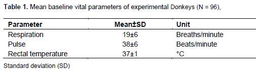
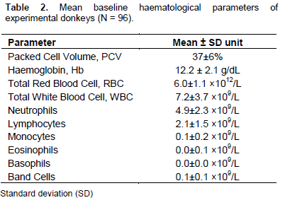
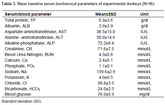
The baseline data obtained on the parameters was not analysed on the basis of gender and age differences for the fact that earlier reports showed no significant influence of variations in age, sex, lactation status and body condition of donkeys, horses and mules on values of normal haemogram and serum biochemical but variation exist between species (Gul et al., 2007). Where variation are observed between the values from this study and earlier reports on donkeys (Tables.1-3), it could be as a result of bio-variations in breeds and environmental conditions (de Aluja et al., 2001; Tesfaye et al., 2014) such as stress imposed by seasonal weather changes which can influence variations in values of normal respiratory rate, pulse rate and rectal temperature (Table 1) of donkeys (Ayo et al., 2008). Haemogram values (Table 2) and biochemical values (Table 3) are also reported as not significantly influenced by age, sex, lactation status and body condition differences in donkeys, horses and mules but variation exist between species (Gul et al., 2007). Findings by Annita et al. (2013) indicated no differences in values of creatinine kinase, albumin, urea and magnesium among age groups of donkeys. Serum biochemical values are reported to be, most of the time, within related ranges globally in donkeys, however, creatinine and aspartate amino-transferase can be low in some breeds of donkeys (Sow et al., 2014). Values of fractions of blood proteins also do not vary between sexes in donkeys (Paulo et al., 2012). Therefore, knowledge of the deviations of laboratory clinical examination findings from the published normal physiological values is an essential tool for accurate diagnoses of diseases of donkeys (Mori et al., 2003, 2004). The results were; respiratory rate = 19 ± 6 breaths/minute; pulse rate = 38 ± 6 beats/minute and rectal temperature = 37 ± 1°C. PCV was 37 ± 6%, Hb = 12.2 ± 2.1 g/dl, red blood cell (RBC) = 6.0 ± 1 × 10¹²/L, white blood cell (WBC) = 7.2 ± 3.7 × 10⁹/L, neutrophils = 4.9 ± 2.3 × 10⁹/L, lymphocytes = 2.1 ± 1.5 × 10⁹/L, monocytes = 0.1 ± 0.2 × 10⁹/L, eosinophils = 0.0 ± 0.1 × 10⁹/L, basophils = 0.0 ± 0.0 × 10⁹/L and band cells = 0.1 ± 0.1 × 10⁹/L. Blood glucose was 76.1 ± 6.9 mg/dl, total protein = 6.9 ± 3.5 g/dl, albumin = 3.9 ± 3.9 g/dl, aspartate aminotransferase = 38.5 ± 15.0 IU/L, alanine aminotransferase = 50.0 ± 14.6 IU/L, alkaline phosphatase = 72.2 ± 8.4 IU/L, creatinine = 71.6 ± 7.5 mmol/L, blood urea nitrogen = 4.3 ± 0.8 mmol/L. Electrolyte values were: calcium = 2.4 ± 0.1 mmol/L, phosphate ion = 1.1 ± 0.1 mmol/L, sodium = 139.6 ± 2.9 mmol/L, potassium = 4.0 ± 0.5 mmol/L, chloride = 98.8 ± 3.5 mmol/L, and bicarbonate = 24.0 ± 2.3 mmol/L.
CONCLUSION
The values generated from this work will serve as additional baseline data for accurate laboratory diagnosis of diseases of donkeys especially for the young animals aged 11 to 15 months in North-Western part of Nigeria.
ACKNOWLEDGEMENT
We are grateful to Dr. J.A. Natala, Head of Department of Veterinary Parasitology and Entomology for approving the use of research animal pen; Technical Staff of Protozoology, Helminthology and Haematology Laboratories, Faculty of Veterinary Medicine, ABU, Zaria for providing guidance in the laboratory during the study as well as the contribution of Mr. Olu of Chemical Pathology Laboratory, ABU Teaching Hospital, Shika, Zaria, Nigeria for his assistance in sourcing the reagents and serum analysis.
CONFLICT OF INTEREST
The authors have no conflict of interest.
REFERENCES
| Aganga AA, Letso M, Aganga AO (2000). Feeding donkeys. Livestock Res. Rural Dev. 12(2). | ||||||||||||||||||||||||
| Aliu YO (2007). Nigerian Veterinary Formulary; Handbook of Essential Veterinary Drugs, Biologics and Pesticide Chemicals. First edition. Veterinary Council of Nigeria. P 67. | ||||||||||||||||||||||||
|
Ayo JO, Dzenda T, Zakari FO (2008). Individual and diurnal variations in rectal temperature, respiration, and heart rate of Pack donkeys during the early rainy season. J. Equine Vet. Sci. 28(5):281-288. Crossref |
||||||||||||||||||||||||
| Blench R, De Jode A, Gherzi E (2013). Donkeys in Nigeria: history, distribution and productivity. A Resource Book of the Animal Traction Network for Eastern and Southern Africa (ATNESA). ACP-EU Technical Centre for Agricultural and Rural Cooperation (CTA), Wageningen, The Netherlands. 244p. | ||||||||||||||||||||||||
|
Charles MH (2007). Internal parasites, external paraspites. In: Charles MH, Margi S (Eds), Laboratory Procedures for Veterinary Technicians. Fifth ed. Mosby Elsevier Inc. St Louis Missouri. pp. 187-252. Pubmed |
||||||||||||||||||||||||
|
De Aluja AS, Mondragón VRL, Castillo MDA, Ochoa P (2001). Hematological and biochemical reference values in the donkey (Equus asinus) in Mexico. In: Matthews NS, Taylor TS (Eds.), Veterinary Care of Donkeys. Veterinary Info. Service, Ithaca, New York. Pubmed |
||||||||||||||||||||||||
|
Elaine A, Margi S (2007). Hematology and hemostasis. In: Charles MH, Margi S (Eds.), Laboratory Procedures for Veterinary Technicians, 5th edition, Mosby Elsevier Inc. St Louis Missouri. pp. 27-73. |
||||||||||||||||||||||||
| Girardi AM, Marques LC, Toledo CZP, Barbosa JC, Maldonado Jr W,Jorge RLN, Nogueira CA (2013). Biochemical profile of Pega donkeys (Equus asinus) breed: Influence of age and sex. Comp. Clin. Pathol. 23(4): 941-947. | ||||||||||||||||||||||||
| Gul ST, Ahmad M, Khan A, Hussain I (2007). Haemato-biochemical observations in apparently healthy equine species. Pak. Vet. J. 27(4). | ||||||||||||||||||||||||
| Joe A (2012). The Donkey Sanctuary. British Veterinary Association. | ||||||||||||||||||||||||
|
Mori E, Fernandes WR, Mirandola RMS, Kubo G, Ferreira RR, Oliveira JV, Gacek F (2003). Reference values on serum biochemical parameters of Brazilian donkeys (Equus asinus) breed. J. Equine Vet. Sci. 23:356-364. Crossref |
||||||||||||||||||||||||
|
Mori E, Mirandola RMS, Ferreira RR, Oliveira JV, Gacek F, Fernandes WR (2004). Reference values on Haematological parameters of Brazilian donkeys (Equus asinus) breed. J. equine Vet. Sci. 24:271-276. Crossref |
||||||||||||||||||||||||
| OIE (2010). Trypanosoma evansi infection (surra).Version adopted by The World Assembly of Deligates of the OIE in May 2010. Chapter 2.1.17. Available at: http://www.oie.int/fileadmin/Home/eng/Health_standards/tahm/2.01.17_TRYPANO.pdf | ||||||||||||||||||||||||
| Paulo HC, Andrea CCS, Miyoshi S, Benito SB (2012). Serum proteins fractions in Brazilian- breed donkeys using Agarose gel electrophoresis. Turk. J. Vet. Anim. Sci. 36(1):9-12. | ||||||||||||||||||||||||
|
Sow A, Sidibe I, Kalandi M, Bathily A, Ndiaye NP, Ouédraogo M, Mouiche MMM, Sawadogo GJ (2014). Biochemical changes induced by natural infection of trypanosomosis in Burkinabese local donkey breeds. Comp. Clin. Pathol. 23(1):103. Crossref |
||||||||||||||||||||||||
| Starkey P (1994). A World-wide view of animal traction highlighting some key issues in eastern and Southern Africa, In: Starkey P, Nwenya E, Stares J (Eds.), Improving Animal Traction Technology. CTA, Mills Litho, Maitland, Cape Town, South Africa. pp. 146-149. | ||||||||||||||||||||||||
|
Tesfaye T, Mamo G, Endebu B, Abayneh T (2014). Comparative serum biochemical profiles of three types of donkeys in Ethiopia. Comp. Clin. Pathol. 23(1):205-212. Crossref |
||||||||||||||||||||||||
| Wayne L, Melvin B (2000). Determining age of horses by their teeth. Extension and Agricultural Information. Extension and Agricultural Information, 1-98 Agriculture Building, Columbia, MO 65211. | ||||||||||||||||||||||||
| Weiser G (2012a). Sample collection, processing and analysis of laboratory service options, In: Mary AT, Glade W, Robin WA, Terry WC (Eds.), Veterinary Hematology and Clinical Chemistry. Second ed. Wiley-BlackWell, UK. pp. 34-50. | ||||||||||||||||||||||||
|
Weiser G (2012b). Laboratory technology for veterinary medicine. In: Mary AT, Glade W, Robin WA, Terry WC (Eds.), Veterinary Hematology and Clinical Chemistry. Second edition, Wiley-BlackWell, UK. pp. 3-33. Pubmed |
||||||||||||||||||||||||
| Wolfensohn S, Lloyd M (2013). Handbook of Laboratory Animal Management and Welfare. Fourth edition, Wiley-Blackwell Publishing Ltd, UK. 371p. | ||||||||||||||||||||||||
| Wosu LO (2002). The Veterinarian's Handbook. First edition, Mike Social Press, Nsukka, Nigeria. 268p. | ||||||||||||||||||||||||
Copyright © 2024 Author(s) retain the copyright of this article.
This article is published under the terms of the Creative Commons Attribution License 4.0


