Full Length Research Paper

ABSTRACT
Bovine fasciolosis is an economically important parasitic disease of cattle in tropical and subtropical countries responsible for considerable economic losses in the cattle industry, mainly through condemnation of fluke-infected liver. A cross sectional study was conducted between October, 2013 and March, 2014 to determine the prevalence and monetary losses associated with bovine fasciolosis and comparing coprology and post mortem techniques of examination among cattle slaughtered at Dangila municipal abattoir in Amhara region. Out of a total of 384 cattle examined, 85 (22.14%) and 116 (30.21%) were positive for fasciolosis through coprological and postmortem examinations, respectively. Fasciola hepatica was found to be the predominant Fasciola species in the study area with prevalence of 42.2% whereas Fasciola gigantica, mixed, and immature or unidentified forms of Fasciola species were found to be 27.6, 19.0 and 11.2%, respectively. However, there was no statistically significant variation (P > 0.05) in the prevalence of fasciolosis based on age of the animal, young (< 5 years) were counted more prevalence of 23.1 and 32.7% in coprology and post mortem examinations, respectively than adult (> 5 years) with prevalence of 21.8% and 29.3% in coprological and post mortem examinations, respectively. Statistically significant variation (P < 0.05) was observed in the prevalence of fasciolosis among animals with different body conditions and months both in coprology and post mortem examinations. The highest prevalence of bovine fasiolosis was recorded in poor body conditions, with prevalences of 37.2 and 46.5% in coprology and post mortem examinations, respectively, while the highest prevalence was found to be in October with 30.7 and 40.6% both in coprology and post mortem examinations, respectively. The total monetary loss incurred due to condemned liver and carcass weight loss was estimated to be 945,270 Ethiopian birr (48,432 USD) per annum, indicating the great impact on the economy. Therefore, more detailed studies on ecology and biology of the snail intermediate host and its effective control measures should be planned.
Key words: Abattoir, cattle, Dangilla, Ethiopia, fasciolosis, monetary loss, prevalence.
INTRODUCTION
Ethiopia is believed to have the largest livestock population in Africa, of roughly 53.99 million animals. Out of thistotal cattle population, the female cattle constitute about 55.48% and the remaining 44.52% are male cattle (Community-supported agriculture (CSA), 2012).
However, this great potential is not properly exploited as a result of diseases, malnutrition and other manage-ment problems. Parasitism is one of the major bottlenecks to livestock development in the tropics (Gupta and Singla, 2013). Among many parasitic problems of animals, fasciolosis, caused by trematodes of the genus, Fasciola, is a major disease, which imposes direct and indirect economic impact on livestock production, particularly of sheep and cattle. Apart from its veterinary and economic importance throughout the world, fasciolosis has recently been shown to be a re-emerging and widespread zoonosis affecting many people (Kassai, 1999; Chhabra and Singla, 2009).
Fasciola hepatica and Fasciola gigantica are the two liver flukes most commonly reported to cause fasciolosis in ruminants. F. hepatica has a cosmopolitan distribution, mainly in temperate zones, while F. gigantica is found in tropical regions of Africa and Asia (Mihreteab et al., 2010). In Ethiopia, F. hepatica and F. gigantica infections occur in areas above 1800 meters above sea level (m.a.s.l.) and below 1200 m.a.s.l, respectively which has been attributed to variations in the climatic and ecological conditions such as rainfall, altitude, temperature and livestock management system (Yilma and Malone, 1998).
The life cycle of liver flukes involves a freshwater snail as an intermediate host. The geographical distribution of trematode species is dependent on the distribution of suitable species of snails. The Galba/ Lymnaea complex in general and Galba (Lymnaea) truncatula and Lymnaea (Radix) natalensis in particular are the most common intermediate hosts for F. hepatica and F. gigantica, respectively. The presence of fasciolosis due to F. hepatica and F. gigantica in Ethiopia has long been known and its prevalence and economic significance has been reported (Tadele and Worku, 2007).
Bovine fasciolosis is an economically important parasitic disease of cattle in tropical and subtropical countries responsible for considerable economic losses in the cattle industry, mainly through mortality, morbidity, reduced growth rate, condemnation of fluke infected liver, increased susceptibility to secondary infections and expense due to control measures. Production loss in livestock industry is estimated at more than 90 million USD annually (Rahmeto et al., 2010).
Although a number of studies have been undertaken with regard to abattoir-based prevalence and evaluation of the economic loss due to fasciolosis in different parts of Ethiopia, very little has been done in Awi zone of the Amhara regional state of the country, and particularly Dangila district. Therefore, this study was conducted with the aims to determine the prevalence of fasciolosis and Fasciola species among cattle slaughtered at Dangila municipal abattoir, compare the diagnostic efficiencies of fecal versus post mortem examinations and assess direct and indirect economic losses caused by fasciolosis in the study area.
MATERIALS AND METHODS
Study area
The study was conducted at Dangila town, located under the administration of Awi zone in Amhara region, Ethiopia. Geogra-phically, it is located on elevation of 2200 m.a.s.l with latitude and longitude of 11° 16’N and 36° 50’E, respectively, and it is 485 km southwest from the capital city, Addis Ababa. With Woina Dega temperate climate, the annual average rainfall and temperature amount is 1576 mm and 17°C, respectively. The area experiences two seasons, the rainy season (Keremt) lasts from May to October and dry season (Bega) from November to April. The town has a total land area of 89,007 hectares with total population of 158,688. The livestock population of the area comprises of 926,870 cattle, 11,399 horses, 46,628 sheep, 859,213 poultry, and 11,323 beehives (CSA, 2012).
Study population
The study was conducted on indigenous zebu male cattle slaughtered at Dangila municipal abattoir brought from different localities and livestock markets in their vicinity.
Study design and sampling method
A cross sectional study was conducted from October, 2013 to March, 2014 to determine the prevalence and monetary loss of bovine fasciolosis by using post postmortem examination of liver of each slaughtered animal and coprological examination during ante mortem before the animals were allowed for slaughter. Simple random sampling method was employed among cattle brought into the abattoir for determining the prevalence of fasciolosis in cattle and the magnitude of direct monetary loss due to liver condemna-tion and indirect carcass weight loss at Dangila municipal abattoir. Information regarding age, body condition and months of the study animals were recorded during ante mortem and post mortem examinations.
To calculate the total sample size, the following parameters were used: 95% level of confidence interval (CI), 5% desired level of precision and with the assumption of 50% expected prevalence of bovine fasciolosis in the study area the sample sizes were determined using the formula given in Thrusfield (1995).
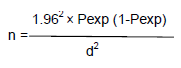
Where n = required sample size, Pexp = expected prevalence, d2 = desired absolute precision. Hence, 384 samples were used for the study.
Study methodology
Coprological examination
Ante mortem inspections were conducted on individual animals in motion and at rest, while they were in the lairage. During ante mortem examination, detail records with regard to their ages and body conditions of the animals were performed. The age of the animals was scored according to De-Lahunta and Habel (1986) and the body condition of the animal was scored according to Mari (1989). Following the judgments passed by Food and Agricultural Organization (FAO) (1994), animals fit for human consumption were allowed for slaughter. Prior to slaughter, fecal samples were collected directly from the rectum of each study animal, using disposable plastic gloves and placed in clean universal bottle and each sample was clearly labeled with animal identification, age and body condition score. Fecal samples were preserved with 10% formalin solution to avoid the eggs developing and hatching. In the laboratory, coprology was used to detect the presence of Fasciola eggs using the standard sedimentation techniques recommended by Hansen and Perry (1994). Abattoir-based prevalence was used to determine positivity of the animals for the disease, but coprology was used to compare the diagnostic efficiency of the disease with post mortem examination.
Post mortem examination
The prevalence of fasciolosis was conducted by post mortem examination technique from liver parenchyma and major bile ducts to recover the young flukes and adult parasites, respectively. The previously (in ante mortem) identified animals and their livers were carefully supervised and examined, so as to avoid mixing up of the organs to be inspected with the fecal samples. Post mortem examination of liver and associated bile duct was carefully performed by visualization and palpation of the entire organ followed by transverse incision of the organ across the thin left lobe based on routine meat inspection by FAO (2003) in order to confirm the presence of the parasites. Livers were inspected by making multiple deep incisions of the lobes and making a deep cut with a number of small sub cuts. Gall bladders were opened using a knife and thoroughly investigated for the presence of Fasciola. For Fasciola species identification, one or more samples of the worms were collected from condemned livers which had active infection. Species identification was conducted on recovered Fasciola based on morphological features of the agents and classified into F. hepatica and F. gigantica, mixed infection by both flukes and unidentified or immature forms of liver flukes (Urquhart et al., 1996).
Monetary loss assessment
The total monetary loss due to fasciolosis in cattle slaughtered from the summation of annual liver condemnation cost (direct loss) and cost due to carcass weight reduction (indirect loss) was assessed.
Direct monetary loss
Direct monetary loss was resulted from condemnation of liver affected by fasciolosis. All livers affected with fasciolosis were totally condemned. The annual loss from liver condemnation was assessed by considering the overall annually slaughtered animals in the abattoir and retail market price of an average zebu liver. Annual slaughtered rate was estimated from the retrospective abattoir records of the last three years, while retail market price of an average size zebu liver was determined from the information collected from butcheries in Dangila town. The average numbers of cattle slaughtered in Dangila municipal abattoir were 3500 cattle per year based on three years recorded data while the mean retail price of one liver is 35 Ethiopian birr. The information obtained was subjected to mathematical computation using the formula set by Ogunrinade and Adegoke (1982).
ALC = MCS × M LC × P
Where; ALC = annual loss from liver condemnation; MCS = mean annual cattle slaughtered at Dangila municipal abattoir; M LC = mean cost of one liver in Dangila town; P = prevalence of the disease at the study abattoir
Indirect monetary loss
Indirect monetary loss was associated with carcass weight reduction due to fasciolosis. Carcass weight loss in individual cattle due to fasciolosis is 10%, while an average carcass weight of Ethiopian zebu was taken as 126 kg (International Lactation Consultant Association (ILCA), 1992). The mean retail price of one kilogram of beef in Dangila town was taken as 80 Ethiopian birr. The annual carcass weight loss due to bovine fasciolosis was assessed using the following formula set by Ogunrinade and Adegoke (1982).
ACW = MCS × CL × BC × P × 126 kg
Where; ACW = annual loss from carcass weight reduction; MCS = mean annual cattle slaughtered at Dangila municipal abattoir; CL = carcass weight loss in individual cattle due to fasciolosis (10%), BC = average price of 1 kg beef at Dangila town; P = prevalence of the disease at the study abattoir, average carcass weight of Ethiopian zebu = 126 kg.
Data analysis
All raw data generated from this study were coded and entered in MS Excel database system. Using statistical package for social science (SPSS) version 16.0 computer program, data were analyzed. The prevalence of fasciolosis was calculated as the number of infected individuals divided by the number of cattle examined × 100. Categorical data were analyzed with the Pearson's Chi-square (χ2) test to measure the association between prevalence of the parasite with the potential risk factors as a statistical tool. Identification of the dominant Fasciola species was calculated using percentage. For all analysis, P < 0.05 was considered as significant differences between the parameters measured. The monetary loss of the disease was analyzed and calculated using the formula set by Ogunrinade and Adegoke (1982).
RESULT
Coprological examination
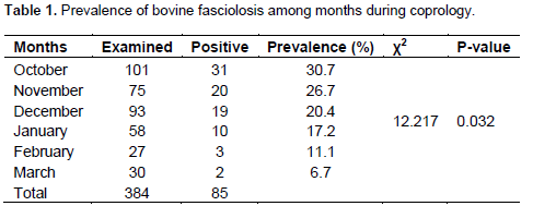
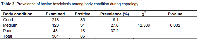
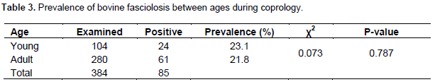
From a total of 384 fecal samples examined for fasciolosis, 22.14% (85/384) were found to be positive by coprological examination (Table 1). The highest preva-lence of bovine fasciolosis was observed during October 30.7% (31/101) and the lowest was seen during March 6.7% (2/30). Statistically analysis showed that there was significant difference (P < 0.05) in prevalence of fascio-losis among months during coprological examination (Table 1). The prevalence of fasciolosis among body conditions of the animals was found to be 16.1% (35/218), 27.6% (34/123) and 37.2% (16/43) in good, medium and poor body conditions, respectively. The highest prevalence was observed in poor body condition as compared to good and medium body conditions. There was statistically significant association (P < 0.05) in prevalence of fasciolosis among the three body condi-tions of the animals (Table 2). Based on age of the animal the prevalence of fasciolosis was found to be 23.1% (24/104) and 21.8% (61/280) in young (< 5 years) and adult (> 5 years), respectively. There was no statistical significant association (P > 0.05) in prevalence of fasciolosis between the two age groups (Table 3).
Post mortem examination
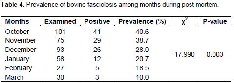
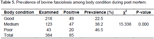
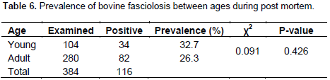
Out of 384 cattle slaughtered, 30.21% (116/384) were found to be positive for fasciolosis by post mortem examination (Table 1). The highest and the lowest prevalence of fasciolosis was recorded 40.6% (41/101) and 10.0% (3/30) in October and March, respectively. Statistical analysis showed that there was significant difference (P < 0.05) on prevalence of fasciolosis among different months (Table 4). During post mortem examination, the prevalence of fasciolosis was found to be highest in poor body condition 46.5% (20/43) when compared to cattle with good 22.5% (49/218) and medium 38.2% (47/123) body conditions. Statistical analysis showed that there was significant difference (P < 0.05) in prevalence of fasciolosis among the three body conditions (Table 5). The prevalence of fasciolosis in association with different age groups, the highest prevalence was recorded in young (< 5 years) 32.7% (34/104) as compared to adult (> 5 years) 29.3% (82/280) during post mortem examination. Statistical analysis showed that there was no significant difference (P > 0.05) in prevalence of fasciolosis between the two age groups of the animals (Table 6).
Fasciola species identification
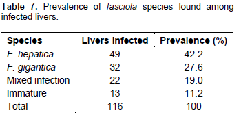
Among 116 livers infected by Fasciola, F. hepatica was found to be the highest prevalence of 42.2% (49/116), while F. gigantica, mixed infestation with the two species and immature flukes were found to be 27.6% (32/116), 19.0% (22/116) and 11.2% (13/116), respectively (Table 7).
Comparison of coprological and post mortem results of fasciolosis
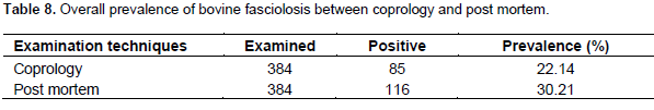
From a total 384 cattle examined for the presence of Fasciola, post mortem finding revealed better results of 116 (30.21%) than coprological findings 85 (22.14%) (Table 8)
Monetary loss analysis due to bovine fasciolosis
The average current cost of one liver and one kilogram beef in Dangila town was 35 and 80 ETB, respectively. Based on this information the total annual monetary loss due to fasciolosis was calculated by using the following formula:
Annual loss from liver condemnation (ALC) = MCS × MLC × P
Where; ALC = Annual loss from liver condemnation; MCS = Mean annual cattle slaughtered at Dangila municipal abattoir (3500); M LC = Mean cost of one liver in Dangila town (35 ETB); P = Prevalence of the disease at the study abattoir (30.21%).
ALC = MCS × M LC × P (31,720 ETB)
Annual loss from carcass weight reduction (ACW) = MCS × CL × BC × P × 126 kg Where; ACW = annual loss from carcass weight reduction; MCS = mean annual cattle slaughtered at Dangila municipal abattoir (3500), CL = carcass weight loss in individual cattle due to fasciolosis (10%), BC = average price of 1kg beef at Dangila town (80 ETB); P = prevalence of fasciolosis at Dangila abattoir (30.21%), average carcass weight of Ethiopian Zebu = 126 kg
ACW = MCS × CL × BC × P × 126 kg (913,550 ETB)
Therefore, the total annual monetary loss due to fasciolosis at Dangila municipal abattoir was found to be the summation of both annual loss from liver condem-nation and annual loss from carcass weight reduction 945,270 ETB (48,432 USD).
DISCUSSION
The overall prevalence of fasciolosis found in the presentstudy was higher during post mortem examination (30.21%) than coprological findings (22.14%). This finding was in line with the prevalence reported by Edilawit et al. (2012) as 25.33 and 15.67% at Wolaita Sodo abattoir and Mulat et al. (2012) as 29.75 and 19.5% at Gondar ELFORA abattoir both in post mortem and coprological examinations, respectively. This may be due to the need of longer period from 8 to 15 weeks after infection for the appearance of Fasciola eggs in the feces. Furthermore, the detection of Fasciola eggs and the appearance of the disease in some areas were difficult to detect during the prepatent period because eggs are expelled intermittently depending on the evacuation of the gall bladder and life cycle of Fasicola (Radostits et al., 2007). Coprological examination also includes numerous steps that increase the chance of losing eggs, as demonstrated by the lower number of positive result recorded in this work, as a result of eggs may remain in the debris while filtering the feces through gauze or may get fixed on the bottom and wall of the container and within the pipette when taking the sediment for microscopic observation.
The prevalence of bovine fasciolosis during coprological examination (22.14%) in this study was lower than the findings of 41.41% in and around Woreta, North Western Ethiopia by Tsegaye et al. (2012); 34.9% by Bekele et al. (2014) in Lemo district, Southern Ethiopia and 31.5% by Ayalew and Endalkachew (2013) at Bahir-Dar municipal abattoir, Northern Ethiopia. The reason behind the variation with the above reports may be due to the expansion of animals’ health post at peasant association level and the intervention of nearby private veterinary drug shop and pharmacies as well as good management practices of the individual owner of the animal. This enables the farmers to have more access for disease control and intervention. Moreover, the variation of season when the research conducted may be the probable reason for the difference prevalence with the above findings.
In the present study, the prevalence of bovine fasciolosis during post mortem examination was found to be 30.21%. This result was in agreement with the findings of Bekele et al. (2014) with prevalence of 30.5% in Hossana municipal abattoir. While this finding was lower as compared to the result by Abie et al. (2012) with prevalence of 54.5% in Jimma municipal abattoir. On the contrary, the result was higher than the findings of 25.33% by Edilawit et al. (2012) in Wolaita Sodo, Ethiopia. The reason behind the variation of the present results with the above findings might be attributed to the differences in ecology of the study areas, management systems of animals, sample size as well as the present study were conducted during the dry period of the year when the infection rates of fasciolsis is expected to be low, since one of the most important factors that influence the occurrence of fasciolosis in an area is the availability of a suitable habitat for the snail intermediate hosts and essential for the development of fluke eggs, miracidia searching for snails and dispersal of cercariae (Urquhart et al., 1996).
The present study according to age groups had no significant difference (P > 0.05). This showed that age groups have no effect for the presence or prevalence of fasciolosis; hence, both animals were equally exposed to infection, but the prevalence of fasciolosis in young was higher than adults both in coprology and post mortem examinations in the study area, which is in line with the finding by Bekele et al. (2014) resulting highest prevalence in young than adult animals. This is clearly justified that the decrease in infection rate (prevalence) as age increase is the result of acquired immunity which is manifested by humeral respond and tissue reaction in bovine liver due to previous challenge. The increased resistance (low prevalence) as age increase is most likely related to the high level of tissue reaction seen in bovine liver, server fibrosis which impedes the passage of immature fluke, acquired resistance, thickening, stenosis and calcification of bile ducts, assumed unfavorable site for adult parasites and consequently fasten their explosion. On the contrary, results indicating inverse correlation of prevalence rate and age of cattle were reported by Tsegaye et al. (2012) in young and adult with prevalence of 40.0 and 42.24%, respectively.
The monthly/seasonal variation in the prevalence of fasciolosis has been studied for 6 dry months in the study area. It was difficult to indicate the effect of seasonal variation on the prevalence of bovine fasciolosis since the study period was too short without incorporating wet months of the season. An accurate description of seasonal occurrence requires long term epidemiological investigation over several years. Even though the study period is short and in dry season, the highest prevalence of fasciolosis was encountered in October and Novem-ber, while lower prevalence in February and March both in coprology and post mortem examinations with a statistical significant difference (P < 0.05). This result was in line with the report of Edilawit et al. (2012) and Yohannes and Abebaw (2012) who showed the highest prevalence during October and November. The probable justification behind it could be, since the wet ecological conditions still prevailed in October and November, it favors the bionomic requirements for breeding of the Lymnaea snails and development of the interamolluscan stages of flukes often reaches the optimum threshold during the wet months of the year. During the dry periods, breeding of snails and development of the larval flukes slow down or stops completely and snails undergo a state of aestivation (Yilma and Malone, 1998).
Species identification on 116 Fasciola infected livers, F. hepatica was found to be the most prevalent (42.2%) as compared to F. gigantica (27.6%), mixed infection by both species (19.0%), and immature flukes (11.2%). This finding is in agreement with reported infection rate of cattle with F. hepatica (63.89%), F. gigantica (24.07%) and mixed (16.5%) by Tadele and Worku (2007), F. hepatica (65.4%), F. gigantica (36.0%), mixed infection (11.5%) and immature Fasciola species (10.1%) by Abie et al. (2012). Furthermore, it is in agreement with the highest prevalence of F. hepatica findings by Mihreteab et al. (2010), Rahmeto et al. (2010), Edilawit et al. (2012) and Bekele et al. (2014). This highest prevalence of F. hepatica species might be associated with the existence of favorable ecological biotopes for the snail, Galba (Lymnaea) truncatula.
The association between the prevalence of fasciolosis and body condition of the animals was found to be statistical significant. The present study was revealed highest prevalence in poor body condition 46.5 and 37.2% both in post mortem and coprological examina-tions, respectively as compared to good and medium body conditions. In support of this finding, a study was conducted in Adwa, Ethiopia by Mihreteab et al. (2010), in Wolaita Sodo, Ethiopia by Edilawit et al. (2012); in Jimma, Ethiopia by Abie et al. (2012) and in Hossana, Southern Ethiopia by Bekele et al. (2014) revealed highest prevalence in poor body conditions of cattle than good and medium body conditions. The probable reason could be due to the fact that animals with poor body condition are usually less resistant and are consequently susceptible to various diseases including fasciolosis and due to reduced performance of the animals created by luck of essential nutrients and poor management by the animal owner.
The direct monetary loss as a result of condemnation of liver of cattle and indirect monetary loss due to carcass weight reduction incurred during this study was estimated to be 31,720 Ethiopian birr (ETB) and 913,550 ETB per annum, respectively. Therefore, the total annual monetary loss due to fasciolsis in the study abattoir is the summation of losses from organ condemnation and carcass weight reduction which is 945,270 ETB (48,432 USD) per annum.
The present finding was higher than the financial losses reported by Mihreteab et al. (2010) 4,672 USD at Adwa municipal abattoir, Rahmeto et al. (2010) 106,400 ETB at Hawassa municipal abattoir. On the contrary, the result was lower than the findings of Edilawit et al. (2012) 1,574,482 Ethiopian birr (87,471 USD) per annum in Wolaita Sodo and Abie et al. (2012) 2,570,396 Ethiopian birr (151,200 USD) per annum in Jimma municipal abattoir. This difference in the estimated monetary losses could be attributed to the increase in the price of liver and meat in the global market in general and in Ethiopia in particular.
CONCLUSION
Bovine fasciolosis is an economically important parasitic disease of cattle in tropical and subtropical countries responsible for considerable economic losses in the cattle industry, mainly through condemnation of fluke infected liver. Although moderate prevalence of bovine fasciolosis was recorded in the study area, the preva-lence was significantly affected by body condition and months of the year and they were identified as important risk factors for the occurrence of fasciolosis in cattle.
Higher prevalence of bovine fasciolosis was recorded in poor than good body conditions of the animal. F. hepatica was found to be the predominant fasciola species causing bovine fasciolsis in the study area. There was substantial agreement between fecal examination and liver inspection in the diagnosis of fasciolosis, indicated that coprological examination for parasite eggs has significant limitation in detecting the presence or absence of fasciolosis, while examination of the liver of animals during post mortem is the most reliable method to detect fluke infection. The total annual monetary losses due to bovine fasciolosis in the study abattoir from liver condemnation (direct loss) and carcass weight reduction (indirect loss) was estimated to be 945,270 Ethiopian birr, indicating that fasciolosis causes significant losses to economy of the study area in particular and the country in general.
ACKNOWLEDGEMENTS
The authors would like to thank College of Agriculture and Veterinary Medicine, Jimma University for the unreserved cooperation and financial support that it provided for this study. The authors are very grateful to all staffs of Dangilla municipal abattoir who helped them in samples collection and contributing significantly in the interview during the research.
REFERENCES
| Abie D, Fentahun B, Ababu B, Muli e M, Murad B, Mekonnen A (2012). An abattoir survey on the prevalence and monetary loss of fasciolosis in cattle in Jimma town, Ethiopia, Glob. Vet. 8:381-385. | ||||
| Ayalew S, Endalkachew N (2013). Prevalence and risk factors of bovine and ovine fasciolosis, and evaluation of direct sedimentation sensitivity method at Bahir-Dar municipal abattoir, Northern Ethiopia. Ethiopian Vet. J. 17:1-17. | ||||
| Bekele C, Sissay M, Mulugeta D (2014). On farm study of bovine fasciolosis in Lemo district and its economic loss due to liver condemnation at Hossana municipal abattoir, Southern Ethiopia. Int. J. Curr. Microbiol. Appl. Sci. 3:1122-1132. | ||||
| Chhabra MB, Singla LD (2009). Food-borne parasitic zoonoses in India: Review of recent reports of human infections. J. Vet. Parasitol. 23(2):103-110 | ||||
| CSA (2012). Central Statistical Agency, Federal Democratic Republic of Ethiopia, central statistical investigation, Statistical abstract. | ||||
| De-Lahunta, A, Habel RE (1986). Teeth applied veterinary anatomy. W.B. Saunders Company. pp. 4-6. | ||||
| Edilawit W, Mekonnen A, Mulugeta T (2012). An abattoir survey on the prevalence and monetary loss of fasciolosis among cattle in Wolaita Sodo town, Ethiopia. Adv. Biol. Res. 6:95-100. | ||||
| FAO (1994). Diseases of domestic animals caused by flukes: Epidemiology, diagnosis and control of Fasciola, Paramphistomum, Dicrocoelium, Eurytrema and Schistosome infections of ruminants in developing countries. FAO/UN, Viale delle Terme di caracalla, Rome, Italy. pp. 49. | ||||
| FAO (2003). Food Agricultural Organization of United Nations. Diagnostic manual on meat inspection for developing countries. | ||||
| Gupta SK, Singla LD (2013). Diagnostic trends in parasitic diseases of animals. In: Veterinary Diagnostics: Current Trends, Gupta RP, Garg SR, Nehra V, Lather D (eds). Satish Serial Publishing House, Delhi, pp 81-112 | ||||
| Hansen I, Perry B (1994). The epidemiology, Diagnoses and Control of Helminth parasite of ruminants. A hand book for Research epidemiology; International Laboratory on Animal Disease (ILARD). Nairobi, Kenya. | ||||
| ILCA. (1992). Annual significance of bovine fasciolosis at Bedele municipal abattoir, Addis Ababa University, Debre Zeit, Ethiopia. | ||||
| Kassai T (1999). Veterinary helminthology. University of veterinary science, Oxford: Butter worth Heinemann. pp. 90-110. | ||||
| Mari Henonen, (1989). Body condition scoring as cattle in Ethiopia. MOA. pp. 45-46. | ||||
| Mihreteab B, Haftom T, Yehenew G (2010). Bovine fasciolosis: Prevalence and its economic loose due to liver condemnation at Adwa Municipal Abattoir, North Ethiopia. Ethiopian J. Appl. Sci.and Technol. 1:39-47. | ||||
| Mulat N, Basaznew B, Mersha C, Achenef M, Tewodros F (2012). Comparison of coprological and postmoretem examinations techniques for the deterimination of prevalence and economic significance of bovine fasciolosis at Gondar ELFORA abattoir. J. Adv. Vet. Res. 2:18-23. | ||||
|
Ogunrinade A, Adegoke GO (1982). Bovine fasciolosis in Nigeria Inter current Parasitic and bacterial infection. Trop. Anim. Health Prod. 14:121-125. Crossref |
||||
| Radostits OM, Gay CC, Hinchcliff KW, Blood DC, Constable PD (2007). A text book of the disease of cattle, sheep, goats, pigs and horse: Veterinary Medicine. 10th edn. New York, Saunders Elsevier. pp. 1516- 1579. | ||||
| Rahmeto A, Fufa A, Mulugeta B, Solomon M, Bekele M, Alemayehu, R. (2010). Fasciolosis: Prevalence, financial losses due to liver condemnation and evaluation of a simple sedimentation diagnostic technique in cattle slaughtered at Hawassa Municipal abattoir, southern Ethiopia. Ethiopian Vet. J. 14:39-51. | ||||
| Tadele T, Worku T (2007). The prevalence and economic significance of bovine fasciolosis at Jimma, Abattoir, Ethiopia. Int. J. Vet. Med. 3:15 | ||||
| Thrusfield, M. (1995). Veterinary epidemiology, 2nd edn. U.K. Black wall science Ltd. pp. 182-198. | ||||
| Tsegaye B, Abebaw H, Girma S (2012). Study on coprological prevalence of bovine fasciolosis in and around Woreta, Northwestern Ethiopia. J. Vet. Med. Anim. Health Vol. 4:89-92. | ||||
| Urquhart GM, Armour J, Duncan JL, Dunn AM, Jennings FW (1996). Veterinary Parasitology. 2 nd ed. Blackwell Science, UK. pp. 103-113. | ||||
|
Yilma J, Malone JB (1998). A geographical information system forecast model for strategic control of fasciolosis in Ethiopia. Vet. Parasitol. 78:103-127. Crossref |
||||
| Yohannes E, Abebaw G (2012). Prevalence of bovine fasciolosis, amplitude of liver condemnation and its economic impact in slaughtered cattle at municipal abattoir of Mekelle, North Ethiopia. Int. J. Curr. Agric. Res. 1:10-14. | ||||
Copyright © 2024 Author(s) retain the copyright of this article.
This article is published under the terms of the Creative Commons Attribution License 4.0


