Full Length Research Paper

ABSTRACT
Chronic emaciation reflects the severity and duration of the aetiologies associated with a disease condition. The primary aim of this investigation was to determine the trend and most significant causes of chronic emaciation and cachexia in off-take cattle in Ibadan metropolis and to evaluate the clinico-pathologic findings. The study was a prospective one. In this particular study, causative factors of chronic emaciation were examined. The purposive sampling technique was used for this study which was carried out at the Veterinary Teaching Hospital of the University of Ibadan, Nigeria, between September, 2019 and December, 2019. Samples were collected from 100 chronically emaciated off-take cattle of different breeds, sexes and ages, not less than two years old. The diagnostic protocol of complete physical examination and comprehensive laboratory investigations such as parasitology, haematology, serum chemistry and urinalysis amongst others, were followed as well. The study revealed six major groups of aetiologies of chronic emaciation and cachexia. The haematologic parameters of packed cell volume (PCV), haemoglobin (Hb) concentration, mean corpuscular volume (MCV), mean corpuscular haemoglobin concentration (MCHC), neutrophil and platelet count were statistically significant (p<0.05) with differences between the mean values of emaciated cases compared to control subjects. The serum chemistry parameters of albumin, globulin, bilirubin, aspartate amino transferase (AST) and gamma glutamyl transferase (GGT) were statistically significant (p<0.05) with differences between the mean values of emaciated cases and control subjects. The increasing prevalence of dicrocoeliasis and paramphistomiasis as revealed in the study should be of epidemiological and clinical relevance to livestock health institutions and large scale animal practitioners in Nigeria.
Key words: Aetiologies, chronic emaciation, investigation, endoparasitic infection, organ disorders, cattle, Ibadan metropolis.
INTRODUCTION
Emaciation is defined as excessive leanness and is not a disease but rather a symptom that can be found in many diseases either singly or in combination with other unspecific clinical signs (Fischer, 1993). The clinical problem of weight loss or emaciation suggests that an individual large animal patient or a herd has lost weight over a known period of time (Smith, 2008) as a consequence of chronic disorder (Freeman, 2012). It may also suggest that the patient has reached a subnormal adult weight and size (Smith, 2008). The subject of chronic emaciation is broad as there are multifactorial causes, and with a progression to syndrome of cachexia The array of reasons includes chronic bacterial infections, such as tuberculosis (Purushothaman, 2001; Belachew, 2017), paratuberculosis (Johne’s Disease) (Radostits et al., 2006; Smith, 2008; Sweeney et al., 2012; Fecteau, 2018), actinomycosis, actinobacillosis, nocardiosis, corynebacteriosis (Radostits et al., 2006; Smith, 2008; Ribeiro, 2019; Smith, 2019) etc.; chronic inflammations such as endocarditis, lymphadenitis, pleuritis, pericarditis and enteritis etc. (Radostits et al., 2006; Smith, 2008); parasitic infections such as trypanosomiasis, toxoplasmosis, chronic babesiosis (Urquhart et al., 1996; Rajavelu, 2001; Love and Hutchinson, 2003; Khan and Line, 2010; Odeniran and Welburn, 2018; Petersen and Grinnage-Pulley, 2020), anaplasmosis, besnoitiosis, (Bussieras and Chermette, 1991; Khan and Line, 2010), sarcosporidiosis (Urquhart et al., 1996; Bussieras and Chermette, 1991; JanuškeviÄius et al., 2018; More, 2019), and chronic helminthosis (Urquhart et al., 1996; Khan and Line, 2010) etc.; hepatic and renal conditions associated with several causes (Haroun and Hussein, 1975; Radostits et al., 2006; Smith, 2008); gastro-intestinal disorders such as infiltrative diseases (Radostits et al., 2006; Smith, 2008; Wittek, 2014); immune mediated condition associated with amyloid deposition in various organs and hyperglobulinemia, consequent upon chronic infection/inflammation or neoplasm (Radostits et al., 2006); congenital disorders such as congenital cardiac malformations (tetralogy of fallot, interventricular septal defect) and congenital renal disease (agenesis, dysplasia, hypoplasia, polycystic kidney disease) (Smith, 2008) etc., most of which create physiologic inefficiencies that require energy beyond the body's ability to supply it; endocrine dysfunction such as diabetes mellitus (Mostaghni and Ivoghli, 1977); neoplasms such as lymphosarcoma (Bertone, 1990; NADIS, 2011); and primary undernutrition (Oetzel, 1988; Smith, 2008) etc.
Skinny animals are seen in primary undernutrition. Females carrying multiple fetuses and lactating animals with metabolic abnormalities secondary to abomasal displacements would also show signs of weight loss (Smith, 2008). Nonetheless, the precipitating causes of chronic emaciation often result from disorders with a protracted course such as cardio-pulmonary disorders, chronic renal disease, chronic hepatopathy, neoplasm, intestinal malassimilation, and some other chronic conditions (Smith, 2008; Khan and Line, 2010). There are risk factors which may be associated with the aetiologies of chronic emaciation in cattle. These may be related to age, breed, genetics, environment or climatic condition, nutrition, farming or management practices (Putt et al., 1988; Radostits et al., 2006) etc.
Anorexia is a significant occurrence and the primary mechanism for weight loss (Khan and Line, 2010). Weight loss results from decreased nutrient intake, when partial anorexia occurs over a long period (Khan and Line, 2010). Anorexia is usually associated with a primary disease condition, and is regulated by cytokines, including interleukin (IL) - 1 and tumor necrosis factor-alpha (TNF-α), released during an inflammatory response (Smith, 2008). These inflammatory cytokines directly cause anorexia, increase lean body mass loss and may increase energy requirements (Fearon et al., 2012; Freeman, 2012). Chronic emaciation or cachexia appears to be a response to increased catabolism with either normal or decreased appetite (Sanderson, 2019).
Diagnostic investigation of chronic emaciation should follow the conventional protocol of comprehensive history taking, careful physical examination (Jackson and Cockcroft, 2002; Radostits et al., 2006; Smith, 2008; Khan and Line, 2010), laboratory examination of relevant samples such as parasitology, haematology, serum chemistry and serology etc. (Anderson et al., 1976; Baker and Silverton, 1998; Khan and Line, 2010; Latimer, 2011; Sivajothi et al., 2014; Jibrin et al., 2018; Bildfell, 2019; eClinpath, 2019), and special diagnostic tests such as Polymerase Chain Reaction (Khan and Line, 2010; Fajinmi et al., 2011; Sivajothi et al., 2014); measuring and recording techniques like nerve conduction velocity (Schubert, 2019) and electrocardiogram (Kittleson, 2019); endoscopy (Franz, 2011); diagnostic imaging such as radiography (Smith, 2008; Lattimer, 2019b), ultrasonography (Braun, 1991; Radostits et al., 2006; Streeter and Step, 2007; Smith, 2008; Buczinski and Bélanger, 2010; Imran and Sharma, 2014; Lattimer, 2020), scintigraphy (Radostits et al., 2006), computed tomography (Nuss et al., 2011), and magnetic resonance imaging (Lattimer, 2019a); cytology (Trevor, 2019); and histopathology (Radostits et al., 2006; Smith, 2008; Khan and Line, 2010; Bildfell, 2019). However, this study was limited to panel screening tests to provide clues of the causes of emaciation in randomly selected off-take cattle in Ibadan metropolis.
An average number of 450 cattle are slaughtered at the central abattoir located at Amosun village in Ibadan. Post mortem records indicate partial condemnation of carcasses with organ involvement; and this is of economic significance. Besides carcass losses and reduced meat quality due to distinct organoleptic changes, it can be imagined that there would have been production losses when these animals were alive (Pelczyńska, 1987; Phiri, 2006). Relatively few studies have been carried out to investigate emaciation in cattle related to some specific diseases such as the one carried out by Raji et al. (2010) to examine pathological lesions of organs of 7812 cattle slaughtered in Zaria abattoir. The main aim of this study therefore, was to investigate the trend of aetiologies of chronic emaciation in various breeds of cattle in Ibadan metropolis, Nigeria, using standard diagnostic protocol to evaluate clinico-pathological abnormalities.
MATERIALS AND METHODS
Scope and design
The study was a prospective one. In this particular study, causative factors of chronic emaciation were investigated. The purposive sampling technique was used for this study. Thus, samples were collected from only chronically emaciated cattle presented at the central slaughter house in Ibadan metropolis. A total number of 100 cattle of different breeds, sexes and ages not less than two years old and body condition score between 1 and 3 were selected for the study. However, there were 50 animal control samples. The control animals were healthy and of body condition score from 6 and above. The sample size was calculated using the statistical programme, G-Power® version 3.1.1; Germany.
Sampling
The various samples for which data were derived included the animal, blood, serum, feces, urine and skin scrapings. The samples were collected over 3 months. Consents to conduct a physical examination and contain representatives from the animals were obtained from the owners after explaining the purpose of the study. Subject data was captured on a preprinted form. Each animal was subjected to a thorough physical examination; and basic parameters including temperature, respiratory rate, pulse and body condition score were taken and recorded. Abnormal systemic findings observed were noted. Blood samples were taken from all the animals; for parasitology and haematologic evaluations. The sampling procedure was as described by Baker and Silverton (1998). Serum samples were retrieved from blood samples collected for chemistry evaluation. The sampling procedure was as described by Baker and Silverton (1998). Feces of animals were collected for various examinations including parasitology, cytology and occult blood examination. The sampling procedure was as described by Baron (1973) and CDC (2020). Urine of each animal was collected for urinalysis by stroking the escutcheon in females, and by massaging of the preputial orifice in males. The voided urine was collected into a clear tube for urinalysis and sediment examination as described by Khan and Line (2010). Scrapings of the skin of animals with lesions were carried out for dermatologic investigations. These were done using sharp scalpel blades to scrape the skin near and around observed lesions, following drops of glycerine oil; the scrapings including hairs and scales were transferred into clean glass slides as described by Khan and Line (2010).
Procedure
Epidemiological data sourcing
Various data were obtained from responses of cattle merchants and physical observation. History related to subjects such as sources, environmental conditions, husbandry practices and abnormal symptoms were sought by oral questioning of cattle merchants. The age, sex and breed of the animals were noted from physical observation. Breeds of all the cattle were identified using morphological features which distinguish them such as horn shape, body size and coat color as described by Kugonza et al. (2011) and Terefe et al. (2015). Age was determined using the technique of dentition as described by Lasisi et al. (2002).
Physical examination
Each animal was subjected to a thorough physical examination beginning with a distance and followed by a close examination as described by Radostits et al. (2006). Abnormalities observed related to animal’s condition were recorded.
Laboratory Investigations
Blood smear examination (thin films)
A clean slide was wiped with alcohol and a tiny drop of blood placed near the end of the slide. Another clean and disinfected slide was used to spread the drop of blood along the sample slide to form a thin smear. The slide was air-dried, fixed briefly in methanol and stained with Giemsa stain. The slide was then examined under the microscope (magnification up to 1000 x) to identify protozoa organisms (such as Babesia Sp, Trypanosoma Sp) and Rickettsial organisms (such as Anaplasma bovis). This method was as described by Khan and Line (2010).
Buffy coat examination
This concentration technique was used for trypanosome identification as described by Khan and Line (2010). Blood was taken up from one end of the hematocrit tube, and the other open end was sealed with a burner. The sealed tube containing heparinized blood was placed in the groove of the rotor plate with the sealed end outwards; and spunned at a high speed (12, 000 rpm) for 5 min. After centrifugation the tube was removed and buffy coat examined for the presence of trypanosomes by making a smear of this area. The slide was air-dried, fixed in methanol and stained with Giemsa stain before examining under microscope (100 x).
Haematology
Haematologic procedure as described by Baker and Silverton (1998) include methods to determine packed cell volume, haemoglobin level, red cell count, red cell indices, white cell count, platelet count; and blood glucose concentration. The PCV was determined using a microhematocrit tube, a specifically designed centrifuge and a reader as described by Baker and Silverton (1998). Haemoglobin concentration was determined by the cyanmethaemoglobin method as described by Baker and Silverton (1998). Red cell count of blood sampled from each animal was performed by a visual method using the improved Neubauer counting chamber as described by Baker and Silverton (1998).
White cell count of sampled blood was achieved by a visual method using the improved Neubauer counting chamber as described by Baker and Silverton (1998). Platelet Count was performed by a visual method using the improved Neubauer counting chamber as described by Baker and Silverton (1998). Blood Glucose Concentrations of test samples were determined using the manual technique of Trinders glucose oxidase method (using phenol and 4 - Aminophenazone) as described by Baker and Silverton (1998).
Serum biochemical analysis
Serum Total Proteins, Albumins and Globulin fractions were determined with the manual biuret method as described by Baker and Silverton (1998). All stages were carried out to the point at which the color was developed with the biuret reagent, at a temperature above 25°C to avoid crystallization of sodium sulphate. This was achieved by precipitating the globulins in a centrifuge tube placed inside wider centrifuge tube containing water warmed to about 30°C and centrifuging both tubes. Serum urea concentration (SUC) was determined using the urease method which uses the Berthelot reaction as described by Baker and Silverton (1998).
Serum creatinine concentration was determined with the manual method in samples collected as described by Baker and Silverton (1998). The technique is the Jaffe reaction which produces a red color with an alkaline picrate solution. Serum gamma glutamyl transferase (GGT) concentration was determined using a commercial Colorimetric Assay kit (Sigma, 2013). GGT activity is determined by a coupled enzyme assay, in which the GGT transfers the g-glutamyl group from the substrate L-g Glutamyl-p-nitroanilide, liberating the chromogen p-nitroanilide (pNA, 418 nm) proportional to the GGT present. One unit of GGT is the amount of enzyme that will generate 1.0 mmole of pNA per minute at 37°C.
Serum aspartate aminotransferase (AST) concentration was determined with the use of a kinetic method as described by Baker and Silverton (1998). AST catalyzes the transfer of the amino group aspartate to 2-oxoglutarate to yield oxaloacetate and glutamate. The oxaloacetate formed in the first reaction is then reacted with reduced nicotinamide adenine dinucleotide (NADH) in the presence of malate dehydrogenase (MDH) to form nicotinamide adenine dinucleotide (NAD). AST is determined by measuring the rate of oxidation of NADH at 340 nm.
Serum creatine kinase (CK) Concentration was determined using a commercial Colorimetric Assay kit (Sigma, 2014). In this assay, creatine kinase activity is determined by a coupled enzyme reaction resulting in the production of reduced nicotinamide adenine dinucleotide phosphate (NADPH), measured at 340 nm, proportionate to the CK activity present in the serum sample. Serum bilirubin concentration in test samples was determined using the method of Powell (1944) as described by Baker and Silverton (1998). The ‘accelerating agent’ urea-sodium benzoate solution causes bilirubin to react with the diazo reagent at low dilution without precipitating proteins. The resulting color when compared with a suitable standard gave a measure of total bilirubin in serum.
Urinalysis
The procedures for urinalysis in the study carried out include; physical examination of urine, use of colorimetric test pads for several semi quantitative chemical evaluation and microscopic examination of urine sediment. These procedures were described by Khan and Line (2010) and Parrah et al. (2013).
Faecal microscopy
Faecal microscopy was done to investigate for metazoans, protozoa and blood cells as described by Khan and Line (2010); and literature of CDC (2020). The technique of zinc-sulphate centrifugation – flotation was used to investigate for the presence of cysts of protozoa and helminth ova in faeces. Investigation for blood cells (leukocytes and red blood cells) in faeces was carried out using Gram staining technique.
Faecal occult blood test
The Haemoccult slid test (Smith – Kline diagnostics, England) was used to investigate the presence of occult blood in faeces. It is a guaiac-based test as described by Baron (1973). One side of a guaiac – impregnated paper was smeared with faeces and a few drops of developer solution (stabilized peroxide reagent) were added to the opposite side of the paper. The appearance of a blue color within 30 sec was considered a positive test.
Method of data analysis
Both qualitative and quantitative forms of analysis were used for the study to analyze obtained data. All parameters were subjected to a two-tail analysis of variance (t-test) to determine the level of their significance as previously described by Norman and Bailey (1997) using Microsoft excel (2010 version). Statistical data analysis was performed with STATA 12, using an unpaired two-sample t-test with the assumption of unequal variances between the clinical cases and the control subjects. Means and standard deviations with confidence intervals for each study variable were calculated for each group. The means obtained were then compared between the two groups and against the standard reference values for each haematologic or serum biochemical values. Level of significance threshold was set at P = 0.05.
RESULTS
Results of laboratory investigations are summarized as follows:
Frequency distribution of blood parasitological findings of emaciated/cachexic cattle
Babesia sp (Nil); Trypanosoma sp (12); Anaplasma bovis (Nil); Haemobartonella bovis (4); Absence of Parasites (84).
Frequency distribution of faecal examination findings in emaciated/cachexic cattle
The presence of Nematodes (48); Presence of Cestodes (6); Presence of Trematodes (28); Presence of Protozoa (Pathogenic Eimeria sp) (42); Presence of Leukocytes (90); Presence of Red Blood Cells (55); Presence of Occult Blood (55); Absence of Abnormal Finding (12). NB: Some of the parasites were present as mixed or concurrent infections.
Frequency distribution of helminths detected in emaciated and cachexic cattle
Cestodes (Moniezia sp (6)); Nematodes (Capillaria sp (2); Strongyle (37); Toxocara vitulorum (17); Trichuris sp (4)); Trematodes (Dicrocoelium sp (12); Fasciola hepatica (6); Paramphistomum sp (16)); Absence of Helminths (24). NB: Some of the helminths were present as mixed infection.
Frequency distribution of mixed parasitic infections detected in faeces of emaciated and cachexic cattle
Dicrocoelium sp + Nematode (2); Dicrocoelium sp +Paramphistomum sp (3); Dicrocoelium sp + Eimeria sp (3); Fasciola hepatica + Paramphistomum sp (3); Fasciola hepatica + Eimeria sp (1); Paramphistomum sp + Nematode (6); Moniezia sp + Nematode (2); Moniezia sp + Nematode (Capillaria sp) + Eimeria sp (2); Moniezia sp + Eimeria sp (2); Mixed Nematode sp with or without Eimeria sp (6); Single Nematode sp + Eimeria sp (18).
Frequency distribution of parameters of urinalysis of emaciated/cachexic cattle
Changes in pH (56); Elevated/Upper Mark Specific Gravity (50); Reduced Specific Gravity (0); Presence of Occult Blood (2); Proteinuria (2); Glycosuria (0); Ketonuria (0); Bilirubinuria (0); Significant Presence of RBC (2); Significant Presence of Leukocytes/Bacteria (2); Presence of Epithelial Cells (2); Significant Presence of Epithelial Casts (2); Presence of Granular Casts (2); Presence of Hyaline Casts (59); Significant Presence of Crystals (Phosphates) (10).
Frequency distribution of concurrent infection of nematode and non-gastrointestinal parasitic/disease conditions in emaciated and cachexic cattle
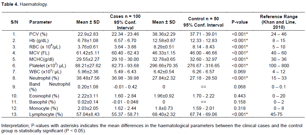
Trypanosoma brucei + Nematode (Strongyle) (2); Trypanosoma congolense + Nematode (Strongyle) (2); Trypanosoma congolense + Nematode (Toxocara vitulorum) (2); Urinary Tract Infection + Paramphistomum sp + Nematode (Toxocara vitulorum) (2). The results are summarized in Tables 1 to 5.
DISCUSSION
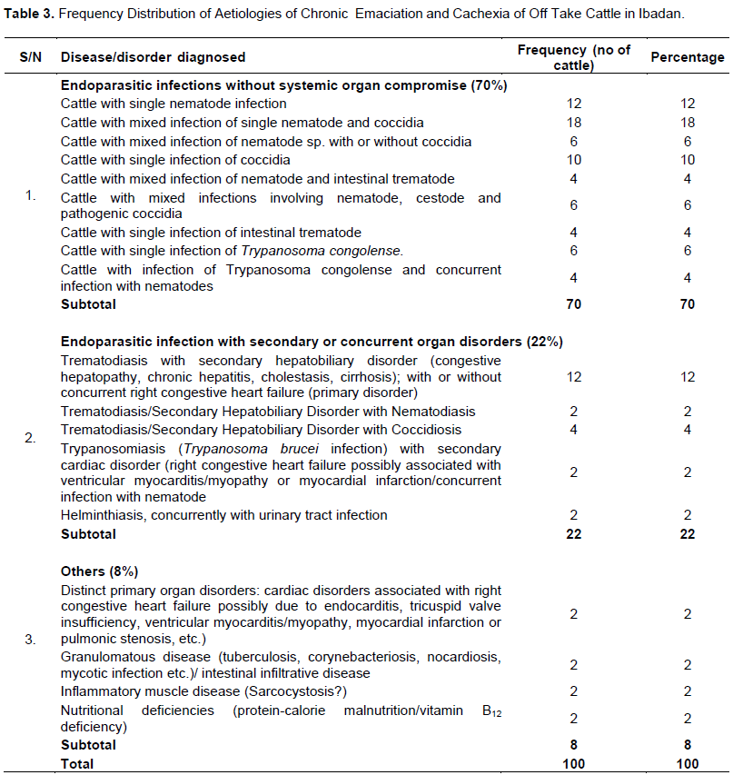
The general history of the emaciated subjects was either not known or obscure because most of the off-take animals were in possession of traders rather than the owners. However, these trypano-intolerant breeds of cattle were raised under an extensive grazing system with associated risk factors (Putt et al., 1988). In the study, 6 groups of aetiologies were broadly identified.
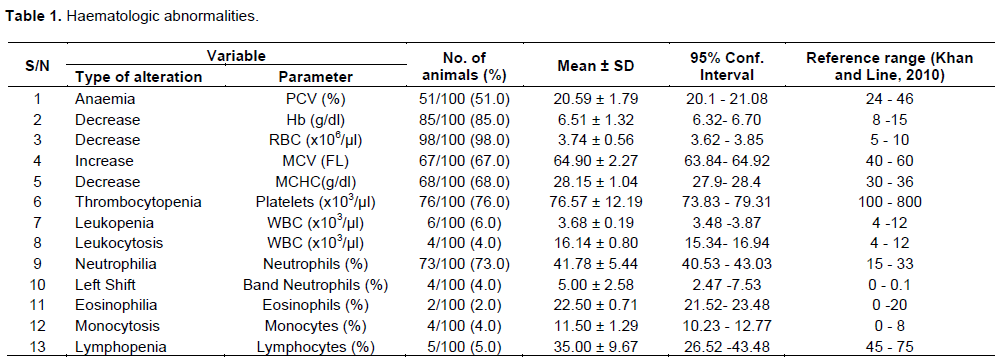
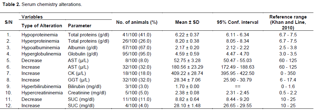
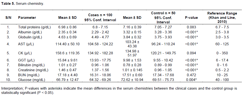
Endoparasitic Infections without Secondary Organ Compromise findings were 70% of the total number of chronic emaciated and cachexic cattle investigated (Table 3). Diagnostic results for intestinal parasites in this group include nonspecific signs such as unthrifty appearance, emaciation, mucopurulent ocular discharges, ruffled hair coat, weakness, dullness, pale mucous membranes, tachycardia, dyspnea, tachypnea, various forms of diarrhea, haematochezia (trichuriasis), melena (coccidiosis and paramphistomiasis), and dehydration. Haematologic correlates were normocytic normochromic anaemia of chronic state (capillariasis, strongyle worm infection, toxocariasis, cestode (Moniezia sp infection), macrocytic hypochromic anaemia (coccidiosis, paramphistomiasis and trichuriasis) as an indication of blood loss, thrombocytopenia (coccidiosis, paramphistomiasis and trichuriasis), as well as relative lymphopenia and neutrophilia in some subjects (Latimer, 2011; Roland et al., 2014). Thrombocytopenia may be due to increased platelet consumption which resulted from intestinal damage and haemorrhage, while the relative lymphopenia and neutrophilia may be associated with endogenous stress due to intestinal inflammation (Latimer, 2011; Roland et al., 2014; Reddy et al., 2015). Serum chemistry findings include hypoalbuminaemia (caused by damage of intestinal mucosa and plasma protein leakage (Love and Hutchinson, 2003), hyperglobulinaemia (possibly due to intestinal inflammation and acute phase proteins (Latimer, 2011) and increased serum urea concentration (SUC), perhaps of prerenal cause (Latimer, 2011). Faecal examination findings include presence of leukocytes (indicating some degree of intestinal inflammation) and numerous red blood cells (for subjects with trichuriasis) on faecal cytology; and presence of occult blood (coccidiosis and paramphistomiasis) on faecal occult blood test.
Diagnoses were based on clinical findings including the detection of ova of nematode species, cestode species and Paramphistomum sp by both direct saline microscopic examination of faeces and zinc sulphate centrifugation-flotation technique; and detection of pathogenic Eimeria sp oocysts with zinc sulphate centrifugation-flotation technique (Fraser et al., 1991). The detection of gastrointestinal parasites in faeces was following work done by Ahmed et al. (2015) in a study of the prevalence of gastrointestinal parasitism of cattle in Banskhali upazilla, Chittagong, Bangladesh. Physical examination findings in 10 subjects which had Trypanosoma congolense (Table 3) include unthrifty appearance, emaciation, pallour, lymphadenopathy, watery faeces (in cases with concurrent nematodiasis) and dehydration etc. Haematologic finding of normocytic normochromic anaemia reflected a chronic state contrary to the usual finding of mild regenerative anaemia in acute condition as may be associated with the complex pathogenesis involving mainly increased erythrophagocytosis, some haemolysis, and dyshaematopoiesis (ILRAD, 1990; Radostits et al., 2006; Latimer, 2011). Other haematologic findings include thrombocytopenia as may have been caused by intravascular coagulation and platelet consumption due to affinity of Trypanosoma congolense for vascular endothelium (Radostits et al., 2006; Latimer, 2011); and relative lymphopenia and neutrophilia as may be associated with endogenous stress due to inflammation (Latimer, 2011).
Serum chemistry findings include hypoalbuminaemia (more severe in the 4 subjects with concurrent nematodiasis), hyperglobulinaemia, and mildly elevated AST, CK and GGT (reflection of systemic inflammation in the subjects (Smith, 2008; Latimer, 2011). Diagnosis of trypanosome infection was established with both blood smear and buffy coat examination as described by Petersen and Grinnage-Pulley (2020).
Endoparasitic Infections with Secondary or Concurrent systemic organ Involvement in 22 cases as previously reported by Akhaine et al. (2020) include 12 emaciated cattle with trematodiasis and secondary hepatobiliary disorder (congestive hepatopathy, chronic hepatitis, cholestasis, cirrhosis), with or without concurrent right Congestive Heart Failure (Primary Disorder); 4 emaciated subjects with trematodiasis and secondary hepatobiliary disorder with Eimeria sp infection; 2 emaciated subjects with trematodiasis and secondary hepatobiliary disorder, with nematode infection; 2 emaciated subjects with trypanosomiasis (Trypanosoma brucei infection) and secondary cardiac disorder (right Congestive Heart Failure possibly associated with ventricular myocarditis/myopathy or myocardial infarction); in addition to 2 emaciated subjects with helminthiasis and concurrent Urinary Tract Infection.
It was discovered that 6% of the emaciated cattle had Fasciola hepatica, while Dicrocoelium sp was detected in 12% (Table 3). Mixed infection of Fasciola hepatica and Paramphistomum sp was detected in 3 subjects, while mixed infection of Dicroecoelium sp and Paramphistomum sp was detected in another three subjects. The general physical examination findings in this group with trematodiasis include unthrifty appearance, arched back posture, emaciation, mucopurulent ocular discharges, ruffled hair coat, weakness, dullness, pale mucous membranes, tachycardia, dyspnea, tachypnea, diarrhea and dehydration, patchy erythema of the skin, ascites and distended abdomen. However, 4 out of the 18 subjects in this group with trematodiasis presented obvious signs of cardiopulmonary compromise such as a distended jugular vein, increased jugular pulse, abducted forelimbs, extended neck and flared nostril, cyanosis, cardiac murmur, edema and ascites, breathing difficulty and tachycardia. These findings may have been associated with right Congestive Heart Failure as a concurrent condition in these subjects. Similar results were reported in four clinical cases of dicrocoeliosis in lamas by Klein et al. (2012). The findings of abducted forelimbs, extended forelimbs, flared nostril, cyanosis, abnormal breathing and tachycardia were suggestive of reduced oxygen diffusion-perfusion in the lungs; while the finding of a right systolic cardiac murmur may be associated with a cardiac abnormality such as endocarditis, tricuspid valve insufficiency, right ventricular myocarditis/myopathy, myocardial infarction or pulmonic stenosis, pulmonary hypertension/cor pulmonale etc. (Radostits et al., 2006). The finding of arched back posture generally in the group may be due to abdominal pain associated with pressure on the liver capsule from parenchymal swelling or directly from the lesions (Radostits et al., 2006). The findings of patchy erythema of the skin and subcutaneous edema were possibly due to hypersensitization associated with phylloerythrin retention, consequent upon compromised hepatic function (Radostits et al., 2006; Foreman, 2020). Ascites as determined by the presence of abdominal fluid waves may have resulted from portal hypertension caused by venous blockage and protein leakage in to the peritoneal cavity; and low albumin concentrations (Radostits et al., 2006; Smith, 2008; Foreman, 2020). Low albumin concentrations and reduced oncotic pressure may be reasons for the observed edema in the emaciated subjects (Foreman, 2020). Congestive Heart Failure may have contributed to hepatic congestion and ascites for the 4 subjects with concurrent right Congestive Heart Failure (Buczinski et al., 2010).
Other diagnostic findings include haematologic and serum chemistry alterations. Haematologic correlate of macrocytic hypochromic anaemia with lowered red cell indices and higher MCV may have been due to liver haemorrhage as a result of immature wandering flukes which destroy liver tissue, and also as a consequence of trauma and feeding activity of adult flukes within the bile ducts (Latimer, 2011; Foreman, 2020); while thrombocytopenia may have resulted from sequestration of thrombocytes associated with splenic enlargement, and possibly from increased platelet consumption due to liver haemorrhage (Khan and Line, 2010; Latimer, 2011; Roland et al., 2014). Relative lymphopenia and neutrophilia was a haematologic finding possibly associated with endogenous stress (Latimer, 2011; Roland et al., 2014). Serum chemistry revealed elevation of Aspartate amino transferase (AST) and Gamma glutamyl transferase (GGT) (for 14 subjects without concurrent Congestive Heart Failure) with normal serum creatine kinase levels (within the normal range for the specie). An elevated AST (a nonspecific enzyme) with a normal CK was suggestive of exclusion of muscle related disorder, and presence of liver pathology such as may have been caused by flukes in 14 subjects (Radostits et al., 2006; Smith, 2008; Latimer, 2011; Foreman, 2020). However, AST and CK were both elevated for the other 4 subjects with concurrent Congestive Heart Failure, suggestive of muscle disorder (possibly cardiac abnormality) (Radostits et al., 2006; Smith, 2008; Latimer, 2011) as part of the systemic disease condition. The damaging effects of Adult flukes residing in the bile ducts include cholecystitis, thickened duct wall and fibrosis etc.; and this may have resulted in enzyme (GGT) leakage from the biliary epithelium with increased serum concentration (Latimer, 2011; Ballweber, 2020; Foreman, 2020). Elevation of liver enzymes such as AST and GGT was similarly reported in clinical cases of fluke infection (dicrocoeliosis) in lamas by Klein et al. (2012). Hypoalbuminaemia and hyperglobulinaemia were critical clinical findings. Hypoproteinemia and hypoalbuminaemia are expected findings with compromised liver which has reduced capacity to metabolize proteins (eClinpath, 2019). Consequently, serum urea and creatinine concentrations were observed to be low normal (Latimer, 2011). Hyperglobulinaemia may have been due to inflammation and acute phase proteins (Latimer, 2011). In subjects with concurrent infections with Eimeria sp or Paramphistomum sp or nematodes, hypoalbuminaemia was severe. Serum glucose was of low average for all the issues possibly due to effects on glucose homeostasis and glycogen storage by the compromised liver (Latimer, 2011). Serum bilirubin concentrations for the subjects in this group were within a normal range, though some were at the upper mark. This finding was not unexpected in liver disease of cattle (Foreman, 2020).
The finding of hyaline casts on urine sediment examination for some of the subjects may have been due to dehydration (Parrah et al., 2013), while the result of significant phosphate crystals in urine of 3 subjects may have been due to high alkaline urine (Parrah et al., 2013). Faecal cytology revealed inflammatory cells (leukocytes) for subjects with concurrent infections with Eimeria sp and nematodes; and also for mixed infection with the rumen fluke, Paramphistomum sp. Faecal occult blood test was positive for all the subjects in this group; this may have resulted from haemorrhage and communication with the intestinal tract; and also, for subjects with concurrent infection with Eimeria sp from small intestinal bleeding (Khan and Line, 2010).
Diagnosis of trematodiasis and associated infection with Eimeria sp or nematodes for some subjects were based on clinical findings including the detection of ova of Fasciola hepatica, Dicrocoelium sp or Paramphistomum sp (in mixed infection) and nematodes; and oocysts of Eimeria sp with zinc sulphate centrifugation-flotation technique (Wamae et al., 1990). Diagnoses of secondary and concurrent systemic organ compromises were based on analysis of physical examination and laboratory findings such as haematology, serum chemistry, urinalysis, faecal cytology and occult blood analysis. The findings of macrocytic hypochromic anaemia associated with haemorrhage and elevated serum GGT associated with biliary disorder were suggestive of the presence of both liver wandering immature and duct residing mature flukes (Foreman, 2020) for subjects in this group. These results further suggest that these cases were perhaps at the subacute stage. Further evaluation of the liver by ultrasonography for imaging, biopsy and guidance for aspiration of bile duct to detect flukes would have supported diagnosis and also revealed the degree of hepatobiliary compromise (Radostits et al., 2006; Streeter and Step, 2007; Smith, 2008; Foreman, 2020). Electrocardiography, echocardiography and measurement of serum cardiac troponin concentrations would have aided evaluation of concurrent cardiac disorder in 4 subjects (Radostits et al., 2006)
Two emaciated subjects in the large group of subjects with endoparasitic infections were diagnosed with trypanosomiasis with secondary systemic organ involvement caused by Trypanosoma brucei (Table 3). Secondary cardiac disorder or right congestive heart failure was diagnosed with findings of increased jugular pulse, right systolic cardiac murmur and distended jugular vein as suggestive of a cardiac disorder by Buczinski et al. (2010); and may be associated with cardiac abnormalities such as myocarditis/myopathy or myocardial infarction and as related to T. brucei infection (Ikede and Losos, 1972). A significant haematologic finding was normocytic normochromic anaemia of chronic inflammation (Latimer, 2011). Serum enzymes such as AST, GGT and CK were mildly elevated possibly due to systemic inflammation which may have been caused by the affinity of Trypanosoma brucei for tissues (Radostits et al., 2006; Petersen and Grinnage-Pulley, 2020). Hypoproteinemia, hypoalbuminaemia and hyperglobulinaemia were important clinical findings.
Two other subjects in this group for endoparasitic infection were diagnosed with helminthiasis (Paramphistomiasis/Toxocariasis) and concurrent Urinary Tract Infection (Table 3). Physical examination findings include emaciation, unthrifty appearance and arched back posture etc. Haematologic findings were macrocytic hypochromic anaemia suggestive of blood loss; thrombocytopenia possibly due to increased platelet consumption as a result of a possible gastrointestinal haemorrhage (Love and Hutchinson, 2003; Latimer, 2011); and relative lymphopenia and neutrophilia which may be associated with endogenous stress due to inflammation and haemorrhage (Roland et al., 2014). Serum chemistry revealed hyperproteinemia, hypoalbuminaemia and hyperglobulinaemia for these subjects. Faecal microscopy revealed ova of Paramphistomum sp and Toxocara vitulorum, while occult blood test was positive. Paramphistomum sp invasion of the rumen may have caused ruminal and/or duodenal/ileal lesions (Love andHutchinson, 2003); and urinalysis revealed active sediment with leukocytes, epithelial and granular casts; and proteinuria. Numerous hyaline casts in urine were suggestive of dehydration. Concurrent infection with Toxocara vitulorum may have contributed to the severity of anaemia and hypoalbuminaemia. Urinary Tract Infection was diagnosed based on the finding of active sediment on urine sediment examination (Parrah et al., 2013). Radiographic procedures, sonographic examination and cystoscopic examination of the bladder usually provide additional valuable information (Sanderson, 2020). The helminths detected may have significantly caused emaciation in these subjects rather than the concurrent Urinary Tract Infection. Distinct primary organ disorders were diagnosed in 2 emaciated subjects, representing 2% of the total number of chronic emaciated and cachexic cattle investigated (Table 3), and specifically, these were subjects with suspected cardiac disorder or right Congestive Heart Failure, and with secondary disorders or effects but without distinct concurrent abnormalities as described for some subjects above. Electrocardiography, echocardiography and measurement of serum cardiac troponin concentrations would have aided evaluation of concurrent cardiac disorder in the subjects (Radostits et al., 2006; Buczinski and Bélanger, 2010).
Granulomatous/Intestinal Infiltrative Disease was suspected in 2 subjects (Table 3), representing 2% of the total number of chronic emaciated and cachexic cattle investigated. These cases may be associated with a range of granulomatous diseases such as paratuberculosis (Radostits et al., 2006; Sweeney et al., 2012; Fecteau, 2018), tuberculosis (Hossain et al., 2018) corynebacteriosis (Shpigel et al., 1993), disseminated or systemic nocardiosis (Ribeiro, 2019), mycotic disease (Fraser et al., 1991; Taboada, 2020); or intestinal infiltrative conditions such as granulomatous enteritis or lymphoma (Wittek, 2014). Diagnostic findings include emaciation, diarrhea, intestinal thickening on rectal palpation, peripheral lymphadenopathy, normocytic normochromic anaemia, thrombocytopenia, leukocytosis with a left shift, monocytosis and absolute lymphocytosis, hypoalbuminaemia, hyperglobulinaemia (may have been caused by acute phase proteins associated with the inflammatory condition (Latimer, 2011)) and mildly elevated serum enzymes such as AST, GGT and CK (a reflection of the general systemic inflammation (Latimer, 2011). The finding of a positive occult blood test for both subjects, and presence of numerous red cells on faecal cytology (1 subject), revealed the diffused and non-diffused involvement or infiltration of the gastro-intestinal segment.
Inflammatory muscle disease was suspected to be caused by the protozoan, Sarcocystis sp in 2 emaciated subjects, representing 2% of the total number of chronic emaciated and cachexic cattle investigated (Table 3). Amongst the diagnostic findings were signs of unthrifty appearance, severe emaciation, weakness, recumbency (1 subject), ruffled hair coat, mucopurulent ocular discharges, pale and dry mucous membranes, sunken eyes, dyspnea, tachycardia, tachypnea, loose and watery faeces, and hyperexcitability as described by JanuškeviÄius et al. (2018) and Moré (2020). Haematologic correlates include normocytic normochromic anaemia as associated with chronic inflammatory condition; thrombocytopenia possibly due to platelet sequestration and destruction associated with a likely splenomegaly; marked leukocytosis with a left shift associated with inflammation; monocytosis reflecting the chronic nature of the disease in the subjects; and marked eosinophilia associated with parasitism or allergic response (Latimer, 2011; Moré, 2020). Serum chemistry revealed hyperproteinemia possibly due to hyperglobulinemia and dehydration, lower mark albumin level, and markedly elevated serum enzymes such as AST, CK, and mildly elevated GGT.
Nutritional deficiencies were diagnosed in 2 emaciated subjects representing 2% of the total number of chronic emaciated and cachexic cattle investigated (Table 3) with diagnostic findings of unthrifty appearance, emaciation, firm and mucoid faeces, distended abdomen (possibly caused by impaction of the forestomach and/or abomasum due to intake of poor quality roughage) and pallor. Other diagnostic findings include macrocytic normochromic anaemia associated with folate or vitamin B12 (cyanocobalamin) deficiency possibly due to increased intake of poor quality roughage or grazing on cobalt deficient soils, and serum chemistry alterations such as hypoproteinemia, hypoalbuminemia, lower mark serum urea concentration (SUC) (< 16mg/dL was suggestive of low protein intake) and lower mark glucose level (Smith and Marston, 1970; Walker and Elliot, 1972; Hedrich et al., 1973; Radostits et al., 2006; Khan and Line, 2010).
Summary of the trend of aetiologies
In order of prevalence, and significance for the four months period of study, endoparasitic causes of emaciation without organ compromise was identified as most prevalent in the study with 70% of subjects affected. This was followed by endoparasitic aetiology of emaciation with secondary or concurrent systemic organ involvement representing 22% of emaciated subjects. Other aetiological classes include primary organ disorder (cardiac disorder), granulomatous disease/lymphoma, inflammatory muscle disease (suspected to be caused by sarcocystis infection) and nutritional deficiencies. These constituted the remaining 8%, with each representing 2% of the emaciated subjects in the study. The study, however, revealed an increasing prevalence of Paramphistomum sp as a cause of emaciation in 16% of subjects, either as single or mixed infection with other parasites; this is relatively most significant in this group (endoparasitic infection without secondary organ compromise), nonetheless the position of nematodes as first in the group, and as should be expected, considering its high prevalence or geographical spread. It is important to note that Dicrocoelium sp was most prevalent or significant (12% of subjects) as a cause of hepatic disorder with or without congestive heart failure compared with Fasciola hepatica (6% of subjects). The general finding of relatively high prevalence of trematodes (a total of 28% of subjects, either as single or mixed infection) is of epidemiological importance. This finding may be associated with increased snail population and seasonal exposure of cattle to infective metacercariae in pastures (Ballweber, 2020).
CONCLUSION
The study has established a trend of aetiologies of emaciation and cachexia in off-take cattle in Ibadan metropolis. Investigation for nutritional cause of emaciation though important (considering the general husbandry (nomadic) system which usually subject most of the cattle to grazing on poor quality roughage particularly during the dry season), was however limited due to inadequate or obscure history of subjects as most respondents were cattle traders, and more so that the quantity and quality of roughage or concentrates fed as well as grazing soil micronutrient composition could not be determined. However, the limited investigation incriminated several causes of emaciation including nutritional deficiencies. Further investigation is however required to evaluate some of the general findings to determine specific causes of emaciation. Nonetheless, the results of the study will be clinically and epidemiologically relevant, particularly in Northern States where these animals are sourced to review their livestock health programs.
Instruments and ethics
All instruments used for the various procedures were up to date as provided by manufacturers, efficiently functional and met with the 141 standard requirements of regulatory bodies – Laboratory and Veterinary councils of Nigeria. Handling of all subjects for various procedures in the study conformed to stated guidelines of safe handling of animals as provided in the animal’s act and Council of International Organizations for Medical Sciences (CIOMS).
CONFLICT OF INTERESTS
The authors have not declared any conflict of interests.
ACKNOWLEDGMENTS
The authors deeply appreciate the invaluable contributions of Staff of parasitology and pathology laboratories of the Veterinary Teaching Hospital, University of Ibadan in facilitating investigation in this research work. They are also grateful for the immense support of the Staff of central abattoir located at Amosun, Ibadan, for the role they played during data collection.
REFERENCES
|
Ahmed R, Biswas KP, Barua M, Alim A Md, Islam K, Islam Z Md (2015). Prevalence of Gastrointestinal Parasitism of Cattle in Banskhali Upazilla, Chittagong, Bangladesh. Journal of Advanced Veterinary and Animal Research 2(4):484-488. |
|
|
Akhaine S, Jeremiah OT, Akanni AS (2020). Aetiologies of Chronic Emaciation: Trematodiasis and Secondary/Concurrent Organ Compromise in Off-Take Cattle in Ibadan Metropolis. Asian Journal of Research in Animal and Veterinary Sciences 6(1):42-56. |
|
|
Anderson PH, Berret S, Patterson DS (1976). The Significance of Elevated Plasma Creatine Phosphokinase Activity in Muscle Disease of Cattle. Journal of Comparative Pathology 86(4):531-538 |
|
|
Baker FJ, Silverton RE (1998). Introduction to Medical Laboratory Technology. 6th ed. Publisher: Hodder Education pp. 320-330 |
|
|
Ballweber LR (2020). Fasciola hepatica in Ruminants. Last Modified Aug. 2014. Accessed 28th February, 2020. |
|
|
Baron DN (1973). A Short Texbook of Chemical Pathology.3th ed. Publisher: Hazell Watson and Viney Ltd Aylesbury, Bucks, Great Britain pp. 202-203. |
|
|
Belachew T (2017). Review on Bovine Tuberculosis. Journal of Dairy and Veterinary Sciences 3(3). |
|
|
Bertone AL (1990). Neoplasms of the Bovine Gastrointestinal Tract. Veterinary Clinics of North America: Food Animal Practice 6(2):515-24. |
|
|
Bildfell R (2019). Overview of Collection and Submission of Laboratory Samples. Accessed 15th June, |
|
|
Braun U 1(991). Ultrasonographic examination of the right kidney in cows. American Journal of Veterinary Research 52(12):1933-9. |
|
|
Buczinski S, Bélanger AM (2010). Bovine Tricuspid Endocarditis as a Cause of Increased Serum Concentration of Cardiac Troponins. Canadian Veterinary Journal 51(2):195-197 |
|
|
Buczinski S, Francoz D, Fecteau G, DiFruscia R (2010). Heart Disease in Cattle with Clinical Signs of Heart Failure: 59 Cases. The Canadian Veterinary Journal 51(10):1123-9. |
|
|
Bussieras J, Chermette R (1991). Veterinary Parasitology- Compendium of Veterinary Parasitology. |
|
|
Center for Disease Control and Prevention (2020). DPDX; Diagnostic Procedures-Stool Specimens. Accessed March 11. |
|
|
eClinpath: Albumin. Accessed 20th April, 2019. |
|
|
eClinpath: Creatine Kinase. Accessed 19th October, 2019. |
|
|
eClinpath:Globulins. Accessed 20th April, 2019. |
|
|
Fajinmi AO, Faleke OO, Magaji AA, Daneji AI, Gweba M (2011). Presence of Trypanosome Species and Determination of Anaemia in Trade Cattle at Sokoto Abattoir, Nigeria. Research Journal of Parasitology 6:31-42. |
|
|
Fearon KCH, Glass DJ, Guttridge DC (2012). Review: "cancer Cachexia"; Mediators, Signaling and Metabolic Pathways. Science Direct. Cell metabolism 16(2):153-166. |
|
|
Fecteau ME (2018). Paratuberculosis in Cattle. Veterinary Clinics of North America: Food Animal Practice 34(1):209-222. |
|
|
Fischer S (1993). Symptoms: Anorexia and Emaciation. Tierarztliche Praxis 21(3):239-242. |
|
|
Foreman JH (2020). Overview of Hepatic Disease in Large Animals. Last modified Aug. 2014. Accessed 28th February. |
|
|
Franz S (2011). Endoscopy in cattle. Tierarztl Prax Ausg G Grosstiere Nutztiere. 39(5):281-8. |
|
|
Fraser CM, Bergeron JA, Mays A, Aiello SE (1991). The Merck Veterinary Manual, 7th ed. Merck & Co., Inc. Rahway, N.J., 1832 pp. |
|
|
Freeman LM (2012). Cachexia and Sarcopenia: Emerging Syndromes of Importance in Dogs and Cats.Journal of Veterinary Internal Medicine. 26(1):3-17. |
|
|
Haroun EM, Hussein MF (1975). Clinico-Pathological Studies on Naturally Occurring Bovine Fascioliasis in the Sudan. Journal of Helminthology 49(3):143-152. |
|
|
Hedrich MF, Elliot JM, Lowe JE (1973). Response in Vitamin B12 Production and Absorption to Increasing Cobalt Intake in Sheep. Journal of Nutrition 103:1646-1651. |
|
|
Hossain MB, Khan MM, Rumi MA, Ahammed M, Bari MS (2018). Comparison of Hemato-Biochemical Parameters Between Apparently Healthy and Bovine Tuberculosis Affected Cattle in Chittagong, Bangladesh. Bangladesh Journal of Veterinary Medicine 16(1):53-57 ISSN: 1729-1793 (Print), 2308-0922 (Online) |
|
|
Ikede BO, Losos GJ (1972). Pathological Changes in Cattle Infected with Trypanosoma brucei. Veterinary Pathology 9(4):272-7. |
|
|
International Laboratory for Research on Animal Dis. (ILRAD) (1990) 8:3 & 4. |
|
|
Imran S, Sharma S (2014). Transcutaneous Ultrasonographic Examination of the Left Kidney in Healthy Cows. Veterinarni Medicina 59(1):29-32 Original Paper29. |
|
|
Jackson PGG, Cockcroft PD (2002). Clinical Examination of Farm Animals. 1st ed. Publisher: John Wiley & Sons. ISBN-13: 978-0632057061, ISBN-10: 0632057068 |
|
|
JanuškeviÄius V, JanuškeviÄienÄ— G, Banys A, DailidaviÄienÄ— J, Aniuliene A (2018). Biochemical Profile of Blood in Naturally Occurring Sarcocystis Infection in Cattle. ACTA VET. BRNO 87:205-211. |
|
|
Jibrin MS, Pilau NN, Garba S, Shehu Z, Ahmad US, Sonfada ML, Dabai YU (2018). Clinical Outbreak of Dermatophilosis in Cattle in Kebbi State Nigeria and Review of Literature. Asian Journal of Research in Animal and Veterinary Sciences 1(3):1-7, Article no. AJRAVS.39720. |
|
|
Khan CM, Line S (2010). The Merck Veterinary Manual, 10th ed. Publisher: Merck & Co., Inc. Whitehouse Station, N.J., 2945 pp. |
|
|
Kittleson MD (2019). Diagnosis of Heart Disease. Accessed 12th May, |
|
|
Klein D, Prosl H, Thaller D, Floeck M (2012). Diagnosis of a Dicrocoelium dendriticum Infection in New World Camelids: A Case Report.Veterinarni Medicina, 57(3):154-162. |
|
|
Kugonza DR, Nabasirye M, Mpairwe D, Hanotte O, Okeyo A (2011). Productivity and morphology of Ankole cattle in three livestock production systems in Uganda. Animal Genetic Resources 48:13-22. |
|
|
Lasisi OT, Ojo NA, Otesile EB (2002). Estimation of Age of Cattle in Nigeria Using Rostral Dentition. Tropical Veterinarian 20(4):204-208. |
|
|
Latimer KS (2011). Duncan & Prasse's Veterinary Laboratory Medicine: Clinical Pathology; 5th ed. Publisher: John Wiley & Sons, Inc., Publication 11(14):64-80,121,177-183,191-193 |
|
|
Lattimer JC (2019a). Magnetic Resonance Imaging. Accessed 20th May, |
|
|
Lattimer JC (2019b). Radiography. Accessed 15th May, |
|
|
Lattimer JC (2020). Ultrasonography. Accessed 11th March, |
|
|
Love SCJ, Hutchinson GW (2003). Pathology and Diagnosis of Internal Parasites in Ruminants in Gross Pathology of Proceedings 350 Post Graduate Foundation in Veterinary Science, University of Sydney, Sydney; 1st April, 309-338. |
|
|
Moré GA (2020). Overview of Sarcocystosis. Accessed 21st February, |
|
|
Mostaghni K, Ivoghli B (1977). Diabetes Mellitus in the Bovine. The Cornell Veterinarian 67(1):24-8 Source: PubMed |
|
|
National Animal Disease Information Service (NADIS) (2011). Lymphatics and Other Tumours in Cattle. Reviewed in 2017 by Scott P. BVM&S DVM&S DIPECBHM CERTCHP DSHP FRCVS. Accessed 12th March, 2020. |
|
|
Norman TJ, Baily MA (1997). Statistical Methods in Biology. 3rd ed. Cambridge University Press,Cambridge, UK. 50 |
|
|
Nuss K, Schnetzler C, Hagen R, Schwarz A, Kircher P (2011). Clinical Application of Computed Tomography in Cattle. Tierarztl Prax Ausg G Grosstiere Nutztiere 39(5):317-24. |
|
|
Odeniran PO, Welburn SC (2018). Bovine and Small Ruminant African Animal Trypanosomiasis in Nigeria- A Review. Veterinary Parasitology: Regional Studies and Reports 13:5-13. |
|
|
Oetzel GR (1988). Protein-Energy Malnutrition in Ruminants. Veterinary Clinics of North America: Food Animal Practice 4(2):317-329. |
|
|
Parrah JD, Moulvi BA, Gazi MA, Makhdoomi DM, Athar H, Din MU, Dar S, Mir AQ (2013). Importance of Urinalysis in Veterinary Practice - A review. Veterinary World 6(9):640-646. |
|
|
Pelczyńska E (1987). Effects of Leanness and Emaciation of Cattle on Quantitative Characteristics of Meat. Pol Arch Weter 7;24 (4):515-528. Polish. PMID: 3697339 |
|
|
Petersen C, Grinnage-Pulley TL (2020) Trypanosomiasis (Blood Parasites). Accessed 22nd February. |
|
|
Phiri AM (2006). Common Conditions Leading to Cattle Carcass and Offal Condemnations at 3 Abattoirs in the Western Province of Zambia and Their Zoonotic Implications to Consumers. Journal of the South African Veterinary Association 77(1):28-32• |
|
|
Purushothaman V (2001). Bacterial and Viral Diseases of Cattle. In: Dhanapalan P., Prathaban S., Ramprabhu R., editors. Training Manual on Ruminant Medicine. 1st ed. Centre of Advanced Studies in Clinical Medicine and Therapeutics, Madras Veterinary College, Tamil Nadu Veterinary and Animal Sciences University, Chennai -600 007. Publisher: TANUVAS; Chennai, 121. |
|
|
Putt SNH, Shaw APM, Woods AJ, Tyler L, James AD (1988). Veterinary epidemiology and economics in Africa; A manual for use in the design and appraisal of livestock health policy. ILCA Manual 3:10-12. |
|
|
Radostits OM, Gay CC, Hinchcliff KW, Constable PD (2006). Veterinary Medicine: A textbook of the diseases of cattle, horses, sheep, pigs and goats. 10th ed. Publisher: Saunders.3-21, 31-38, 73, 115-116,193, 198, 281-282, 294, 337, 378, 383, 399, 421-434, 439-440, 467,544, 654, 661, 787-1123, 1223, 1466, 1476, 1483, 1531,1534, 1697, 1701, 1706,1782 |
|
|
Rajavelu G (2001). Blood Protozoan Parasites of Ruminants. In: Dhanapalan P., Prathaban S., Ramprabhu R., editors. Training Manual on Ruminant Medicine. 1st ed. Centre of Advanced Studies in Clinical Medicine and Therapeutics, Madras Veterinary College, Tamil Nadu Veterinary and Animal Sciences University, Chennai - 600 007. Publisher: TANUVAS; Chennai 96-105. |
|
|
Raji MA, Salami SO, Ameh JA (2010). Pathological conditions and lesions observed in slaughtered cattle in Zaria abattoir. Journal of Clinical Pathology and Forensic Medicine 1:9-12. |
|
|
Reddy BS, Sivajothi S, Rayulu VC (2015). Clinical Coccidiosis in Adult Cattle. Journal of Parasitic Diseases 39(3):557-559. |
|
|
Ribeiro MG (2019). Overview of Nocardiosis. Accessed 14th June. |
|
|
Roland L, Drillich M, Iwersen M (2014). Haematology as a Diagnostic Tool in Bovine Medicine. Journal of Veterinary Investigation. 26(5):592-598. |
|
|
Sanderson SL (2019). Nutrition in Disease Management in Small Animals. Accessed 14th June. |
|
|
Sanderson SL (2020). Overview of the Urinary System. Accessed 24th February. |
|
|
Schubert T (2019). Overview of the Nervous System. Accessed 19th April, . |
|
|
Shpigel NY, Elad D, Yeruham I, Winkler M, Saran A (1993). An Outbreak of Corynebacterium pseudotuberculosis Infection in an Isreali Dairy Herd. The Veterinary Record 133(4):89-94. |
|
|
Sigma-Aldrich. Product Information on Gamma Glutamyl Transferase (GGT) (2013). Activity Colorimetric Assay Kit. Accessed 15th June, 2019. |
|
|
Sigma-Aldrich. Product Information on Assay of Creatine Kinase (CK). (2014). Accessed 15th June, 2019. |
|
|
Sivajothi S, Rayulu VC, Reddy BS (2014). Detection of Trypanosoma Evansi by Different Methods in Bovines in Andhra Pradesh. The Journal of Advances in Parasitology 1(3):35-38. |
|
|
Smith BP (2008). Large Animal Internal Medicine. 4th ed. Publisher: Mosby: 3-14, 156, 158, 159, 160, 163, 167, 668, 782, 784, 848, 851, 881, 893. |
|
|
Smith GW (2019). Overview of Actinomycosis. Accessed 14th June. |
|
|
Smith RM, Marston HR (1970). Production, Absorption, Distribution and Excretion of Vitamin B12 in Sheep. British Journal of Nutrition 24:857-877. |
|
|
Streeter RN, Step DL (2007). Diagnostic Ultrasonography in ruminants. Veterinary Clinics of North America: Food Animal Practice 23(3):541-74,vii. |
|
|
Sweeney RW, Collins MT, Koets AP, McGuirk SM, Roussel AJ (2012). Paratuberculosis (Johne's Disease) in Cattle and Other Susceptible Species. Journal of Veterinary Internal Medicine. 26(6). |
|
|
Taboada J (2020). Overview of Fungal Infections. Accessed 22nd February, |
|
|
Terefe E, Haile A, Mulatu W, Dessie T, Mwai O (2015). Phenotypic characteristics and trypanosome prevalence of Mursi cattle breed in the Bodi and Mursi districts of South Omo Zone, southwest Ethiopia. Tropical Animal Health and Production 47:485-493. |
|
|
Trevor JW (2019). Cytology. Accessed 2nd May, |
|
|
Urquhart GM, Armour J, Duncan JL, Dunn AM, Jennings FW (1996). Veterinary Parasitology, 2nd ed. Publisher: Blackwell Science, Cambridge, Mass. 241p |
|
|
Walker CK, Elliot JM (1972). Lactational Trends in Vitamin B12 Status on Conventional and Restricted-Rations. Journal of Dairy Science 55:474-479. |
|
|
Wamae LW, Ongare JO, Ihiga MAK (1990). Epidemiology of Fasciolosis on a Ranch in the Central Rift Valley, Kenya. Tropical Animal Health and Production 22:132-134. |
|
|
Wittek T (2014). Overview of Malassimilation Syndromes in Large Animals: Last Modified, May, 2014. Accessed 14th June. |
|
Copyright © 2024 Author(s) retain the copyright of this article.
This article is published under the terms of the Creative Commons Attribution License 4.0


